
Article Figures & Data
Figures
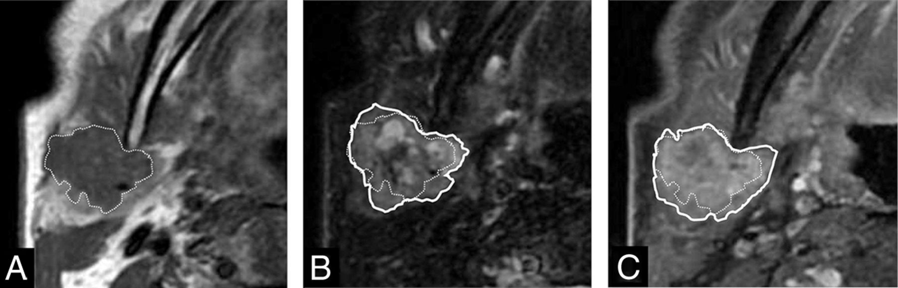
- FIG 1.
Corona sign on FS-T2WI and CE-FS-T1WI. Invasive CXPA (salivary duct carcinoma) of the right parotid gland in a 77-year-old man. MRI shows a homogeneous low-intensity tumor on axial T1WI (A), mixed high- and low-intensity signals on axial FS-T2WI (B), and irregularly enhanced signal on axial CE-FS-T1WI (C). The tumor size on FS-T2WI and CE-FS-T1WI (solid line) was larger than that on T1WI (dotted line). We defined these MRI findings as corona signs on FS-T2WI and CE-FS-T1WI.
- FIG 2.
Black ring sign. Invasive CXPA (high-grade mucoepidermoid carcinoma) of the right submandibular gland in a 76-year-old man. MRI shows a nodule with a thick low-intensity rim (A and B, arrows) and an intra-ring component with mixed high- and low-intensity signals on axial and coronal T2WI (A and B). We defined this MRI finding as the black ring sign.
- FIG 3.
MR imaging–pathology correlation: noninvasive type. Noninvasive CXPA (salivary duct carcinoma) of the left parotid gland in a 56-year-old man. MRI showed a heterogeneous high-intensity tumor on axial T2WI (A, arrow). The corona signs on FS-T2WI and CE-FS-T1WI and the black ring sign were absent. Photomicrograph shows ductal and myoepithelial cells in the chondromyxoid stroma. A part of the tumor contained ductal and myoepithelial cells with atypical hyperchromatic nuclei. The malignant component was completely surrounded by the fibrous capsule of the pre-existing pleomorphic adenoma (B, H&E, original magnification ×20). Note higher magnification of the malignant component (C, H&E, original magnification ×100).
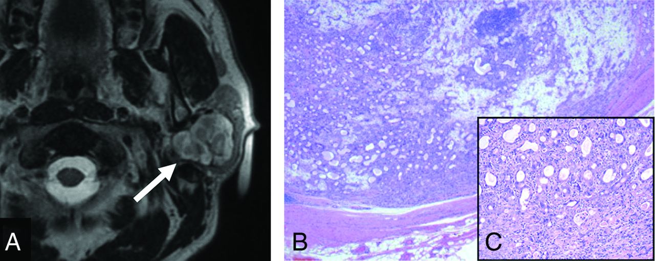
- FIG 4.
MR imaging–pathology correlation: invasive type. Invasive CXPA (undifferentiated carcinoma, large-cell type) of the right submandibular gland in a 72-year-old man. A huge mass replaced the right submandibular gland. MRI shows heterogeneous high intensity on T2WI and FS-T2WI (A and B, dotted arrows). Encapsulated nodules with thick low-intensity rims are present inside the tumor on axial T2WI and FS-T2WI (A and B, solid arrows). Macroscopic findings show a solid and white-yellow tumor with nodules in the nodule pattern. The nodule within the tumor corresponds to a black ring sign on MRI (C). Photomicrograph shows ductal and myoepithelial cells with atypical hyperchromatic nuclei (D, H&E, original magnification ×20). The malignant component invaded beyond the capsule (D, arrows) and infiltrated the surrounding fatty tissue. Most nodules within the tumor showed extensive hyalinization/fibrosis with myxoid stroma (D), and the black ring sign matched the hyalinization/fibrosis. The corona sign on FS-T2WI and CE-FS-T1WI reflects pathologically extracapsular tumor cells and/or inflammatory cells.
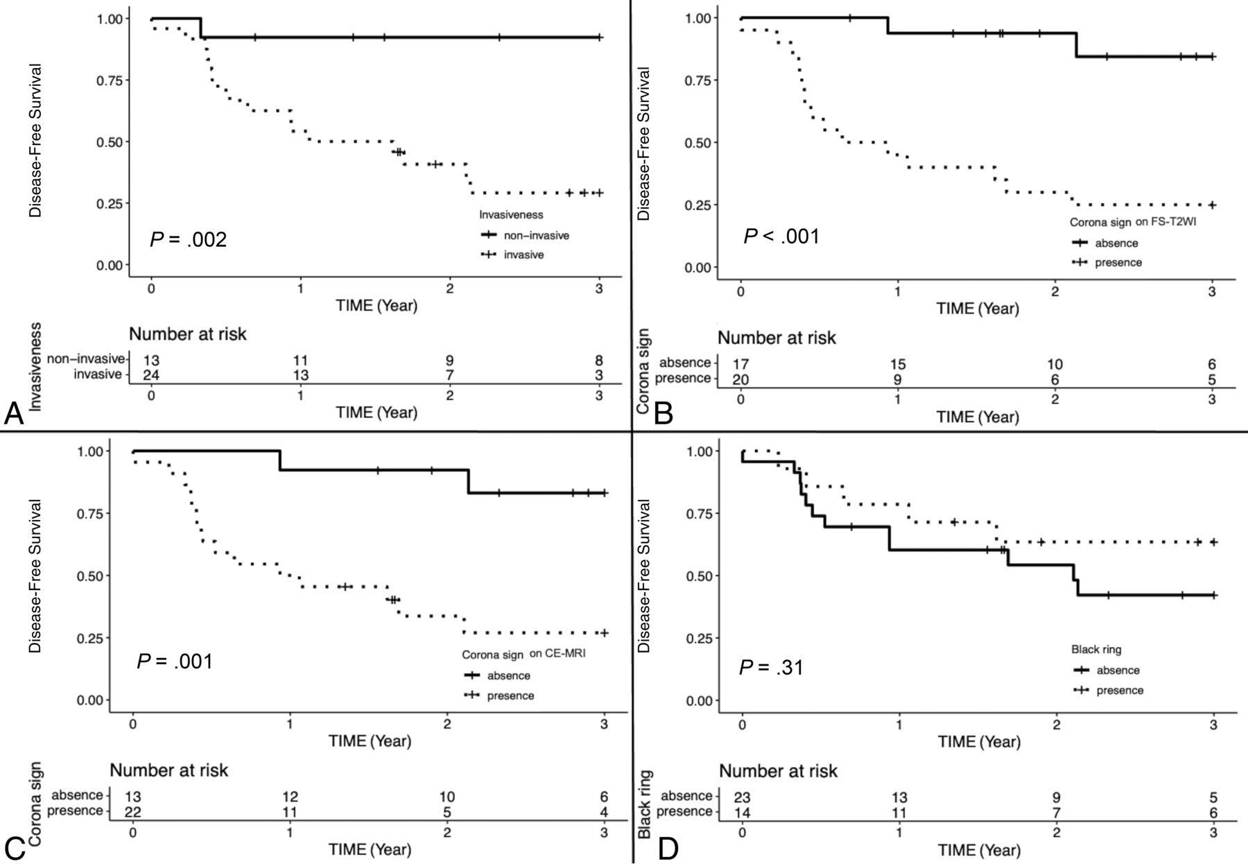
- FIG 5.
Kaplan-Meier disease-free survival curves and number-at-risk table of invasiveness (A), corona signs on FS-T2WI and CE-FS-T1WI (B and C), and the black ring sign (D).


Tables
Total Invasive Noninvasive P Value Age (mean) (yr) 64.7 (SD, 13.4) 65.7 (SD, 12.4) 62.7 (SD, 15.3) .30 Sex Male 26 19 7 .44 Female 11 6 5 Location Parotid gland 25 16 9 .68 Submandibular 7 6 1 Minor salivary 5 3 2 Laterality Left 17 9 8 .16 Right 20 16 4 Swelling Yes 32 20 12 .15 No 5 5 0 Pain Yes 13 10 3 .48 No 24 15 9 Infection Yes 2 2 0 1 No 35 23 12 Immobility Yes 12 10 2 .26 No 25 15 10 Nerve palsy Yes 6 6 0 .15 No 31 19 12 Skin infiltration Yes 1 1 0 1 No 36 24 12 Pathology Total Invasive Noninvasive P Value Salivary duct 18 11 7 .53 Myoepithelial 6 5 1 Adenocarcinoma 3 3 0 Squamous 3 1 2 Mucoepidermoid 2 2 0 Undifferentiated 1 1 0 Unknown 4 2 2 Total Invasive Noninvasive P Value OR 95% CI Border Totally ill-defined 2 2 0 .002 14.41 (2.23–171.2)a Partially ill-defined 19 17 2 Well-defined 16 6 10 Capsule None 9 9 0 <.001 38.18 (4.06–1956.7)b Partial 12 11 1 Total 16 5 11 Corona sign on FS-T2WI Present 21 19 2 .001 14.40 (2.23–171.2) Absent 16 6 10 Corona sign on CE-FS-T1WIc Present 22 19 3 .007 9.31 (1.55–76.4) Absent 13 5 8 Black ring sign Present 15 14 1 .011 13.11 (1.49–642.2) Absent 22 11 11 Rater 1 vs 2 Rater 1 vs 3 Rater 2 vs 3 Border 0.12 0.10 0.45 Capsule 0.19 0.38 0.16 Corona sign on FS-T2WI 0.78 0.79 0.67 Corona sign on CE-FS-T1WI 0.65 0.78 0.65 Black ring sign 0.89 0.84 0.84 ↵a Raters 1, 2, and 3 had 6, 10, and 18 years of experience, respectively.


Jump to section
Related Articles
Cited By...
- No citing articles found.