Advertisement
  • Main menu
  • User menu
  • Search
  • English ▼
    • English
    • Afrikaans
    • Albanian
    • Amharic
    • Arabic
    • Armenian
    • Azerbaijani
    • Basque
    • Belarusian
    • Bengali
    • Bosnian
    • Bulgarian
    • Catalan
    • Cebuano
    • Chichewa
    • Chinese (Simplified)
    • Chinese (Traditional)
    • Corsican
    • Croatian
    • Czech
    • Danish
    • Dutch
    • Esperanto
    • Estonian
    • Filipino
    • Finnish
    • French
    • Frisian
    • Galician
    • Georgian
    • German
    • Greek
    • Gujarati
    • Haitian Creole
    • Hausa
    • Hawaiian
    • Hebrew
    • Hindi
    • Hmong
    • Hungarian
    • Icelandic
    • Igbo
    • Indonesian
    • Irish
    • Italian
    • Japanese
    • Javanese
    • Kannada
    • Kazakh
    • Khmer
    • Korean
    • Kurdish (Kurmanji)
    • Kyrgyz
    • Lao
    • Latin
    • Latvian
    • Lithuanian
    • Luxembourgish
    • Macedonian
    • Malagasy
    • Malay
    • Malayalam
    • Maltese
    • Maori
    • Marathi
    • Mongolian
    • Myanmar (Burmese)
    • Nepali
    • Norwegian
    • Pashto
    • Persian
    • Polish
    • Portuguese
    • Punjabi
    • Romanian
    • Russian
    • Samoan
    • Scottish Gaelic
    • Serbian
    • Sesotho
    • Shona
    • Sindhi
    • Sinhala
    • Slovak
    • Slovenian
    • Somali
    • Spanish
    • Sudanese
    • Swahili
    • Swedish
    • Tajik
    • Tamil
    • Telugu
    • Thai
    • Turkish
    • Ukrainian
    • Urdu
    • Uzbek
    • Vietnamese
    • Welsh
    • Xhosa
    • Yiddish
    • Yoruba
    • Zulu

Main menu

  • Home
  • Content
    • Current Issue
    • Accepted Manuscripts
    • Article Preview
    • Past Issue Archive
    • Video Articles
    • AJNR Case Collection
    • Case of the Week Archive
    • Case of the Month Archive
    • Classic Case Archive
  • Special Collections
    • AJNR Awards
    • Low-Field MRI
    • Alzheimer Disease
    • ASNR Foundation Special Collection
    • Photon-Counting CT
    • View All
  • Multimedia
    • AJNR Podcasts
    • AJNR SCANtastic
    • Trainee Corner
    • MRI Safety Corner
    • Imaging Protocols
  • For Authors
    • Submit a Manuscript
    • Submit a Video Article
    • Submit an eLetter to the Editor/Response
    • Manuscript Submission Guidelines
    • Statistical Tips
    • Fast Publishing of Accepted Manuscripts
    • Graphical Abstract Preparation
    • Imaging Protocol Submission
    • Author Policies
  • About Us
    • About AJNR
    • Editorial Board
    • Editorial Board Alumni
  • More
    • Become a Reviewer/Academy of Reviewers
    • Subscribers
    • Permissions
    • Alerts
    • Feedback
    • Advertisers
    • ASNR Home

User menu

  • Alerts
  • Log in

Search

  • Advanced search
American Journal of Neuroradiology

American Journal of Neuroradiology

ASHNR American Society of Functional Neuroradiology ASHNR American Society of Pediatric Neuroradiology ASSR
  • Alerts
  • Log in

Advanced Search

English ▼
  • English
  • Afrikaans
  • Albanian
  • Amharic
  • Arabic
  • Armenian
  • Azerbaijani
  • Basque
  • Belarusian
  • Bengali
  • Bosnian
  • Bulgarian
  • Catalan
  • Cebuano
  • Chichewa
  • Chinese (Simplified)
  • Chinese (Traditional)
  • Corsican
  • Croatian
  • Czech
  • Danish
  • Dutch
  • Esperanto
  • Estonian
  • Filipino
  • Finnish
  • French
  • Frisian
  • Galician
  • Georgian
  • German
  • Greek
  • Gujarati
  • Haitian Creole
  • Hausa
  • Hawaiian
  • Hebrew
  • Hindi
  • Hmong
  • Hungarian
  • Icelandic
  • Igbo
  • Indonesian
  • Irish
  • Italian
  • Japanese
  • Javanese
  • Kannada
  • Kazakh
  • Khmer
  • Korean
  • Kurdish (Kurmanji)
  • Kyrgyz
  • Lao
  • Latin
  • Latvian
  • Lithuanian
  • Luxembourgish
  • Macedonian
  • Malagasy
  • Malay
  • Malayalam
  • Maltese
  • Maori
  • Marathi
  • Mongolian
  • Myanmar (Burmese)
  • Nepali
  • Norwegian
  • Pashto
  • Persian
  • Polish
  • Portuguese
  • Punjabi
  • Romanian
  • Russian
  • Samoan
  • Scottish Gaelic
  • Serbian
  • Sesotho
  • Shona
  • Sindhi
  • Sinhala
  • Slovak
  • Slovenian
  • Somali
  • Spanish
  • Sudanese
  • Swahili
  • Swedish
  • Tajik
  • Tamil
  • Telugu
  • Thai
  • Turkish
  • Ukrainian
  • Urdu
  • Uzbek
  • Vietnamese
  • Welsh
  • Xhosa
  • Yiddish
  • Yoruba
  • Zulu
  • Home
  • Content
    • Current Issue
    • Accepted Manuscripts
    • Article Preview
    • Past Issue Archive
    • Video Articles
    • AJNR Case Collection
    • Case of the Week Archive
    • Case of the Month Archive
    • Classic Case Archive
  • Special Collections
    • AJNR Awards
    • Low-Field MRI
    • Alzheimer Disease
    • ASNR Foundation Special Collection
    • Photon-Counting CT
    • View All
  • Multimedia
    • AJNR Podcasts
    • AJNR SCANtastic
    • Trainee Corner
    • MRI Safety Corner
    • Imaging Protocols
  • For Authors
    • Submit a Manuscript
    • Submit a Video Article
    • Submit an eLetter to the Editor/Response
    • Manuscript Submission Guidelines
    • Statistical Tips
    • Fast Publishing of Accepted Manuscripts
    • Graphical Abstract Preparation
    • Imaging Protocol Submission
    • Author Policies
  • About Us
    • About AJNR
    • Editorial Board
    • Editorial Board Alumni
  • More
    • Become a Reviewer/Academy of Reviewers
    • Subscribers
    • Permissions
    • Alerts
    • Feedback
    • Advertisers
    • ASNR Home
  • Follow AJNR on Twitter
  • Visit AJNR on Facebook
  • Follow AJNR on Instagram
  • Join AJNR on LinkedIn
  • RSS Feeds

AJNR Awards, New Junior Editors, and more. Read the latest AJNR updates

Research ArticleHead and Neck Imaging

Percutaneous CT-Guided Core Needle Biopsies of Head and Neck Masses: Review of 184 Cases at a Single Academic Institution, Common and Special Techniques, Diagnostic Yield, and Safety

R.W. Jordan, D.P. Shlapak, J.C. Benson, F.E. Diehn, D.K. Kim, V.T. Lehman, G.B. Liebo, A.A. Madhavan, J.M. Morris, P.P. Morris, J.T. Verdoorn and C.M. Carr
American Journal of Neuroradiology January 2022, 43 (1) 117-124; DOI: https://doi.org/10.3174/ajnr.A7348
R.W. Jordan
aFrom the Division of Neuroradiology, Department of Radiology, Mayo Clinic, Rochester, Minnesota
  • Find this author on Google Scholar
  • Find this author on PubMed
  • Search for this author on this site
  • ORCID record for R.W. Jordan
D.P. Shlapak
aFrom the Division of Neuroradiology, Department of Radiology, Mayo Clinic, Rochester, Minnesota
  • Find this author on Google Scholar
  • Find this author on PubMed
  • Search for this author on this site
  • ORCID record for D.P. Shlapak
J.C. Benson
aFrom the Division of Neuroradiology, Department of Radiology, Mayo Clinic, Rochester, Minnesota
  • Find this author on Google Scholar
  • Find this author on PubMed
  • Search for this author on this site
  • ORCID record for J.C. Benson
F.E. Diehn
aFrom the Division of Neuroradiology, Department of Radiology, Mayo Clinic, Rochester, Minnesota
  • Find this author on Google Scholar
  • Find this author on PubMed
  • Search for this author on this site
  • ORCID record for F.E. Diehn
D.K. Kim
aFrom the Division of Neuroradiology, Department of Radiology, Mayo Clinic, Rochester, Minnesota
  • Find this author on Google Scholar
  • Find this author on PubMed
  • Search for this author on this site
  • ORCID record for D.K. Kim
V.T. Lehman
aFrom the Division of Neuroradiology, Department of Radiology, Mayo Clinic, Rochester, Minnesota
  • Find this author on Google Scholar
  • Find this author on PubMed
  • Search for this author on this site
  • ORCID record for V.T. Lehman
G.B. Liebo
aFrom the Division of Neuroradiology, Department of Radiology, Mayo Clinic, Rochester, Minnesota
  • Find this author on Google Scholar
  • Find this author on PubMed
  • Search for this author on this site
  • ORCID record for G.B. Liebo
A.A. Madhavan
aFrom the Division of Neuroradiology, Department of Radiology, Mayo Clinic, Rochester, Minnesota
  • Find this author on Google Scholar
  • Find this author on PubMed
  • Search for this author on this site
  • ORCID record for A.A. Madhavan
J.M. Morris
aFrom the Division of Neuroradiology, Department of Radiology, Mayo Clinic, Rochester, Minnesota
  • Find this author on Google Scholar
  • Find this author on PubMed
  • Search for this author on this site
  • ORCID record for J.M. Morris
P.P. Morris
aFrom the Division of Neuroradiology, Department of Radiology, Mayo Clinic, Rochester, Minnesota
  • Find this author on Google Scholar
  • Find this author on PubMed
  • Search for this author on this site
  • ORCID record for P.P. Morris
J.T. Verdoorn
aFrom the Division of Neuroradiology, Department of Radiology, Mayo Clinic, Rochester, Minnesota
  • Find this author on Google Scholar
  • Find this author on PubMed
  • Search for this author on this site
  • ORCID record for J.T. Verdoorn
C.M. Carr
aFrom the Division of Neuroradiology, Department of Radiology, Mayo Clinic, Rochester, Minnesota
  • Find this author on Google Scholar
  • Find this author on PubMed
  • Search for this author on this site
  • ORCID record for C.M. Carr
  • Article
  • Figures & Data
  • Supplemental
  • Info & Metrics
  • Responses
  • References
  • PDF
Loading

Article Figures & Data

Figures

  • Tables
  • FIG 1.
    • Download figure
    • Open in new tab
    • Download powerpoint
    FIG 1.

    Common biopsy approaches to H&N lesions at different levels of the suprahyoid (A) and infrahyoid neck (B). Note that to avoid the parotid gland, one should use a retromandibular approach for more inferior retropharyngeal, parapharyngeal, pharyngeal mucosal, and masticator space lesions; more superior lesions are better accessed via paramaxillary and subzygomatic approaches. Reprinted with permission of Mayo Foundation for Medical Education and Research, all rights reserved. PG indicates parotid gland; T, tumor; LT, lingual tonsil.
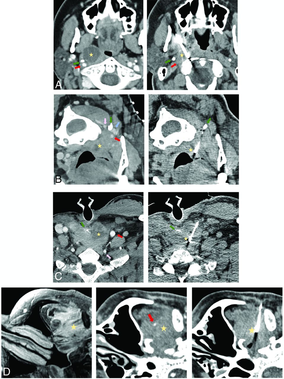
  • FIG 2.
    • Download figure
    • Open in new tab
    • Download powerpoint
    FIG 2.
    • Download figure
    • Open in new tab
    • Download powerpoint
    FIG 2.

    Common approaches used in the current series biopsy. A, A paramaxillary approach targeting a 1.9-cm partially cystic right retropharyngeal lymph node (yellow star). Left, Preprocedural contrast-enhanced CT (venous phase) to better visualize the right ICA (red arrow) and better assess lesion margins due to peripheral enhancement. Right, Intraprocedural CT with a 14-ga needle trough through the lesion (part of the cystic component was initially aspirated). The styloid process (green arrow) is a useful landmark because it is immediately anterior to the ICA. Recurrent human papillomavirus (HPV) + SCC. This approach is commonly used to access retropharyngeal, parapharyngeal, pharyngeal mucosal, masticator, and deep parotid space lesions. B, A retromandibular approach in a patient in a prone position targeting a 1.1-cm partially cystic left retropharyngeal lymph node (yellow star). Left, Contrast-enhanced CT (venous phase) to better visualize the left ICA (red arrow), vertebral artery (pink arrow), and internal jugular (IJ) vein (blue arrow) and highlight more solid lesion portions. Right, Intraprocedural CT with a 16-ga needle trough through the solid portion of the lesion. The C1 transverse process (green arrow) is a useful landmark because it “marks a safe pass” between the vertebral artery posterior to the needle and the ICA/IJ vein anteriorly. Recurrent HPV + SCC. This approach can be used for more inferior retropharyngeal, parapharyngeal, pharyngeal mucosal, masticator, and deep parotid lesions, as well as sublingual, submandibular, perivertebral, and carotid space lesions. C, Anterior approach targeting a 3.4-cm partially cystic nodule at the tracheoesophageal groove (yellow star). Left, Preprocedural contrast-enhanced CT (venous phase) to opacify the left common carotid artery (red arrow) and vertebral artery (pink arrow) and delineate the solid component of the target lesion. Right, Intraprocedural CT with an 18-ga needle trough through the solid component of the lesion. A partially visualized esophageal stent (green arrow) is a useful landmark showing the approximate location of the infiltrated esophagus, lateral to the stent. Recurrent SCC. This approach is used with a neutral supine position for more midline anterior neck lesions such as the visceral space, level VI, and the supraclavicular lymph nodes. D, A subzygomatic approach targeting an infiltrating left masticator space mass seen on MR imaging (left image, yellow star). Middle, Preprocedural contrast-enhanced CT (venous phase) to opacify the left internal maxillary artery (red arrow). Right, Intraprocedural CT with an 18-ga needle trough through the infiltrating lesion with the biopsy device clearly posterior to the expected location of the internal maxillary artery in correlation with the middle image. Recurrent SCC with perineural spread. By far, masticator space lesions are the most common targets for this approach.
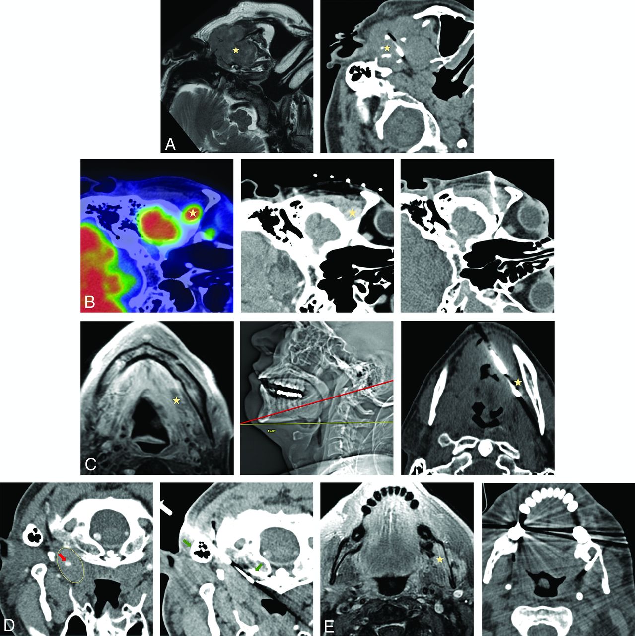
  • FIG 3.
    • Download figure
    • Open in new tab
    • Download powerpoint
    FIG 3.

    Less commonly used biopsy approaches. A, Preauricular approach targeting a 4.2-cm expansile left mandibular lesion with an extraosseous soft-tissue component on MR imaging (left image, yellow star). No preprocedural contrast was administered given the size of the lesion and the lack of proximity of the crucial neurovascular structures. Right, Intraprocedural CT with a 16-ga needle trough through the lesion. Plasmacytoma. This approach is used for superficial lesions in the masticator or superficial parotid space, particularly when a zygoma is partially destructed. B, A suprazygomatic approach targeting hypermetabolic on PET/CT 1.5 -cm right suprazygomatic masticator mass (left image, yellow star). Middle, Preprocedural contrast-enhanced CT (venous phase) to delineate the lesion (yellow star) that enhances more than background tissues. Right, Intraprocedural CT with a 16-ga needle trough through the expected location of the lesion in correlation with bony landmarks and previous contrast enhancement on the middle image. Recurrent adenoid cystic carcinoma. This approach is essentially exclusively used for superficial suprazygomatic masticator space (temporal fossa) lesions. C, Submental approach targeting diffusely heterogeneously enhancing on MR imaging left sublingual/floor of the mouth soft-tissue (left image, yellow star). Middle, Intraprocedural scout CT image showing patient positioning and a cranial gantry tilt of 15° (red line, biopsy plane after 15° gantry tilt) to bypass a mental protuberance. Right, Intraprocedural axial CT with a 14-ga needle trough through the expected location of the target (yellow star). Preprocedural contrast was not administered due to regional vasculature paucity in correlation with prior imaging. Recurrent adenoid cystic carcinoma. Sublingual space and floor of the mouth lesions can be accessed via this approach; to avoid the mental protuberance, cranial gantry tilt and/or substantial neck extension might be needed. D, A retromastoid approach with the patient in the prone position targeting a 2.6-cm left retropharyngeal mass (left image, yellow ellipse), encasing the ICA on preprocedural limited CTA (red arrow). Right, intraprocedural CT with a 16-ga needle advanced between mastoid tip and the C1 anterior arch (right image, green arrows); these osseous landmarks align a safe trajectory into the lesion portion posterior to the ICA in correlation with preprocedural CTA. Recurrent human papillomavirus + SCC. This approach can be used for more lateral retropharyngeal and anterior perivertebral lesions. E, A transosseous approach targeting an enhancing 2.5-cm left masticator mass expanding the mandibular canal on MR imaging (left image, yellow star). Right, Intraprocedural CT scan with a 16-ga needle traversing the buccal cortex of the mandibular ramus. Preprocedural contrast was not administered given lack of critical regional vasculature in correlation with MR imaging. Recurrent adenoid cystic carcinoma with inferior alveolar perineural spread. This approach can be used to access lesions along the inner cortex of the mandible and zygoma.

  • FIG 4.
    • Download figure
    • Open in new tab
    • Download powerpoint
    FIG 4.

    A, PET/CT with hypermetabolic left retropharyngeal 6-mm lymph node (yellow arrow). B, This lesion is centrally cystic/necrotic on follow-up contrast-enhanced CT (yellow arrow) and immediately medial to the ICA (red arrow). C, Intraprocedural CT without contrast with an 18-ga needle advanced via a paramaxillary approach anterior to the target retropharyngeal lymph node (yellow arrow) and medial to the expected location of the unopacified ICA (red arrow). Before further needle advancement, 60 mL of Omnipaque-300 was slowly infused at a rate of 0.6 mL/s to continuously opacify the ICA (red arrows) for adequate visualization before (D) and during (E) lesion sampling (yellow arrows). Pathology reported poorly differentiated metastatic carcinoma in a patient with a history of oropharyngeal human papillomavirus + SCC.

Tables

  • Figures
    • View popup
    Table 1:

    Biopsy approaches

    ApproachNo.%
    Paramaxillarya4323%
    Retromandibulara3016%
    Anteriora2413%
    Subzygomatica2111%
    Submandibulara2011%
    Posteriora1810%
    Laterala148%
    Preauricular53%
    Submental42%
    Suprazygomatic32%
    Retromastoid11%
    Transosseous11%
    Total184100%
    • ↵a More common techniques.

    • View popup
    Table 2:

    Target lesion space

    Head and Neck SpaceNo.%
    Masticator3620%
    Parotid2413%
    Cervical lymph nodesa2313%
    Retropharyngeal2313%
    Sublingual1910%
    Perivertebral148%
    Pharyngeal mucosal137%
    Visceralb95%
    Parapharyngeal63%
    Carotid63%
    Submandibular42%
    Otherc42%
    Transpatial32%
    Total184100%
    • ↵a Cervical lymph node levels II–VI and supraclavicular.

    • ↵b Biopsies involving the supraglottic larynx, glottis, subglottic larynx, tracheoesophageal groove, and thyroid gland.

    • ↵c Biopsies of the periorbital region, anterior maxilla, preauricular, postauricular, and intramuscular regions.

    • View popup
    Table 3:

    Follow-up of “negative for malignancy” biopsies

    Patients’ Characteristics and Lesion ManagementNo.%
    Initially “negative for malignancy” biopsies43/18423%
    Patients with a known malignancy29/4368%
     Rebiopsied/excised7/2920%
     Ultimately +malignancy4/757%
     Stable/resolved (mean f/u time: 32 mo, 2–120 mo)17/2959%
     No f/u5/2917%
    Patients without known malignancy14/4332%
     Rebiopsied/excised5/1436%
     Ultimately + malignancy0/50%
     Stable/resolved (mean f/u time: 25.6 mo, 1–72 mo)7/1450%
     No f/u2/1414%
    Total false-negative rate4/1842%
    Total diagnostic yield167/18491%
    • Note:—f/u indicates follow-up.

PreviousNext
Back to top

In this issue

American Journal of Neuroradiology: 43 (1)
American Journal of Neuroradiology
Vol. 43, Issue 1
1 Jan 2022
  • Table of Contents
  • Index by author
  • Complete Issue (PDF)
Advertisement
Print
Download PDF
Email Article
Cite this article
0 Responses
Respond to this article
Share
Bookmark this article
Percutaneous CT-Guided Core Needle Biopsies of Head and Neck Masses: Review of 184 Cases at a Single Academic Institution, Common and Special Techniques, Diagnostic Yield, and Safety
R.W. Jordan, D.P. Shlapak, J.C. Benson, F.E. Diehn, D.K. Kim, V.T. Lehman, G.B. Liebo, A.A. Madhavan, J.M. Morris, P.P. Morris, J.T. Verdoorn, C.M. Carr
American Journal of Neuroradiology Jan 2022, 43 (1) 117-124; DOI: 10.3174/ajnr.A7348
del.icio.us logo Twitter logo Facebook logo Mendeley logo
  • Tweet Widget
Purchase

Jump to section

  • Article
    • Abstract
    • ABBREVIATIONS:
    • MATERIALS AND METHODS
    • RESULTS
    • DISCUSSION
    • CONCLUSIONS
    • Footnotes
    • References
  • Figures & Data
  • Supplemental
  • Info & Metrics
  • Responses
  • References
  • PDF

Related Articles

  • PubMed
  • Google Scholar

Cited By...

  • Ultrasound Fusion-Guided Core Needle Biopsy for Deep Head and Neck Space Lesions: Technical Feasibility, Histopathologic Yield, and Safety
  • Crossref
  • Google Scholar

This article has not yet been cited by articles in journals that are participating in Crossref Cited-by Linking.

More in this TOC Section

Head and Neck Imaging

  • Hydrops Herniation into the Semicircular Canals
  • ASL Sensitivity for Head and Neck Paraganglioma
  • Post SRS Peritumoral Hyperintense Signal of VSs
Show more Head and Neck Imaging

Neurointervention

  • Cavernous dural AVF treated by transfacial route
  • A Retrospective Study in Tentorial DAVFs
  • Proximal Protection Devices for Carotid Stenting
Show more Neurointervention

Similar Articles

Advertisement

Indexed Content

  • Current Issue
  • Accepted Manuscripts
  • Article Preview
  • Past Issues
  • Editorials
  • Editor's Choice
  • Fellows' Journal Club
  • Letters to the Editor
  • Video Articles

Cases

  • Case Collection
  • Archive - Case of the Week
  • Archive - Case of the Month
  • Archive - Classic Case

More from AJNR

  • Trainee Corner
  • Imaging Protocols
  • MRI Safety Corner
  • Book Reviews

Multimedia

  • AJNR Podcasts
  • AJNR Scantastics

Resources

  • Turnaround Time
  • Submit a Manuscript
  • Submit a Video Article
  • Submit an eLetter to the Editor/Response
  • Manuscript Submission Guidelines
  • Statistical Tips
  • Fast Publishing of Accepted Manuscripts
  • Graphical Abstract Preparation
  • Imaging Protocol Submission
  • Evidence-Based Medicine Level Guide
  • Publishing Checklists
  • Author Policies
  • Become a Reviewer/Academy of Reviewers
  • News and Updates

About Us

  • About AJNR
  • Editorial Board
  • Editorial Board Alumni
  • Alerts
  • Permissions
  • Not an AJNR Subscriber? Join Now
  • Advertise with Us
  • Librarian Resources
  • Feedback
  • Terms and Conditions
  • AJNR Editorial Board Alumni

American Society of Neuroradiology

  • Not an ASNR Member? Join Now

© 2025 by the American Society of Neuroradiology All rights, including for text and data mining, AI training, and similar technologies, are reserved.
Print ISSN: 0195-6108 Online ISSN: 1936-959X

Powered by HighWire
Email this Article

Thank you for your interest in spreading the word on American Journal of Neuroradiology.

NOTE: We only request your email address so that the person you are recommending the page to knows that you wanted them to see it, and that it is not junk mail. We do not capture any email address.

Enter multiple addresses on separate lines or separate them with commas.
Percutaneous CT-Guided Core Needle Biopsies of Head and Neck Masses: Review of 184 Cases at a Single Academic Institution, Common and Special Techniques, Diagnostic Yield, and Safety
(Your Name) has sent you a message from American Journal of Neuroradiology
(Your Name) thought you would like to see the American Journal of Neuroradiology web site.
CAPTCHA
This question is for testing whether or not you are a human visitor and to prevent automated spam submissions.
Cite this article
R.W. Jordan, D.P. Shlapak, J.C. Benson, F.E. Diehn, D.K. Kim, V.T. Lehman, G.B. Liebo, A.A. Madhavan, J.M. Morris, P.P. Morris, J.T. Verdoorn, C.M. Carr
Percutaneous CT-Guided Core Needle Biopsies of Head and Neck Masses: Review of 184 Cases at a Single Academic Institution, Common and Special Techniques, Diagnostic Yield, and Safety
American Journal of Neuroradiology Jan 2022, 43 (1) 117-124; DOI: 10.3174/ajnr.A7348

Citation Manager Formats

  • BibTeX
  • Bookends
  • EasyBib
  • EndNote (tagged)
  • EndNote 8 (xml)
  • Medlars
  • Mendeley
  • Papers
  • RefWorks Tagged
  • Ref Manager
  • RIS
  • Zotero

We use cookies on this site to enhance your user experience. By clicking any link on this page you are giving your consent for us to set cookies.