Advertisement

Main menu

  • Home
  • Content
    • Current Issue
    • Accepted Manuscripts
    • Article Preview
    • Past Issue Archive
    • Video Articles
    • AJNR Case Collection
    • Case of the Week Archive
    • Case of the Month Archive
    • Classic Case Archive
  • Special Collections
    • AJNR Awards
    • Low-Field MRI
    • Alzheimer Disease
    • ASNR Foundation Special Collection
    • Photon-Counting CT
    • View All
  • Multimedia
    • AJNR Podcasts
    • AJNR SCANtastic
    • Trainee Corner
    • MRI Safety Corner
    • Imaging Protocols
  • For Authors
    • Submit a Manuscript
    • Submit a Video Article
    • Submit an eLetter to the Editor/Response
    • Manuscript Submission Guidelines
    • Statistical Tips
    • Fast Publishing of Accepted Manuscripts
    • Graphical Abstract Preparation
    • Imaging Protocol Submission
    • Author Policies
  • About Us
    • About AJNR
    • Editorial Board
    • Editorial Board Alumni
  • More
    • Become a Reviewer/Academy of Reviewers
    • Subscribers
    • Permissions
    • Alerts
    • Feedback
    • Advertisers
    • ASNR Home

User menu

  • Alerts
  • Log in

Search

  • Advanced search
American Journal of Neuroradiology

American Journal of Neuroradiology

ASHNR American Society of Functional Neuroradiology ASHNR American Society of Pediatric Neuroradiology ASSR
  • Alerts
  • Log in

Advanced Search

  • Home
  • Content
    • Current Issue
    • Accepted Manuscripts
    • Article Preview
    • Past Issue Archive
    • Video Articles
    • AJNR Case Collection
    • Case of the Week Archive
    • Case of the Month Archive
    • Classic Case Archive
  • Special Collections
    • AJNR Awards
    • Low-Field MRI
    • Alzheimer Disease
    • ASNR Foundation Special Collection
    • Photon-Counting CT
    • View All
  • Multimedia
    • AJNR Podcasts
    • AJNR SCANtastic
    • Trainee Corner
    • MRI Safety Corner
    • Imaging Protocols
  • For Authors
    • Submit a Manuscript
    • Submit a Video Article
    • Submit an eLetter to the Editor/Response
    • Manuscript Submission Guidelines
    • Statistical Tips
    • Fast Publishing of Accepted Manuscripts
    • Graphical Abstract Preparation
    • Imaging Protocol Submission
    • Author Policies
  • About Us
    • About AJNR
    • Editorial Board
    • Editorial Board Alumni
  • More
    • Become a Reviewer/Academy of Reviewers
    • Subscribers
    • Permissions
    • Alerts
    • Feedback
    • Advertisers
    • ASNR Home
  • Follow AJNR on Twitter
  • Visit AJNR on Facebook
  • Follow AJNR on Instagram
  • Join AJNR on LinkedIn
  • RSS Feeds

AJNR Awards, New Junior Editors, and more. Read the latest AJNR updates

LetterLetter

Regarding “CTA Evaluation of Basilar Septations: An Entity Better Characterized as Aberrant Basilar Fenestrations”

J. Hynes and E. Kavanagh
American Journal of Neuroradiology September 2021, 42 (9) E71-E73; DOI: https://doi.org/10.3174/ajnr.A7206
J. Hynes
aDepartment of RadiologyMater Misericordiae University HospitalDublin, Ireland
  • Find this author on Google Scholar
  • Find this author on PubMed
  • Search for this author on this site
  • ORCID record for J. Hynes
E. Kavanagh
bDepartment of RadiologyMater Misericordiae University HospitalDublin, Ireland
cDepartment of RadiologyCappagh National Orthopaedic HospitalDublin, Ireland
dUniversity College DublinBelfield, Ireland
  • Find this author on Google Scholar
  • Find this author on PubMed
  • Search for this author on this site
  • ORCID record for E. Kavanagh
  • Article
  • Figures & Data
  • Info & Metrics
  • Responses
  • References
  • PDF
Loading

We read with great interest the recent article by Small et al,1 “CTA Evaluation of Basilar Septations: An Entity Better Characterized as Aberrant Basilar Fenestrations,” a retrospective review describing luminal abnormalities of the basilar artery. We wish to highlight a recent case at our institution that we believe constitutes an interesting example of a further variation in the luminal morphology of the basilar artery.

A 71-year-old male patient presented with transient sensory disturbance of the right upper limb, clinically considered to be suggestive of a transient ischemic attack. The patient underwent noncontrast CT of the head, CT angiography of the head and neck, and subsequently MR imaging of the brain. Imaging did not demonstrate an acute infarct or vascular occlusion; however, note was made of abnormal morphology of the vertebrobasilar system with what was interpreted as a small communicating vessel joining the distal V4 segments of the vertebral arteries (Fig 1). It is possible, however, that this, in fact, represents a further transversely orientated variant of basilar fenestration morphology, not conforming directly to the classification described by Small et al. We have been unable to identify additional previously described examples of this in the literature.2⇓-4 Incidentally,the patient’s imaging also demonstrated a right-sided persistent trigeminal artery (Fig 2).

FIG 1.
  • Download figure
  • Open in new tab
  • Download powerpoint
FIG 1.

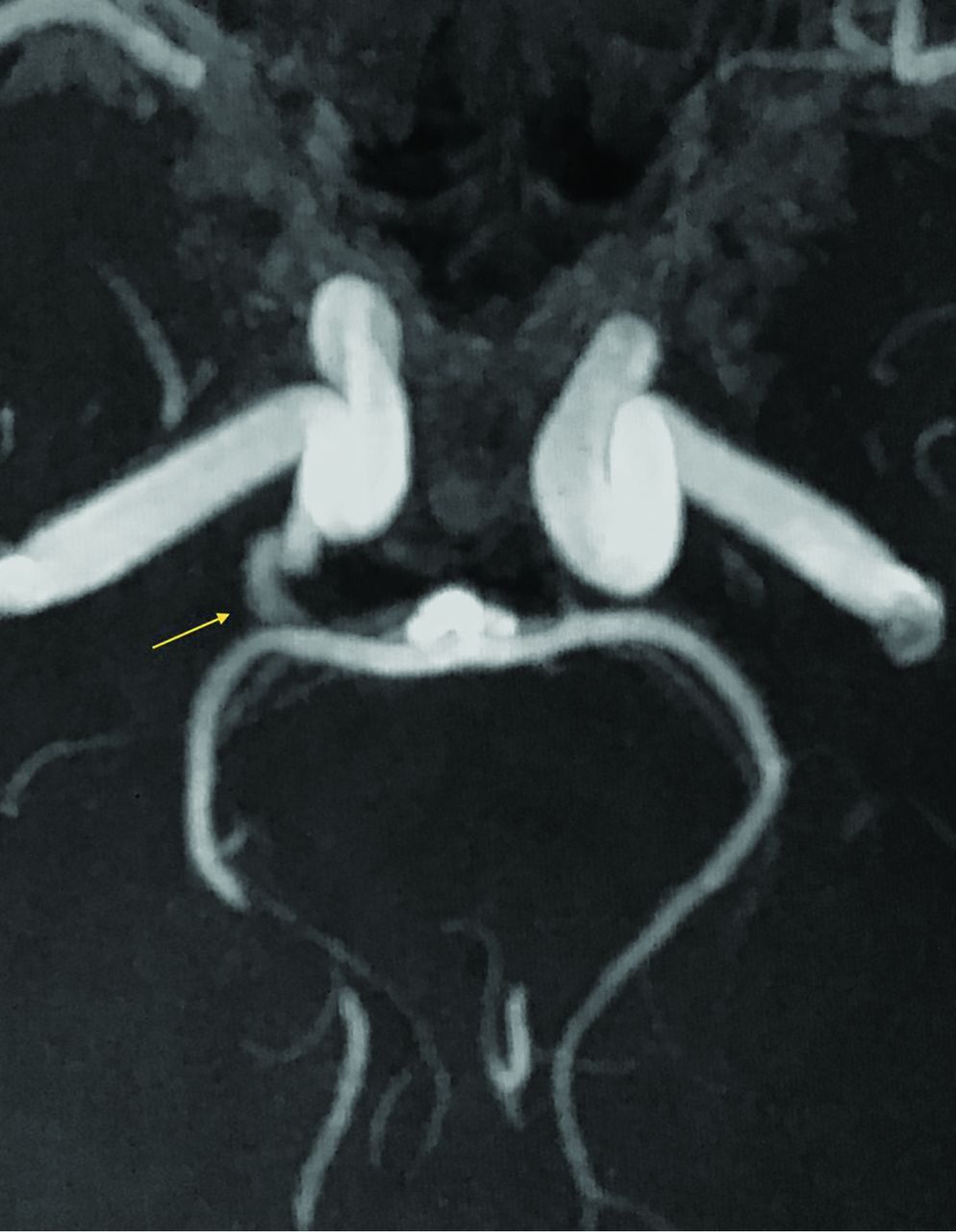
MIP TOF-MRA image demonstrates a small vascular communication (arrow) between the distal vertebral arteries.

FIG 2.
  • Download figure
  • Open in new tab
  • Download powerpoint
FIG 2.

MIP TOF-MRA image demonstrates a right-sided persistent trigeminal artery (arrow).

References

  1. 1.↵
    1. Small JE,
    2. Macey MB,
    3. Wakhloo AK, et al
    . CTA evaluation of basilar septations: an entity better characterized as aberrant basilar fenestrations. AJNR Am J Neuroradiol 2021 Feb 18. [Epub ahead of print] doi:10.3174/ajnr.A7008 pmid:33602748
    CrossRefPubMed
  2. 2.↵
    1. Pérez-Carrillo GJ,
    2. Hogg JP
    . Intracranial vascular lesions and anatomical variants all residents should know. Curr Probl Diagn Radiol 2010;39:91–109 doi:10.1067/j.cpradiol.2009.07.005 pmid:20307787
    CrossRefPubMed
  3. 3.↵
    1. Case D,
    2. Kumpe D,
    3. Roark C, et al
    . Neuroangiography: review of anatomy, periprocedural management, technique, and tips. Semin Intervent Radiol 2020;37:166–74 doi:10.1055/s-0040-1709171 pmid:32419729
    CrossRefPubMed
  4. 4.↵
    1. Thurlow PC,
    2. Andrus JM,
    3. Wholey MH
    . Common cervical and cerebral vascular variants. Interv Cardiol Clin 2014;3:123–34 doi:10.1016/j.iccl.2013.09.002 pmid:28582148
    CrossRefPubMed
  • © 2021 by American Journal of Neuroradiology
PreviousNext
Back to top

In this issue

American Journal of Neuroradiology: 42 (9)
American Journal of Neuroradiology
Vol. 42, Issue 9
1 Sep 2021
  • Table of Contents
  • Index by author
  • Complete Issue (PDF)
Advertisement
Print
Download PDF
Email Article

Thank you for your interest in spreading the word on American Journal of Neuroradiology.

NOTE: We only request your email address so that the person you are recommending the page to knows that you wanted them to see it, and that it is not junk mail. We do not capture any email address.

Enter multiple addresses on separate lines or separate them with commas.
Regarding “CTA Evaluation of Basilar Septations: An Entity Better Characterized as Aberrant Basilar Fenestrations”
(Your Name) has sent you a message from American Journal of Neuroradiology
(Your Name) thought you would like to see the American Journal of Neuroradiology web site.
CAPTCHA
This question is for testing whether or not you are a human visitor and to prevent automated spam submissions.
Cite this article
J. Hynes, E. Kavanagh
Regarding “CTA Evaluation of Basilar Septations: An Entity Better Characterized as Aberrant Basilar Fenestrations”
American Journal of Neuroradiology Sep 2021, 42 (9) E71-E73; DOI: 10.3174/ajnr.A7206

Citation Manager Formats

  • BibTeX
  • Bookends
  • EasyBib
  • EndNote (tagged)
  • EndNote 8 (xml)
  • Medlars
  • Mendeley
  • Papers
  • RefWorks Tagged
  • Ref Manager
  • RIS
  • Zotero
0 Responses
Respond to this article
Share
Bookmark this article
Regarding “CTA Evaluation of Basilar Septations: An Entity Better Characterized as Aberrant Basilar Fenestrations”
J. Hynes, E. Kavanagh
American Journal of Neuroradiology Sep 2021, 42 (9) E71-E73; DOI: 10.3174/ajnr.A7206
del.icio.us logo Twitter logo Facebook logo Mendeley logo
  • Tweet Widget
  • Facebook Like
  • Google Plus One
Purchase

Jump to section

  • Article
    • References
  • Figures & Data
  • Info & Metrics
  • Responses
  • References
  • PDF

Related Articles

  • No related articles found.
  • PubMed
  • Google Scholar

Cited By...

  • No citing articles found.
  • Crossref
  • Google Scholar

This article has not yet been cited by articles in journals that are participating in Crossref Cited-by Linking.

More in this TOC Section

  • Letter to the Editor regarding “Automated Volumetric Software in Dementia: Help or Hindrance to the Neuroradiologist?”
  • Reply:
  • Brain AVM’s Nidus: What if We Hadn’t Understood Anything?
Show more LETTERS

Similar Articles

Advertisement

Indexed Content

  • Current Issue
  • Accepted Manuscripts
  • Article Preview
  • Past Issues
  • Editorials
  • Editor's Choice
  • Fellows' Journal Club
  • Letters to the Editor
  • Video Articles

Cases

  • Case Collection
  • Archive - Case of the Week
  • Archive - Case of the Month
  • Archive - Classic Case

More from AJNR

  • Trainee Corner
  • Imaging Protocols
  • MRI Safety Corner
  • Book Reviews

Multimedia

  • AJNR Podcasts
  • AJNR Scantastics

Resources

  • Turnaround Time
  • Submit a Manuscript
  • Submit a Video Article
  • Submit an eLetter to the Editor/Response
  • Manuscript Submission Guidelines
  • Statistical Tips
  • Fast Publishing of Accepted Manuscripts
  • Graphical Abstract Preparation
  • Imaging Protocol Submission
  • Evidence-Based Medicine Level Guide
  • Publishing Checklists
  • Author Policies
  • Become a Reviewer/Academy of Reviewers
  • News and Updates

About Us

  • About AJNR
  • Editorial Board
  • Editorial Board Alumni
  • Alerts
  • Permissions
  • Not an AJNR Subscriber? Join Now
  • Advertise with Us
  • Librarian Resources
  • Feedback
  • Terms and Conditions
  • AJNR Editorial Board Alumni

American Society of Neuroradiology

  • Not an ASNR Member? Join Now

© 2025 by the American Society of Neuroradiology All rights, including for text and data mining, AI training, and similar technologies, are reserved.
Print ISSN: 0195-6108 Online ISSN: 1936-959X

Powered by HighWire