Advertisement

Main menu

  • Home
  • Content
    • Current Issue
    • Accepted Manuscripts
    • Article Preview
    • Past Issue Archive
    • Video Articles
    • AJNR Case Collection
    • Case of the Week Archive
    • Case of the Month Archive
    • Classic Case Archive
  • Special Collections
    • AJNR Awards
    • Low-Field MRI
    • Alzheimer Disease
    • ASNR Foundation Special Collection
    • Photon-Counting CT
    • View All
  • Multimedia
    • AJNR Podcasts
    • AJNR SCANtastic
    • Trainee Corner
    • MRI Safety Corner
    • Imaging Protocols
  • For Authors
    • Submit a Manuscript
    • Submit a Video Article
    • Submit an eLetter to the Editor/Response
    • Manuscript Submission Guidelines
    • Statistical Tips
    • Fast Publishing of Accepted Manuscripts
    • Graphical Abstract Preparation
    • Imaging Protocol Submission
    • Author Policies
  • About Us
    • About AJNR
    • Editorial Board
    • Editorial Board Alumni
  • More
    • Become a Reviewer/Academy of Reviewers
    • Subscribers
    • Permissions
    • Alerts
    • Feedback
    • Advertisers
    • ASNR Home

User menu

  • Alerts
  • Log in

Search

  • Advanced search
American Journal of Neuroradiology

American Journal of Neuroradiology

ASHNR American Society of Functional Neuroradiology ASHNR American Society of Pediatric Neuroradiology ASSR
  • Alerts
  • Log in

Advanced Search

  • Home
  • Content
    • Current Issue
    • Accepted Manuscripts
    • Article Preview
    • Past Issue Archive
    • Video Articles
    • AJNR Case Collection
    • Case of the Week Archive
    • Case of the Month Archive
    • Classic Case Archive
  • Special Collections
    • AJNR Awards
    • Low-Field MRI
    • Alzheimer Disease
    • ASNR Foundation Special Collection
    • Photon-Counting CT
    • View All
  • Multimedia
    • AJNR Podcasts
    • AJNR SCANtastic
    • Trainee Corner
    • MRI Safety Corner
    • Imaging Protocols
  • For Authors
    • Submit a Manuscript
    • Submit a Video Article
    • Submit an eLetter to the Editor/Response
    • Manuscript Submission Guidelines
    • Statistical Tips
    • Fast Publishing of Accepted Manuscripts
    • Graphical Abstract Preparation
    • Imaging Protocol Submission
    • Author Policies
  • About Us
    • About AJNR
    • Editorial Board
    • Editorial Board Alumni
  • More
    • Become a Reviewer/Academy of Reviewers
    • Subscribers
    • Permissions
    • Alerts
    • Feedback
    • Advertisers
    • ASNR Home
  • Follow AJNR on Twitter
  • Visit AJNR on Facebook
  • Follow AJNR on Instagram
  • Join AJNR on LinkedIn
  • RSS Feeds

AJNR Awards, New Junior Editors, and more. Read the latest AJNR updates

LetterLetter

Susceptibility Artifacts in the Anterior Cranial Fossa Mimicking Hemorrhage in Patients with Anosmia

J.J. Manov, M. Alfonso and G. Saigal
American Journal of Neuroradiology September 2021, 42 (9) E64-E65; DOI: https://doi.org/10.3174/ajnr.A7184
J.J. Manov
aDepartment of RadiologyUniversity of Miami Miller School of MedicineMiami, Florida
  • Find this author on Google Scholar
  • Find this author on PubMed
  • Search for this author on this site
  • ORCID record for J.J. Manov
M. Alfonso
aDepartment of RadiologyUniversity of Miami Miller School of MedicineMiami, Florida
  • Find this author on Google Scholar
  • Find this author on PubMed
  • Search for this author on this site
  • ORCID record for M. Alfonso
G. Saigal
aDepartment of RadiologyUniversity of Miami Miller School of MedicineMiami, Florida
  • Find this author on Google Scholar
  • Find this author on PubMed
  • Search for this author on this site
  • ORCID record for G. Saigal
  • Article
  • Figures & Data
  • Info & Metrics
  • Responses
  • References
  • PDF
Loading

There has been a steady stream of requests for MR imaging studies of the brain for investigating anosmia in patients with coronavirus disease 2019 (COVID-19) at our institution in Miami, which was briefly the American center of the pandemic. We read with great interest the report of Aragão et al1 in the September 2020 issue of AJNR, titled “Anosmia in COVID-19 Associated with Injury to the Olfactory Bulbs Evident on MRI,” and we have occasionally encountered similar findings in patients with anosmia. In interpreting these examinations, we have assumed (given the limited history available to us) that these patients are either known or presumed to have been infected with COVID-19. As the novelty of these findings has lessened, members of our group have begun to realize similar findings are present in many patients who were referred for unrelated indications. Furthermore, we have ascribed the striking symmetry of the high signal to the presence of methemoglobin. The lack of morphologic (ie, swelling) changes in the areas of high signal and the lack of involvement of the olfactory bulb have led us to question the validity of these findings.

The letter by Dr Mamourian2 published in the February 2021 issue of AJNR closely echoes the suspicion of members of our group that the observed areas of high signal were artifactual, likely secondary to magnetic susceptibility artifacts from the air-bone interfaces present in the anterior cranial fossa.

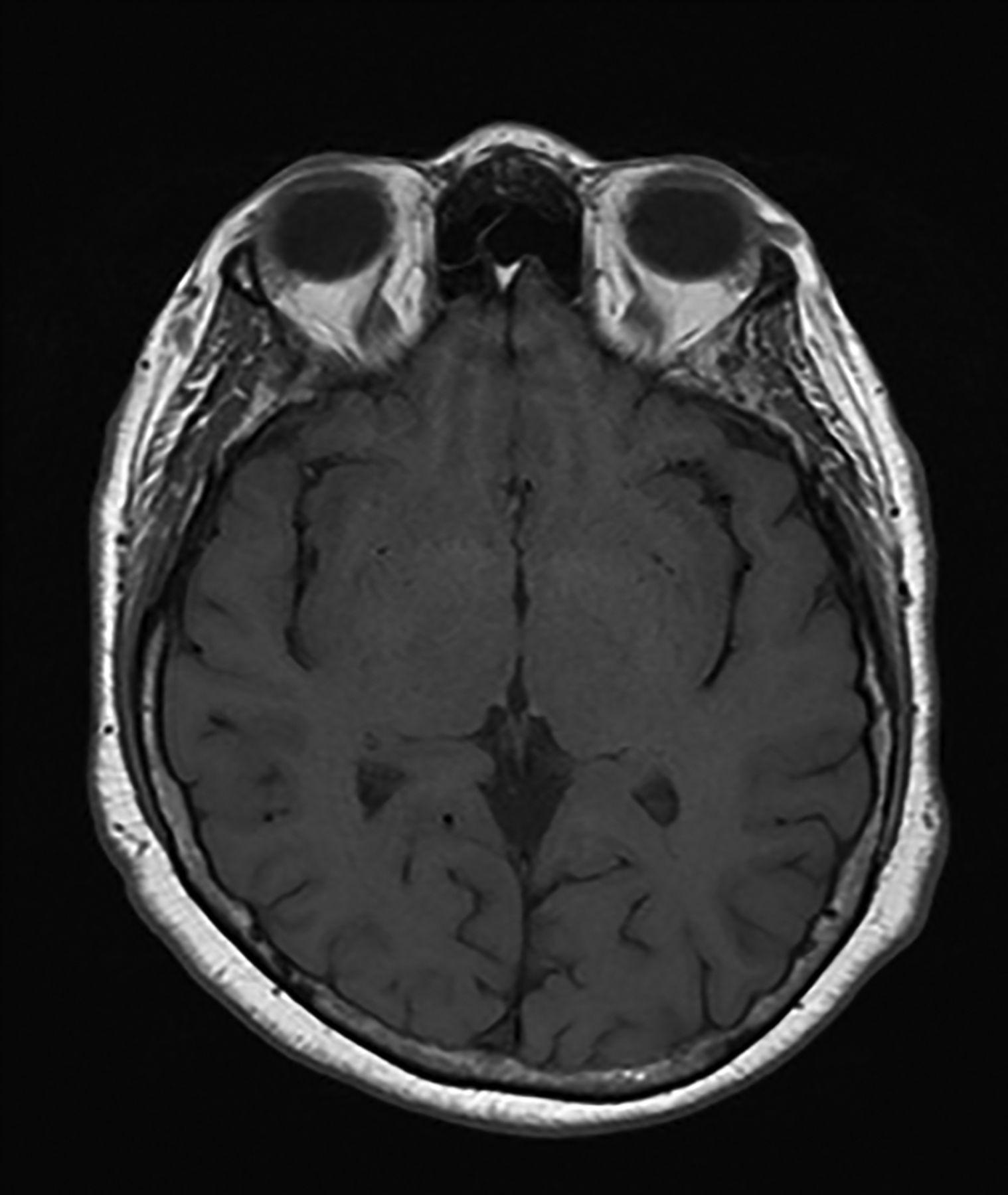
In March 2020, a patient underwent 3T MR imaging of her brain at our public hospital for investigation of anosmia. Areas of high T1 signal were present in the anterior cranial fossa bilaterally, localizing symmetrically to the olfactory sulcus and medial orbital gyrus. No abnormality was observed in the olfactory bulb itself (Fig 1). Upon review of the patient’s medical records, it was discovered the patient’s anosmia greatly predated (by many years) the COVID-19 pandemic and that the patient had antibodies negative for COVID-19. This information heightened our suspicion that the T1 shortening observed in this patient and others with similar findings represented magnetic susceptibility artifacts. It was hypothesized that these findings would be absent or less prominent on a repeat examination with a lower strength magnet, as this is a characteristic of magnetic susceptibility artifacts. After a discussion with the referring physician, the patient returned a week later for a repeat examination on a 1.5T scanner, and the previously observed findings were absent. This strongly indicates an artifactual origin of these findings (Fig 2).

FIG 1.
  • Download figure
  • Open in new tab
  • Download powerpoint
FIG 1.

T1-weighted spin-echo sequence obtained on a 3T magnet demonstrating shortening in the olfactory sulcus and medial orbital gyrus.

FIG 2.
  • Download figure
  • Open in new tab
  • Download powerpoint
FIG 2.

T1-weighted spin-echo sequence obtained on a 1.5T magnet with an absence of the previously observed signal.

While COVID-19 has surely become the most prominent and is an increasingly important cause of anosmia in the American population, other causes of anosmia exist. Learning of purported MR imaging findings in the brain correlating with their symptoms might cause patients distress or a worsening of symptoms. It is incumbent upon neuroradiologists to exercise caution in the description of novel findings in novel diseases.

References

  1. 1.↵
    1. Aragão MD,
    2. Leal MC,
    3. Cartaxo FO, et al
    . Anosmia in COVID-19 associated with injury to the olfactory bulbs evident on MRI. AJNR Am J Neuroradiol 2020;41:1703–06 doi:10.3174/ajnr.a6675 pmid:32586960
    Abstract/FREE Full Text
  2. 2.↵
    1. Mamourian A
    . Seeing what we expect to see in COVID-19. AJNR Am J Neuroradiol 2021;42:E1 doi:10.3174/ajnr.A6912 pmid:33272953
    FREE Full Text
  • © 2021 by American Journal of Neuroradiology
PreviousNext
Back to top

In this issue

American Journal of Neuroradiology: 42 (9)
American Journal of Neuroradiology
Vol. 42, Issue 9
1 Sep 2021
  • Table of Contents
  • Index by author
  • Complete Issue (PDF)
Advertisement
Print
Download PDF
Email Article

Thank you for your interest in spreading the word on American Journal of Neuroradiology.

NOTE: We only request your email address so that the person you are recommending the page to knows that you wanted them to see it, and that it is not junk mail. We do not capture any email address.

Enter multiple addresses on separate lines or separate them with commas.
Susceptibility Artifacts in the Anterior Cranial Fossa Mimicking Hemorrhage in Patients with Anosmia
(Your Name) has sent you a message from American Journal of Neuroradiology
(Your Name) thought you would like to see the American Journal of Neuroradiology web site.
CAPTCHA
This question is for testing whether or not you are a human visitor and to prevent automated spam submissions.
Cite this article
J.J. Manov, M. Alfonso, G. Saigal
Susceptibility Artifacts in the Anterior Cranial Fossa Mimicking Hemorrhage in Patients with Anosmia
American Journal of Neuroradiology Sep 2021, 42 (9) E64-E65; DOI: 10.3174/ajnr.A7184

Citation Manager Formats

  • BibTeX
  • Bookends
  • EasyBib
  • EndNote (tagged)
  • EndNote 8 (xml)
  • Medlars
  • Mendeley
  • Papers
  • RefWorks Tagged
  • Ref Manager
  • RIS
  • Zotero
0 Responses
Respond to this article
Share
Bookmark this article
Susceptibility Artifacts in the Anterior Cranial Fossa Mimicking Hemorrhage in Patients with Anosmia
J.J. Manov, M. Alfonso, G. Saigal
American Journal of Neuroradiology Sep 2021, 42 (9) E64-E65; DOI: 10.3174/ajnr.A7184
del.icio.us logo Twitter logo Facebook logo Mendeley logo
  • Tweet Widget
  • Facebook Like
  • Google Plus One
Purchase

Jump to section

  • Article
    • References
  • Figures & Data
  • Info & Metrics
  • Responses
  • References
  • PDF

Related Articles

  • No related articles found.
  • PubMed
  • Google Scholar

Cited By...

  • No citing articles found.
  • Crossref (1)
  • Google Scholar

This article has been cited by the following articles in journals that are participating in Crossref Cited-by Linking.

  • Reply:
    M.F.V.V. Aragao, P.H.P. deAndrade, V.S. da Nobrega Segundo, M.M. Marques, H.J.G. de Azevedo, R.J.V. de Mello, M. de Carvalho Leal, M.L. Soares, M.M. Valenca
    American Journal of Neuroradiology 2021 42 9

More in this TOC Section

  • Letter to the Editor regarding “Automated Volumetric Software in Dementia: Help or Hindrance to the Neuroradiologist?”
  • Reply:
  • Brain AVM’s Nidus: What if We Hadn’t Understood Anything?
Show more LETTERS

Similar Articles

Advertisement

Indexed Content

  • Current Issue
  • Accepted Manuscripts
  • Article Preview
  • Past Issues
  • Editorials
  • Editor's Choice
  • Fellows' Journal Club
  • Letters to the Editor
  • Video Articles

Cases

  • Case Collection
  • Archive - Case of the Week
  • Archive - Case of the Month
  • Archive - Classic Case

More from AJNR

  • Trainee Corner
  • Imaging Protocols
  • MRI Safety Corner

Multimedia

  • AJNR Podcasts
  • AJNR Scantastics

Resources

  • Turnaround Time
  • Submit a Manuscript
  • Submit a Video Article
  • Submit an eLetter to the Editor/Response
  • Manuscript Submission Guidelines
  • Statistical Tips
  • Fast Publishing of Accepted Manuscripts
  • Graphical Abstract Preparation
  • Imaging Protocol Submission
  • Evidence-Based Medicine Level Guide
  • Publishing Checklists
  • Author Policies
  • Become a Reviewer/Academy of Reviewers
  • News and Updates

About Us

  • About AJNR
  • Editorial Board
  • Editorial Board Alumni
  • Alerts
  • Permissions
  • Not an AJNR Subscriber? Join Now
  • Advertise with Us
  • Librarian Resources
  • Feedback
  • Terms and Conditions
  • AJNR Editorial Board Alumni

American Society of Neuroradiology

  • Not an ASNR Member? Join Now

© 2025 by the American Society of Neuroradiology All rights, including for text and data mining, AI training, and similar technologies, are reserved.
Print ISSN: 0195-6108 Online ISSN: 1936-959X

Powered by HighWire