
Article Figures & Data
Figures
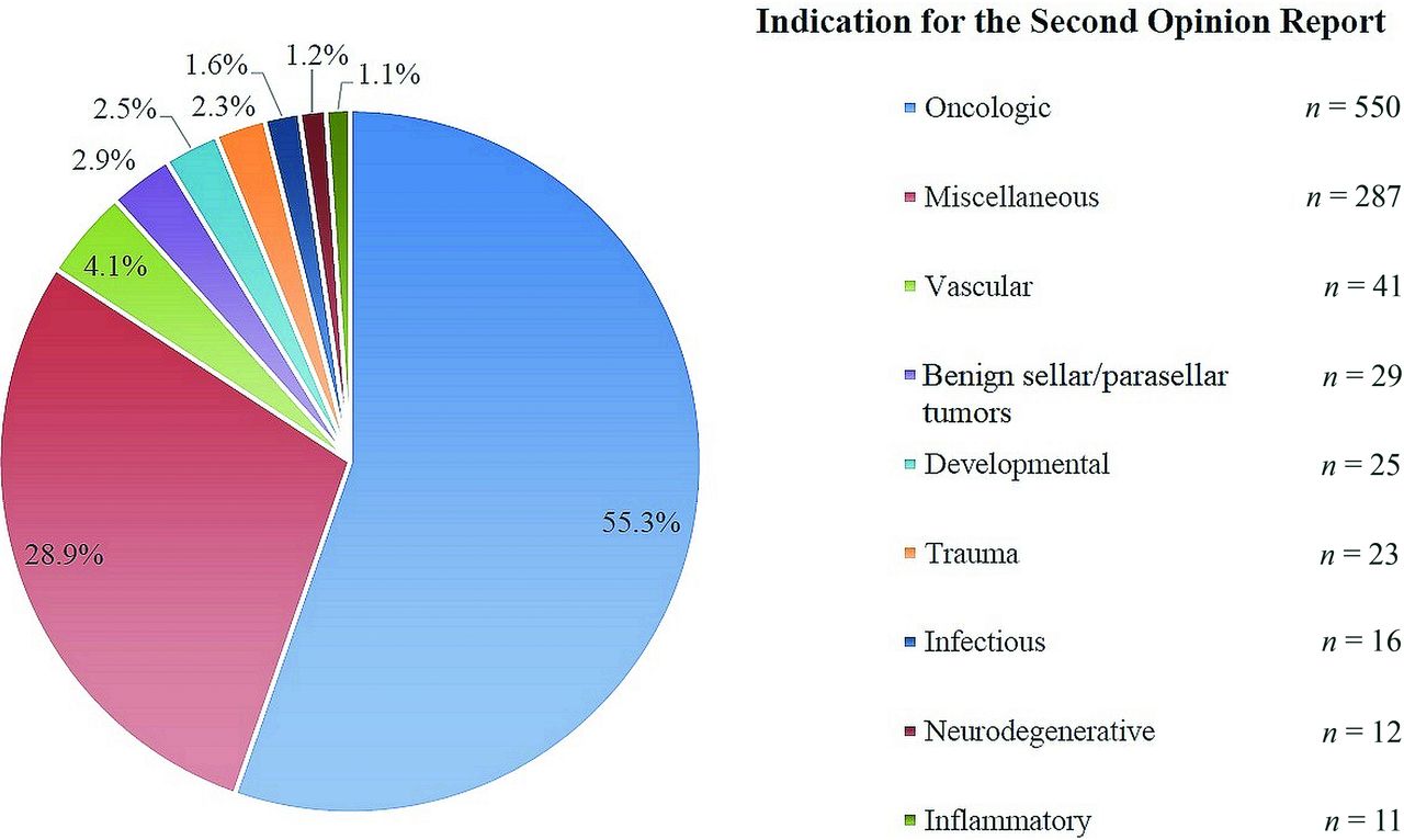
- FIGURE.
Frequencies of the different imaging indications for the 994 second opinion reports included in this study.

Tables
- Table 1:
Patient and NHN examination characteristics for which a second opinion report was requested (n = 994)
Variable No. (%) Sex Female 541 (54.4) Male 453 (45.6) Hospital status Inpatient 76 (7.6) Outpatient 918 (92.4) No. of imaging examinations evaluated for the second opinion reporta 1 864 (86.9) 2 121 (12.2) 3 8 (0.8) 4 1 (0.1) Imaging modalities for the second opinion reportb CT 224 (19.8) MR imaging 901 (79.5) Ultrasonography 7 (0.6) X-ray 2 (0.2) ↵a A second opinion report is not necessarily based on 1 sole imaging examination—eg, both MR imaging and CT can be re-evaluated for the second opinion report.
↵b Because some second opinion reports involved an evaluation of multiple imaging modalities, the numbers of imaging modalities used for second opinion reports (n = 1134) are higher than the number of reports included in this study.
- Table 2:
Specifications of the 134 recommendations made in 121 of 994 NHN second opinion reportsa
Variable No. (%) Strength of the recommendation Hard 78 (58.2) Soft 56 (41.8) Recommendation made due to apparent quality issues of the original imaging examination Yes 19 (14.2) No 115 (85.8) Referring clinicians’ compliance to the recommendation Followed 88 (65.7) Not followed 42 (31.3) Unknown 4 (3.0) ↵a The total number of recommendations made is higher than the number of reports because 1 report may contain several recommendations at once.
- Table 3:
Distribution of recommendations according to 5 groups for recommendations that were followed, that were not followed, and for which it remained unclear whether the recommendation was followed (n = 134)
Followed (%) Not Followed (%) Unknown (%) Total (%) RAI 75 (67.6) 36 (32.4) 0 (0.0) 111 (100.0) Biopsya 6 (75.0) 2 (25.0) 0 (0.0) 8 (100.0) Referral to another specialty 4 (80.0) 1 (20.0) 0 (0.0) 5 (100.0) Recommendation to compare with other previous imaging examination 2 (33.3) 3 (50.0) 1 (16.7) 6 (100.0) Otherb 1 (25.0) 0 (0.0) 3 (75.0) 4 (100.0) - Table 4:
Distribution of categorized experience of the radiologist for recommendations that were followed, that were not followed, and for which it remained unclear whether the recommendation was followed (n = 134)
Followed (%) Not Followed (%) Unknown (%) Total (%) Experience of the radiologist who signed the second opinion reporta 0–5 years 18 (64.3) 8 (28.6) 2 (7.1) 28 (100.0) 6–15 years 44 (67.7) 19 (29.2) 2 (3.1) 65 (100.0) >16 years 26 (63.4) 0 (0.0) 15 (36.6) 41 (100.0) a Calculated from the end of residency.
- Table 5:
Distribution of diagnostic outcome for recommendations that were followed, that were not followed, and for which it remained unclear whether the recommendation was followed (n = 136)a
Followed (%) Not Followed (%) Unknown (%) Total (%) Benign 44 (84.6) 6 (11.5) 2 (3.8) 52 (100.0) Malignant 21 (75.0) 5 (17.9) 2 (7.1) 28 (100.0) Unknown 24 (42.9) 31 (55.4) 1 (1.8) 56 (100.0) ↵a Because some reports contained 1 recommendation asking to check on 2 different structures, the total number is higher than the total count of recommendations (eg, 1 MR imaging focused on the orbits and 1 MR imaging focused on the cerebellopontine angle). The diagnostic outcome was defined as having established either a benign or a malignant diagnosis for a perceived lesion based on the results of the recommended additional diagnostic tests, which clarified perceived lesion nature, or on the second opinion report itself if it already provided a definitive diagnosis.
- Table 6:
Reference standards of the established benign and malignant diagnoses in the study sample (n = 80)
Benign (%) Malignant (%) Imaging examination 31 (59.6) 7 (25.0) Biopsy 9 (17.3) 15 (53.6) Operation 5 (9.6) 6 (21.4) Follow-up diagnostic testsa 4 (7.7) 0 (0.0) Application of the MDS-PSP criteria 2 (3.8) 0 (0.0) Clinical evaluation 1 (1.9) 0 (0.0) Note:—MDS-PSP indicates Movement Disorder Society progressive supranuclear palsy criteria.
↵a Either performed by a clinical specialist or lab results or imaging examinations.


Jump to section
Related Articles
Cited By...
- No citing articles found.