Advertisement

Main menu

  • Home
  • Content
    • Current Issue
    • Accepted Manuscripts
    • Article Preview
    • Past Issue Archive
    • Video Articles
    • AJNR Case Collection
    • Case of the Week Archive
    • Case of the Month Archive
    • Classic Case Archive
  • Special Collections
    • Advancing NeuroMRI with High-Relaxivity Contrast Agents
    • AJNR Awards
    • Low-Field MRI
    • Alzheimer Disease
    • ASNR Foundation Special Collection
    • Photon-Counting CT
    • View All
  • Multimedia
    • AJNR Podcasts
    • AJNR SCANtastic
    • Trainee Corner
    • MRI Safety Corner
    • Imaging Protocols
  • For Authors
    • Submit a Manuscript
    • Submit a Video Article
    • Submit an eLetter to the Editor/Response
    • Manuscript Submission Guidelines
    • Statistical Tips
    • Fast Publishing of Accepted Manuscripts
    • Graphical Abstract Preparation
    • Imaging Protocol Submission
    • Author Policies
  • About Us
    • About AJNR
    • Editorial Board
    • Editorial Board Alumni
  • More
    • Become a Reviewer/Academy of Reviewers
    • Subscribers
    • Permissions
    • Alerts
    • Feedback
    • Advertisers
    • ASNR Home

User menu

  • Alerts
  • Log in

Search

  • Advanced search
American Journal of Neuroradiology

American Journal of Neuroradiology

ASHNR American Society of Functional Neuroradiology ASHNR American Society of Pediatric Neuroradiology ASSR
  • Alerts
  • Log in

Advanced Search

  • Home
  • Content
    • Current Issue
    • Accepted Manuscripts
    • Article Preview
    • Past Issue Archive
    • Video Articles
    • AJNR Case Collection
    • Case of the Week Archive
    • Case of the Month Archive
    • Classic Case Archive
  • Special Collections
    • Advancing NeuroMRI with High-Relaxivity Contrast Agents
    • AJNR Awards
    • Low-Field MRI
    • Alzheimer Disease
    • ASNR Foundation Special Collection
    • Photon-Counting CT
    • View All
  • Multimedia
    • AJNR Podcasts
    • AJNR SCANtastic
    • Trainee Corner
    • MRI Safety Corner
    • Imaging Protocols
  • For Authors
    • Submit a Manuscript
    • Submit a Video Article
    • Submit an eLetter to the Editor/Response
    • Manuscript Submission Guidelines
    • Statistical Tips
    • Fast Publishing of Accepted Manuscripts
    • Graphical Abstract Preparation
    • Imaging Protocol Submission
    • Author Policies
  • About Us
    • About AJNR
    • Editorial Board
    • Editorial Board Alumni
  • More
    • Become a Reviewer/Academy of Reviewers
    • Subscribers
    • Permissions
    • Alerts
    • Feedback
    • Advertisers
    • ASNR Home
  • Follow AJNR on Twitter
  • Visit AJNR on Facebook
  • Follow AJNR on Instagram
  • Join AJNR on LinkedIn
  • RSS Feeds

AJNR Awards, New Junior Editors, and more. Read the latest AJNR updates


Improved Turnaround Times | Median time to first decision: 12 days

Research ArticleHead and Neck Imaging
Open Access

Altered Blood Flow in the Ophthalmic and Internal Carotid Arteries in Patients with Age-Related Macular Degeneration Measured Using Noncontrast MR Angiography at 7T

M.L. Hibert, Y.I. Chen, N. Ohringer, W.J. Feuer, N.K. Waheed, J.S. Heier, M.W. Calhoun, P.J. Rosenfeld and J.R. Polimeni
American Journal of Neuroradiology September 2021, 42 (9) 1653-1660; DOI: https://doi.org/10.3174/ajnr.A7187
M.L. Hibert
aFrom the Athinoula A. Martinos Center for Biomedical Imaging (M.L.H., Y.I.C., N. O., J.R.P.), Massachusetts General Hospital, Charlestown, Massachusetts
  • Find this author on Google Scholar
  • Find this author on PubMed
  • Search for this author on this site
  • ORCID record for M.L. Hibert
Y.I. Chen
aFrom the Athinoula A. Martinos Center for Biomedical Imaging (M.L.H., Y.I.C., N. O., J.R.P.), Massachusetts General Hospital, Charlestown, Massachusetts
bDepartment of Radiology (Y.I.C., J.R.P.), Harvard Medical School, Boston, Massachusetts
  • Find this author on Google Scholar
  • Find this author on PubMed
  • Search for this author on this site
  • ORCID record for Y.I. Chen
N. Ohringer
aFrom the Athinoula A. Martinos Center for Biomedical Imaging (M.L.H., Y.I.C., N. O., J.R.P.), Massachusetts General Hospital, Charlestown, Massachusetts
  • Find this author on Google Scholar
  • Find this author on PubMed
  • Search for this author on this site
  • ORCID record for N. Ohringer
W.J. Feuer
cDepartment of Ophthalmology (W.J.F., P.J.R.), Bascom Palmer Eye Institute, University of Miami Miller School of Medicine, Miami, Florida
  • Find this author on Google Scholar
  • Find this author on PubMed
  • Search for this author on this site
  • ORCID record for W.J. Feuer
N.K. Waheed
dNew England Eye Center (N.K.W.), Tufts Medical Center, Boston, Massachusetts
  • Find this author on Google Scholar
  • Find this author on PubMed
  • Search for this author on this site
  • ORCID record for N.K. Waheed
J.S. Heier
eOphthalmic Consultants of Boston (J.S.H.), Boston, Massachusetts
  • Find this author on Google Scholar
  • Find this author on PubMed
  • Search for this author on this site
  • ORCID record for J.S. Heier
M.W. Calhoun
fOcuDyne Inc (M.W.C.), Roseville, Minnesota
  • Find this author on Google Scholar
  • Find this author on PubMed
  • Search for this author on this site
  • ORCID record for M.W. Calhoun
P.J. Rosenfeld
cDepartment of Ophthalmology (W.J.F., P.J.R.), Bascom Palmer Eye Institute, University of Miami Miller School of Medicine, Miami, Florida
  • Find this author on Google Scholar
  • Find this author on PubMed
  • Search for this author on this site
  • ORCID record for P.J. Rosenfeld
J.R. Polimeni
aFrom the Athinoula A. Martinos Center for Biomedical Imaging (M.L.H., Y.I.C., N. O., J.R.P.), Massachusetts General Hospital, Charlestown, Massachusetts
bDepartment of Radiology (Y.I.C., J.R.P.), Harvard Medical School, Boston, Massachusetts
gHarvard-Massachusetts Institute of Technology Division of Health Sciences and Technology (J.R.P.), Massachusetts Institute of Technology, Cambridge, Massachusetts
  • Find this author on Google Scholar
  • Find this author on PubMed
  • Search for this author on this site
  • ORCID record for J.R. Polimeni
  • Article
  • Figures & Data
  • Supplemental
  • Info & Metrics
  • Responses
  • References
  • PDF
Loading

Article Figures & Data

Figures

  • Tables
  • FIG 1.
    • Download figure
    • Open in new tab
    • Download powerpoint
    FIG 1.

    Positioning of PC-MRA slices using the 3D-TOF-MRA data as an anatomic guide. A, Red lines depict cross-sections of the triple-oblique positioning and location of PC-MRA slices centered on and perpendicular to each OA (green arrows), identified from the reference 3D-TOF image. The OA cross-section can be visualized in the center of the magnitude (B and C) and phase (D and E) images from a PC-MRA scan (yellow box represents an enlarged region from B and D in C and E, respectively).

  • FIG 2.
    • Download figure
    • Open in new tab
    • Download powerpoint
    FIG 2.

    Analysis procedure for correcting EC bias and defining the vessel ROI. A, A PC-MRA magnitude image is used to place a 12-mm-diameter ROI for EC correction in vessel-free tissue adjacent to the vessel ROI (blue). The red line in the inset TOF image indicates the PC-MRA slice position. The vessel ROI (yellow) is shown within the yellow box, which depicts the region enlarged in B and C. Enlarged PC-MRA magnitude images show the vessel cross-section alone (B) and with the vessel ROI superimposed (C). D, Time-series of EC-corrected flow data in units of centimeters/second from 1 example PC-MRA acquisition with a single VENC value in 1 subject.

  • FIG 3.
    • Download figure
    • Open in new tab
    • Download powerpoint
    FIG 3.

    Boxplots of unnormalized flow velocities (A and B) and volumetric flow rates (C and D) in the OAs (A and C) and ICAs (B and D). Vessels were divided into 4 groups: healthy controls, early AMD, intermediate AMD, and late AMD. The asterisk signifies P < .05 compared with controls.

  • FIG 4.
    • Download figure
    • Open in new tab
    • Download powerpoint
    FIG 4.

    Linear trend test plot, which demonstrates that the rate of decline in OA volumetric flow with disease progression is statistically significant.
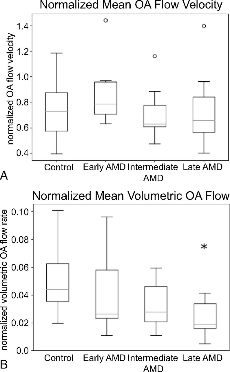
  • FIG 5.
    • Download figure
    • Open in new tab
    • Download powerpoint
    FIG 5.

    Boxplots of flow velocity (A) and volumetric flow (B) in the OA normalized to upstream ICA flow rates. Normalized flow values represent the OA flow as a ratio of ICA flow. Vessels are divided into 4 groups: healthy controls, early AMD, intermediate AMD, and late AMD. The asterisk signifies P < .05 compared with controls.

  • FIG 6.
    • Download figure
    • Open in new tab
    • Download powerpoint
    FIG 6.

    Resistance indices for the OA (A) and ICA (B). Vessels are divided into 4 groups: healthy controls, early AMD, intermediate AMD, and late AMD. The asterisk signifies P < .05 compared with controls.

Tables

  • Figures
  • Summary of vessel diameter and blood flow measures stratified by disease stagea

    Control (Mean) (SD) (n = 20 OA, 24 ICA)Early AMD (Mean) (SD) (n = 9 OA, 15 ICA)Interm. AMD(Mean) (SD) (n = 7 OA, 8 ICA)Late AMD (Mean) (SD) (n = 12 OA, 17 ICA)All AMD (Mean) (SD) (n = 28 OA, 40 ICA)
    OA diameter (mm)1.1 (0.19)0.9 (0.25)P = .111.0 (0.26)P = .420.8 (0.25)P < .01b0.9 (0.26)P < .01b
    OA volume flow (mL/min)7.0 (3.0)6.5 (4.2)P = .745.6 (3.1)P = .373.8 (1.8)P < .01b5.1 (3.2)P = .07
    OA flow velocity (cm/s)c13.1 (4.0)16.0 (4.5)P < .01b11.9 (4.3)P = .9115.0 (5.7)P = .03b14.6 (5.1)P = .02b
    OA RI0.70 (0.10)0.82 (0.10)P < .01b0.81 (0.06)P < .01b0.80 (0.07)P < .01b0.81 (0.08)P < .01b
    ICA diameter (mm)4.3 (0.51)4.2 (0.52)P = .384.6 (0.63)P = .474.2 (0.44)P = .554.3 (0.52)P = .67
    ICA volume flow (mL/min)c163.1 (44.8)154.3 (44.2)P = .98167.0 (23.4)P = .49166.6 (27.7)P = .19162.2 (33.8)P = .46
    ICA flow velocity (cm/s)c18.3 (3.5)18.7 (3.7)P = .0717.8 (4.5)P = .5820.6 (5.2)P < .01b19.4 (4.6)P < .01b
    ICA RI0.61 (0.09)0.66 (0.09)P = .180.70 (0.04)P < .01b0.65 (0.11)P = .290.67 (0.09)P = .07
    • Note:—RI indicates resistance index; Interm., intermediate.

    • a Values represent group mean and group SD, and reported P values indicate significance relative to controls based on a GEE analysis followed by post hoc least significant difference comparisons between disease groups and controls. The sample size for ophthalmic artery measurements was 48 hemisphere measurements from 30 subjects. The sample size for the ICA measurements was 64 hemispheres of 33 subjects.

    • ↵b Tests that were significant at the P < .05 level (uncorrected).

    • ↵c Adjusted for age, which was statistically significant (P < .05) in the GEE model.

PreviousNext
Back to top

In this issue

American Journal of Neuroradiology: 42 (9)
American Journal of Neuroradiology
Vol. 42, Issue 9
1 Sep 2021
  • Table of Contents
  • Index by author
  • Complete Issue (PDF)
Advertisement
Print
Download PDF
Email Article

Thank you for your interest in spreading the word on American Journal of Neuroradiology.

NOTE: We only request your email address so that the person you are recommending the page to knows that you wanted them to see it, and that it is not junk mail. We do not capture any email address.

Enter multiple addresses on separate lines or separate them with commas.
Altered Blood Flow in the Ophthalmic and Internal Carotid Arteries in Patients with Age-Related Macular Degeneration Measured Using Noncontrast MR Angiography at 7T
(Your Name) has sent you a message from American Journal of Neuroradiology
(Your Name) thought you would like to see the American Journal of Neuroradiology web site.
CAPTCHA
This question is for testing whether or not you are a human visitor and to prevent automated spam submissions.
Cite this article
M.L. Hibert, Y.I. Chen, N. Ohringer, W.J. Feuer, N.K. Waheed, J.S. Heier, M.W. Calhoun, P.J. Rosenfeld, J.R. Polimeni
Altered Blood Flow in the Ophthalmic and Internal Carotid Arteries in Patients with Age-Related Macular Degeneration Measured Using Noncontrast MR Angiography at 7T
American Journal of Neuroradiology Sep 2021, 42 (9) 1653-1660; DOI: 10.3174/ajnr.A7187

Citation Manager Formats

  • BibTeX
  • Bookends
  • EasyBib
  • EndNote (tagged)
  • EndNote 8 (xml)
  • Medlars
  • Mendeley
  • Papers
  • RefWorks Tagged
  • Ref Manager
  • RIS
  • Zotero
0 Responses
Respond to this article
Share
Bookmark this article
Altered Blood Flow in the Ophthalmic and Internal Carotid Arteries in Patients with Age-Related Macular Degeneration Measured Using Noncontrast MR Angiography at 7T
M.L. Hibert, Y.I. Chen, N. Ohringer, W.J. Feuer, N.K. Waheed, J.S. Heier, M.W. Calhoun, P.J. Rosenfeld, J.R. Polimeni
American Journal of Neuroradiology Sep 2021, 42 (9) 1653-1660; DOI: 10.3174/ajnr.A7187
del.icio.us logo Twitter logo Facebook logo Mendeley logo
  • Tweet Widget
  • Facebook Like
  • Google Plus One
Purchase

Jump to section

  • Article
    • Abstract
    • ABBREVIATIONS:
    • MATERIALS and METHODS
    • RESULTS
    • DISCUSSION
    • CONCLUSIONS
    • Acknowledgments
    • Footnotes
    • References
  • Figures & Data
  • Supplemental
  • Info & Metrics
  • Responses
  • References
  • PDF

Related Articles

  • PubMed
  • Google Scholar

Cited By...

  • Quantifying brain-wide cerebrospinal fluid flow dynamics using slow-flow-sensitized phase-contrast MRI
  • Ophthalmic artery stenosis on three-dimensional rotational angiography: interrater agreement, prevalence, and risk factors
  • Regarding "Altered Blood Flow in the Ophthalmic and Internal Carotid Arteries in Patients with Age-Related Macular Degeneration Measured Using Noncontrast MR Angiography at 7T"
  • Subretinal drusenoid deposits are strongly associated with coexistent high-risk vascular diseases
  • Ophthalmic artery angioplasty for age-related macular degeneration
  • Crossref
  • Google Scholar

This article has not yet been cited by articles in journals that are participating in Crossref Cited-by Linking.

More in this TOC Section

  • Diagnostic Performance of US in Neck Node NIRADS 2
  • Hydrops Herniation into the Semicircular Canals
  • ASL Sensitivity for Head and Neck Paraganglioma
Show more HEAD AND NECK IMAGING

Similar Articles

Advertisement

Indexed Content

  • Current Issue
  • Accepted Manuscripts
  • Article Preview
  • Past Issues
  • Editorials
  • Editor's Choice
  • Fellows' Journal Club
  • Letters to the Editor
  • Video Articles

Cases

  • Case Collection
  • Archive - Case of the Week
  • Archive - Case of the Month
  • Archive - Classic Case

More from AJNR

  • Trainee Corner
  • Imaging Protocols
  • MRI Safety Corner
  • Book Reviews

Multimedia

  • AJNR Podcasts
  • AJNR Scantastics

Resources

  • Turnaround Time
  • Submit a Manuscript
  • Submit a Video Article
  • Submit an eLetter to the Editor/Response
  • Manuscript Submission Guidelines
  • Statistical Tips
  • Fast Publishing of Accepted Manuscripts
  • Graphical Abstract Preparation
  • Imaging Protocol Submission
  • Evidence-Based Medicine Level Guide
  • Publishing Checklists
  • Author Policies
  • Become a Reviewer/Academy of Reviewers
  • News and Updates

About Us

  • About AJNR
  • Editorial Board
  • Editorial Board Alumni
  • Alerts
  • Permissions
  • Not an AJNR Subscriber? Join Now
  • Advertise with Us
  • Librarian Resources
  • Feedback
  • Terms and Conditions
  • AJNR Editorial Board Alumni

American Society of Neuroradiology

  • Not an ASNR Member? Join Now

© 2025 by the American Society of Neuroradiology All rights, including for text and data mining, AI training, and similar technologies, are reserved.
Print ISSN: 0195-6108 Online ISSN: 1936-959X

Powered by HighWire