Advertisement

Main menu

  • Home
  • Content
    • Current Issue
    • Accepted Manuscripts
    • Article Preview
    • Past Issue Archive
    • Video Articles
    • AJNR Case Collection
    • Case of the Week Archive
    • Case of the Month Archive
    • Classic Case Archive
  • Special Collections
    • AJNR Awards
    • Low-Field MRI
    • Alzheimer Disease
    • ASNR Foundation Special Collection
    • Photon-Counting CT
    • View All
  • Multimedia
    • AJNR Podcasts
    • AJNR SCANtastic
    • Trainee Corner
    • MRI Safety Corner
    • Imaging Protocols
  • For Authors
    • Submit a Manuscript
    • Submit a Video Article
    • Submit an eLetter to the Editor/Response
    • Manuscript Submission Guidelines
    • Statistical Tips
    • Fast Publishing of Accepted Manuscripts
    • Graphical Abstract Preparation
    • Imaging Protocol Submission
    • Author Policies
  • About Us
    • About AJNR
    • Editorial Board
    • Editorial Board Alumni
  • More
    • Become a Reviewer/Academy of Reviewers
    • Subscribers
    • Permissions
    • Alerts
    • Feedback
    • Advertisers
    • ASNR Home

User menu

  • Alerts
  • Log in

Search

  • Advanced search
American Journal of Neuroradiology

American Journal of Neuroradiology

ASHNR American Society of Functional Neuroradiology ASHNR American Society of Pediatric Neuroradiology ASSR
  • Alerts
  • Log in

Advanced Search

  • Home
  • Content
    • Current Issue
    • Accepted Manuscripts
    • Article Preview
    • Past Issue Archive
    • Video Articles
    • AJNR Case Collection
    • Case of the Week Archive
    • Case of the Month Archive
    • Classic Case Archive
  • Special Collections
    • AJNR Awards
    • Low-Field MRI
    • Alzheimer Disease
    • ASNR Foundation Special Collection
    • Photon-Counting CT
    • View All
  • Multimedia
    • AJNR Podcasts
    • AJNR SCANtastic
    • Trainee Corner
    • MRI Safety Corner
    • Imaging Protocols
  • For Authors
    • Submit a Manuscript
    • Submit a Video Article
    • Submit an eLetter to the Editor/Response
    • Manuscript Submission Guidelines
    • Statistical Tips
    • Fast Publishing of Accepted Manuscripts
    • Graphical Abstract Preparation
    • Imaging Protocol Submission
    • Author Policies
  • About Us
    • About AJNR
    • Editorial Board
    • Editorial Board Alumni
  • More
    • Become a Reviewer/Academy of Reviewers
    • Subscribers
    • Permissions
    • Alerts
    • Feedback
    • Advertisers
    • ASNR Home
  • Follow AJNR on Twitter
  • Visit AJNR on Facebook
  • Follow AJNR on Instagram
  • Join AJNR on LinkedIn
  • RSS Feeds

AJNR Awards, New Junior Editors, and more. Read the latest AJNR updates

LetterLetter
Open Access

Leptomeningeal Enhancement Due to COVID-19 on 3D-FLAIR and T1 Black-Blood MR Imaging Sequences

G. Velonakis, E. Karavasilis, D.K. Filippiadis, M.P, Almyroudi and E. Korompoki
American Journal of Neuroradiology July 2021, 42 (7) E39-E41; DOI: https://doi.org/10.3174/ajnr.A7104
G. Velonakis
aResearch Unit of Radiology – 2nd Department of RadiologyMedical School, National and Kapodistrian University of AthensAthens, Greece
  • Find this author on Google Scholar
  • Find this author on PubMed
  • Search for this author on this site
  • ORCID record for G. Velonakis
E. Karavasilis
aResearch Unit of Radiology – 2nd Department of RadiologyMedical School, National and Kapodistrian University of AthensAthens, Greece
  • Find this author on Google Scholar
  • Find this author on PubMed
  • Search for this author on this site
  • ORCID record for E. Karavasilis
D.K. Filippiadis
aResearch Unit of Radiology – 2nd Department of RadiologyMedical School, National and Kapodistrian University of AthensAthens, Greece
  • Find this author on Google Scholar
  • Find this author on PubMed
  • Search for this author on this site
  • ORCID record for D.K. Filippiadis
M.P, Almyroudi
aResearch Unit of Radiology – 2nd Department of RadiologyMedical School, National and Kapodistrian University of AthensAthens, Greece
  • Find this author on Google Scholar
  • Find this author on PubMed
  • Search for this author on this site
  • ORCID record for M.P, Almyroudi
E. Korompoki
aResearch Unit of Radiology – 2nd Department of RadiologyMedical School, National and Kapodistrian University of AthensAthens, Greece
  • Find this author on Google Scholar
  • Find this author on PubMed
  • Search for this author on this site
  • ORCID record for E. Korompoki
  • Article
  • Figures & Data
  • Info & Metrics
  • Responses
  • References
  • PDF
Loading

We read with great interest the literature review by Gulko et al1 that included 22 articles reporting the MR imaging brain findings of 126 patients with coronavirus disease 2019 (COVID 19). We agree with the authors that the most frequent diagnosis on MR imaging is acute and subacute infarct, which was reported in 32 of 126 patients. However, because of the different imaging techniques used in the original articles, the frequency of leptomeningeal involvement may be underestimated.

We observed great differences in the reported frequency of leptomeningeal enhancement in the articles included in this review. Helms et al2 reported enhancement in leptomeningeal spaces in 8 patients (68%). On the other hand, leptomeningeal enhancement was absent (0%) in several other studies with similar patient samples.1 This discrepancy may be attributed to the different imaging techniques used to evaluate leptomeningeal involvement. The contrast-enhanced 3D-FLAIR sequence was used by Helms et al,2 while only conventional T1WI postcontrast sequences were performed in several other studies.

In our practice, 3D-FLAIR sequences or 3D T1 black-blood imaging or both are used to clearly demonstrate leptomeningeal enhancement. Leptomeningeal involvement seems to be a relatively common manifestation of CNS involvement in patients with COVID-19. Herein, we present a 68-year-old woman with COVID-19 who underwent brain MR imaging 2 weeks after extubation, because of confusion and muscle weakness in her limbs. Turbo spin-echo 3D-T1 black-blood and 3D-FLAIR sequences postcontrast administration revealed leptomeningeal enhancement in the left angular gyrus (Fig 1) and on both sides of the right parieto-occipital sulcus (Fig 2). Enhancement was not prominent on conventional (turbo-field echo) 3D-T1 images.

FIG 1.
  • Download figure
  • Open in new tab
  • Download powerpoint
FIG 1.

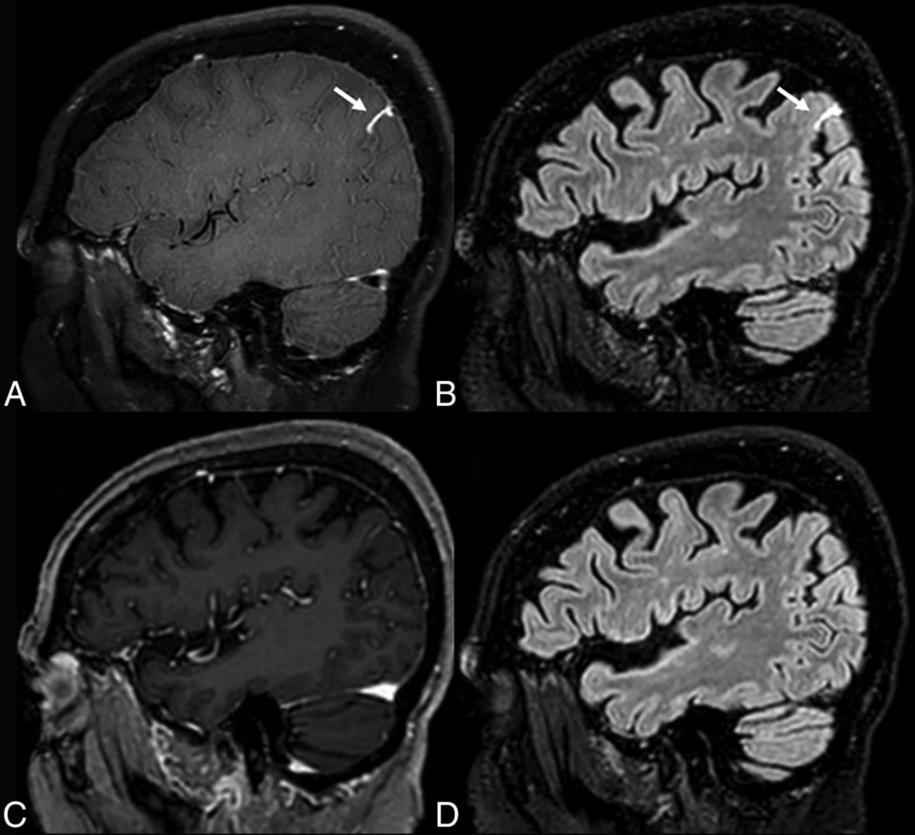
Sagittal 3D turbo spin-echo black-blood T1-weighted (A) and 3D-FLAIR (B) images, both postcontrast administration, reveal leptomeningeal enhancement in the left angular gyrus (arrows, A and B). Enhancement is not prominent on T1 contrast-enhanced images (C). There are no hyperintensities in this area on the sagittal 3D-FLAIR image without contrast (D).

FIG 2.
  • Download figure
  • Open in new tab
  • Download powerpoint
FIG 2.

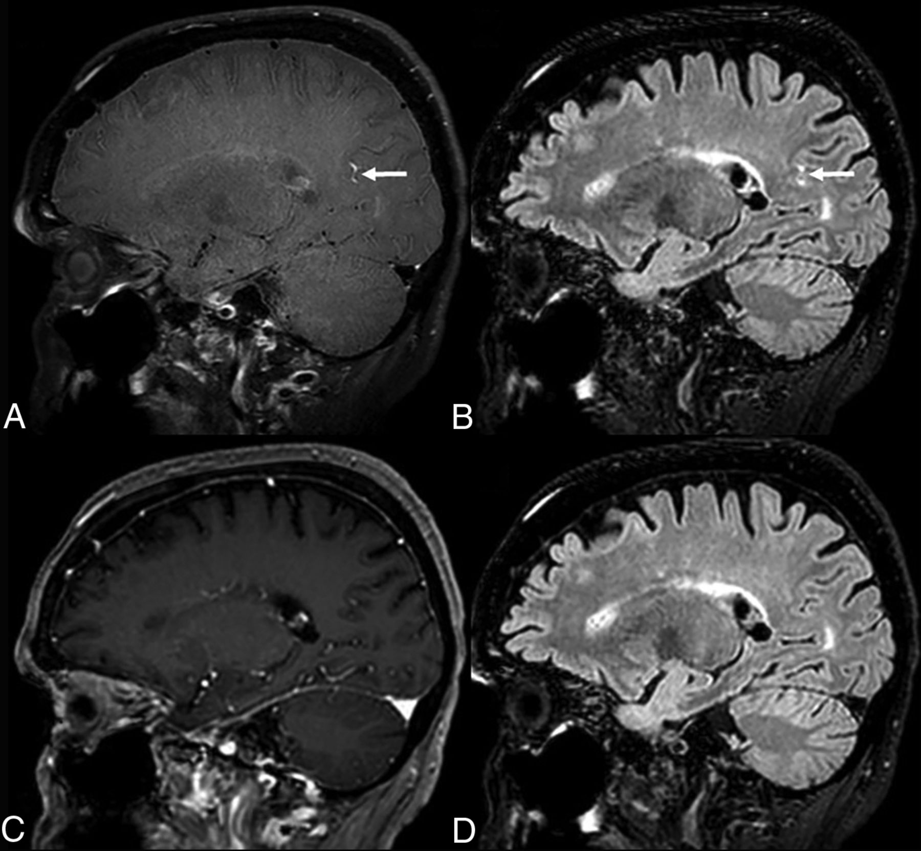
Leptomeningeal enhancement on both sides of the right parieto-occipital sulcus is more evident on sagittal 3D-T1 turbo spin-echo black-blood (A) and 3D-FLAIR (B) images (arrows, A and B), than on T1 postcontrast (C). No hyperintensities on 3D-FLAIR before contrast administration (D) are noticed in the right parieto-occipital sulcus.

Leptomeningeal enhancement on FLAIR images is known to occur frequently in MS and non-MS inflammatory neurologic conditions, including encephalitis. In their systematic review Zurawski et al3 suggested that contrast-enhanced FLAIR provided as much as 10-fold sensitivity compared with conventional T1 sequences in the detection of low concentration of contrast in the subarachnoid space.

The increased sensitivity of T1 black-blood imaging in detecting leptomeningeal enhancement has also been reported. Oh et al4 compared contrast-enhanced gradient recalled-echo, contrast-enhanced spin-echo, and black-blood imaging for the detection of leptomeningeal carcinomatosis. According to their results, black-blood imaging had a significantly higher sensitivity than contrast-enhanced gradient recalled-echo and contrast-enhanced spin-echo (97.43%, 64.1%, and 66.67%, respectively) in identifying foci of leptomeningeal enhancement.

The possible underlying mechanism of focal leptomeningeal enhancement may include inflammation-related focal disruption of the blood-meninges barrier and associated fibrosis. Pathologist analysis in patients with MS showed leptomeningeal inflammation, which may lead to the up-regulation of reactive fibroblasts and collagen deposition causing local meningeal fibrosis.3 Consequently, both fibrosis and inflammation may result in the trapping of low concentration of gadolinium-based contrast medium within the subarachnoid spaces.

Although contrast-enhanced 3D-FLAIR and T1 black-blood imaging are sensitive in detecting leptomeningeal enhancement, they are not routinely performed in patients with COVID-19 in many institutions. Including one or both sequences in MR imaging protocols for patients with COVID-19 with neurologic symptoms may help to avoid underestimation of leptomeningeal and CNS involvement.

Indicates open access to non-subscribers at www.ajnr.org

References

  1. 1.↵
    1. Gulko E,
    2. Oleksk ML,
    3. Gomes W, et al
    . MRI brain findings in 126 patients with COVID-19: initial observations from a descriptive literature review. AJNR Am J Neuroradiol 2020;41:2199–203 doi:10.3174/ajnr.A6805 pmid:32883670
    Abstract/FREE Full Text
  2. 2.↵
    1. Helms J,
    2. Kremer S,
    3. Merdji H, et al
    . Neurologic features in severe SARS-CoV-2 infection. N Engl J Med 2020;382:2268–70 doi:10.1056/NEJMc2008597 pmid:32294339
    CrossRefPubMed
  3. 3.↵
    1. Zurawski J,
    2. Lassmann H,
    3. Bakshi R
    . Use of magnetic resonance imaging to visualize leptomeningeal inflammation in patients with multiple sclerosis: a review. JAMA Neurol 2017;74:100–09 doi:10.1001/jamaneurol.2016.4237 pmid:27893883
    CrossRefPubMed
  4. 4.↵
    1. Oh J,
    2. Choi SH,
    3. Lee E, et al
    . Application of 3D fast spin-echo T1 black-blood imaging in the diagnosis and prognostic prediction of patients with leptomeningeal carcinomatosis. AJNR Am J Neuroradiol 2018;39:1453–59 doi:10.3174/ajnr.A5721 pmid:30002052
    Abstract/FREE Full Text
  • © 2021 by American Journal of Neuroradiology
PreviousNext
Back to top

In this issue

American Journal of Neuroradiology: 42 (7)
American Journal of Neuroradiology
Vol. 42, Issue 7
1 Jul 2021
  • Table of Contents
  • Index by author
  • Complete Issue (PDF)
Advertisement
Print
Download PDF
Email Article

Thank you for your interest in spreading the word on American Journal of Neuroradiology.

NOTE: We only request your email address so that the person you are recommending the page to knows that you wanted them to see it, and that it is not junk mail. We do not capture any email address.

Enter multiple addresses on separate lines or separate them with commas.
Leptomeningeal Enhancement Due to COVID-19 on 3D-FLAIR and T1 Black-Blood MR Imaging Sequences
(Your Name) has sent you a message from American Journal of Neuroradiology
(Your Name) thought you would like to see the American Journal of Neuroradiology web site.
CAPTCHA
This question is for testing whether or not you are a human visitor and to prevent automated spam submissions.
Cite this article
G. Velonakis, E. Karavasilis, D.K. Filippiadis, M.P, Almyroudi, E. Korompoki
Leptomeningeal Enhancement Due to COVID-19 on 3D-FLAIR and T1 Black-Blood MR Imaging Sequences
American Journal of Neuroradiology Jul 2021, 42 (7) E39-E41; DOI: 10.3174/ajnr.A7104

Citation Manager Formats

  • BibTeX
  • Bookends
  • EasyBib
  • EndNote (tagged)
  • EndNote 8 (xml)
  • Medlars
  • Mendeley
  • Papers
  • RefWorks Tagged
  • Ref Manager
  • RIS
  • Zotero
0 Responses
Respond to this article
Share
Bookmark this article
Leptomeningeal Enhancement Due to COVID-19 on 3D-FLAIR and T1 Black-Blood MR Imaging Sequences
G. Velonakis, E. Karavasilis, D.K. Filippiadis, M.P, Almyroudi, E. Korompoki
American Journal of Neuroradiology Jul 2021, 42 (7) E39-E41; DOI: 10.3174/ajnr.A7104
del.icio.us logo Twitter logo Facebook logo Mendeley logo
  • Tweet Widget
  • Facebook Like
  • Google Plus One
Purchase

Jump to section

  • Article
    • References
  • Figures & Data
  • Info & Metrics
  • Responses
  • References
  • PDF

Related Articles

  • No related articles found.
  • PubMed
  • Google Scholar

Cited By...

  • No citing articles found.
  • Crossref (1)
  • Google Scholar

This article has been cited by the following articles in journals that are participating in Crossref Cited-by Linking.

  • Concurrent Diffuse Dural and Leptomeningeal Enhancements in Brain Magnetic Resonance Imaging Following a Mild COVID-19 Infection: A Novel Case Report and Review of Literature
    Rashed Bawand, Masoud Ghiasian, Mustapha Samadyan
    Current Medical Imaging Formerly Current Medical Imaging Reviews 2023 20

More in this TOC Section

  • Letter to the Editor regarding “Automated Volumetric Software in Dementia: Help or Hindrance to the Neuroradiologist?”
  • Reply:
  • Brain AVM’s Nidus: What if We Hadn’t Understood Anything?
Show more LETTERS

Similar Articles

Advertisement

Indexed Content

  • Current Issue
  • Accepted Manuscripts
  • Article Preview
  • Past Issues
  • Editorials
  • Editor's Choice
  • Fellows' Journal Club
  • Letters to the Editor
  • Video Articles

Cases

  • Case Collection
  • Archive - Case of the Week
  • Archive - Case of the Month
  • Archive - Classic Case

More from AJNR

  • Trainee Corner
  • Imaging Protocols
  • MRI Safety Corner

Multimedia

  • AJNR Podcasts
  • AJNR Scantastics

Resources

  • Turnaround Time
  • Submit a Manuscript
  • Submit a Video Article
  • Submit an eLetter to the Editor/Response
  • Manuscript Submission Guidelines
  • Statistical Tips
  • Fast Publishing of Accepted Manuscripts
  • Graphical Abstract Preparation
  • Imaging Protocol Submission
  • Evidence-Based Medicine Level Guide
  • Publishing Checklists
  • Author Policies
  • Become a Reviewer/Academy of Reviewers
  • News and Updates

About Us

  • About AJNR
  • Editorial Board
  • Editorial Board Alumni
  • Alerts
  • Permissions
  • Not an AJNR Subscriber? Join Now
  • Advertise with Us
  • Librarian Resources
  • Feedback
  • Terms and Conditions
  • AJNR Editorial Board Alumni

American Society of Neuroradiology

  • Not an ASNR Member? Join Now

© 2025 by the American Society of Neuroradiology All rights, including for text and data mining, AI training, and similar technologies, are reserved.
Print ISSN: 0195-6108 Online ISSN: 1936-959X

Powered by HighWire