Advertisement

Main menu

  • Home
  • Content
    • Current Issue
    • Accepted Manuscripts
    • Article Preview
    • Past Issue Archive
    • Video Articles
    • AJNR Case Collection
    • Case of the Week Archive
    • Case of the Month Archive
    • Classic Case Archive
  • Special Collections
    • AJNR Awards
    • Low-Field MRI
    • Alzheimer Disease
    • ASNR Foundation Special Collection
    • Photon-Counting CT
    • View All
  • Multimedia
    • AJNR Podcasts
    • AJNR SCANtastic
    • Trainee Corner
    • MRI Safety Corner
    • Imaging Protocols
  • For Authors
    • Submit a Manuscript
    • Submit a Video Article
    • Submit an eLetter to the Editor/Response
    • Manuscript Submission Guidelines
    • Statistical Tips
    • Fast Publishing of Accepted Manuscripts
    • Graphical Abstract Preparation
    • Imaging Protocol Submission
    • Author Policies
  • About Us
    • About AJNR
    • Editorial Board
    • Editorial Board Alumni
  • More
    • Become a Reviewer/Academy of Reviewers
    • Subscribers
    • Permissions
    • Alerts
    • Feedback
    • Advertisers
    • ASNR Home

User menu

  • Alerts
  • Log in

Search

  • Advanced search
American Journal of Neuroradiology

American Journal of Neuroradiology

ASHNR American Society of Functional Neuroradiology ASHNR American Society of Pediatric Neuroradiology ASSR
  • Alerts
  • Log in

Advanced Search

  • Home
  • Content
    • Current Issue
    • Accepted Manuscripts
    • Article Preview
    • Past Issue Archive
    • Video Articles
    • AJNR Case Collection
    • Case of the Week Archive
    • Case of the Month Archive
    • Classic Case Archive
  • Special Collections
    • AJNR Awards
    • Low-Field MRI
    • Alzheimer Disease
    • ASNR Foundation Special Collection
    • Photon-Counting CT
    • View All
  • Multimedia
    • AJNR Podcasts
    • AJNR SCANtastic
    • Trainee Corner
    • MRI Safety Corner
    • Imaging Protocols
  • For Authors
    • Submit a Manuscript
    • Submit a Video Article
    • Submit an eLetter to the Editor/Response
    • Manuscript Submission Guidelines
    • Statistical Tips
    • Fast Publishing of Accepted Manuscripts
    • Graphical Abstract Preparation
    • Imaging Protocol Submission
    • Author Policies
  • About Us
    • About AJNR
    • Editorial Board
    • Editorial Board Alumni
  • More
    • Become a Reviewer/Academy of Reviewers
    • Subscribers
    • Permissions
    • Alerts
    • Feedback
    • Advertisers
    • ASNR Home
  • Follow AJNR on Twitter
  • Visit AJNR on Facebook
  • Follow AJNR on Instagram
  • Join AJNR on LinkedIn
  • RSS Feeds

AJNR Awards, New Junior Editors, and more. Read the latest AJNR updates

Research ArticlePediatrics
Open Access

Quantitative Susceptibility Mapping of Venous Vessels in Neonates with Perinatal Asphyxia

A.M. Weber, Y. Zhang, C. Kames and A. Rauscher
American Journal of Neuroradiology July 2021, 42 (7) 1327-1333; DOI: https://doi.org/10.3174/ajnr.A7086
A.M. Weber
aFrom the Division of Neurology (A.M.W., A.R.)
bDepartment of Pediatrics and University of British Columbia MRI Research Centre (A.M.W., C.K., A.R.)
  • Find this author on Google Scholar
  • Find this author on PubMed
  • Search for this author on this site
  • ORCID record for A.M. Weber
Y. Zhang
eDepartment of Radiology (Y.Z.), Children’s Hospital of Chongqing Medical University, Chongqing, China
fMinistry of Education Key Laboratory of Child Development and Disorders (Y.Z.), Chongqing, China
gKey Laboratory of Pediatrics in Chongqing (Y.Z.), Chongqing, China
hChongqing International Science and Technology Cooperation Center for Child Development and Disorders (Y.Z.), Chongqing, P.R. China
  • Find this author on Google Scholar
  • Find this author on PubMed
  • Search for this author on this site
  • ORCID record for Y. Zhang
C. Kames
bDepartment of Pediatrics and University of British Columbia MRI Research Centre (A.M.W., C.K., A.R.)
cDepartment of Physics and Astronomy (C.K., A.R.)
  • Find this author on Google Scholar
  • Find this author on PubMed
  • Search for this author on this site
  • ORCID record for C. Kames
A. Rauscher
aFrom the Division of Neurology (A.M.W., A.R.)
bDepartment of Pediatrics and University of British Columbia MRI Research Centre (A.M.W., C.K., A.R.)
cDepartment of Physics and Astronomy (C.K., A.R.)
dDepartment of Radiology (A.R.), University of British Columbia, Vancouver, British Columbia, Canada
  • Find this author on Google Scholar
  • Find this author on PubMed
  • Search for this author on this site
  • ORCID record for A. Rauscher
  • Article
  • Figures & Data
  • Supplemental
  • Info & Metrics
  • Responses
  • References
  • PDF
Loading

Article Figures & Data

Figures

  • Tables
  • FIG 1.
    • Download figure
    • Open in new tab
    • Download powerpoint
    FIG 1.

    Sample internal veins selected after thresholding out the lower 99.95% χ values. As can be seen in these sagittal, coronal, and axial views from a sample healthy term neonate, the major veins that were left over include the straight sinus, inferior sagittal sinus, and the internal cerebral vein. Note the weak contrast between gray and white matter and the basal ganglia due to the low myelin and iron content of the neonate brain.
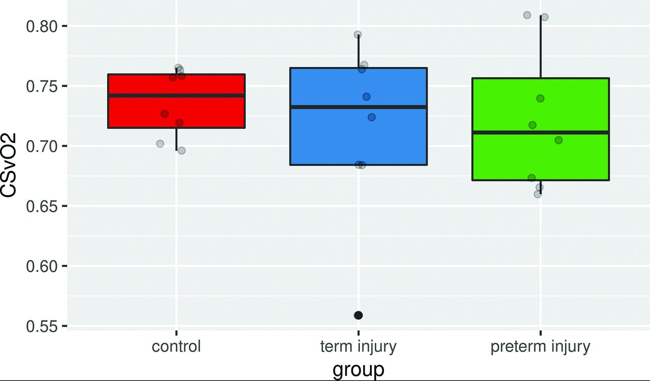
  • FIG 2.
    • Download figure
    • Open in new tab
    • Download powerpoint
    FIG 2.

    Boxplot of CSvO2 percentages by group. Gray circles are the ROI measurements from each subject.

Tables

  • Figures
    • View popup
    Table 1:

    Demographic data and clinical characteristics of the 3 groupsa

    CharacteristicsHealthy Controls (n = 8)Term Neonates with HIE
    (n = 8)
    P Value between Controls and Term HIEPreterm Neonates with HIE (n = 8)P Value between Term and Preterm HIE
    Gestational age (mean) (wk)39.3 (SD, 0.6)40.0 (SD, 0.8).05633.5 (SD, 2.1)<.001
    Corrected gestational age at MRI (mean) (wk)41.3 (SD, 1.1)41.9 (SD, 0.7).16635.7 (SD, 2.6)<.001
    Time interval between age at birth and age at MR imaging (mean) (wk)2.0 (SD, 0.7)1.9 (SD, 0.6).8192.2 (SD, 0.9).404
    Birth weight (mean) (g)3306.3 (SD, 353.1)3430.6 (SD, 471.4).5601926.3 (SD, 350.3)<.001
    No. of male neonates (No.) (%)4 (50)5 (62.5)1.0004 (50)1.000
    Apgar grade (median) (IQR)10 (10–10)6.5 (4–7)<.0016 (2–7).543
    Umbilical cord around the neck
    (No.) (%)
    2 (25)1 (12.5)1.0001 (12.5)1.000
    Oxygen inhalation (No.) (%)04 (50)NA7 (87.5)1.000
    Pulse oximetry (%)95–10090–95<.00190–95.122
    Meconium-stained amniotic fluid (No.) (%)03 (37.5)NA1 (12.5).569
    Premature rupture of fetal membranes (No.) (%)03 (37.5)NA5 (62.5).619
    Placental abruption (No.) (%)00NA2 (25)NA
    Cord prolapse (No.) (%)00NA1 (12.5)NA
    Fetal intrauterine distress (No.) (%)07 (87.5)NA2 (25).119
    Neonatal asphyxia resuscitation
    (No.) (%)
    08 (100)NA8 (100)NA
    Respiratory failure and ventilation (No.) (%)02 (25)NA5 (62.5).315
    Obtundation (No.) (%)07 (87.5)NA6 (75)1.000
    Stuporous (No.) (%)01 (12.5)NA2 (25)1.000
    Inhibited primitive reflexes (No.) (%)07 (87.5)NA6 (75)1.000
    Disappeared primitive reflexes
    (No.) (%)
    01 (12.5)NA2 (25)1.000
    Hypotonia (No.) (%)02 (25)NA4 (50).608
    Flaccid (No.) (%)01 (12.5)NA2 (25)1.000
    Seizures (No.) (%)02 (25)NA3 (37.5)1.000
    Therapeutic hypothermia (No.) (%)02 (25)NA1 (12.5)1.000
    • Note:—IQR indicates interquartile range; NA, not applicable.

    • ↵a P values were from the χ2 test or Fisher exact test for categoric variables or the Student t test for continuous variables.

    • View popup
    Table 2:

    Comparison of CSvO2 percentages in fetuses and neonates in the literature

    StudyCSvO2 (mean) (%)MethodSubjectsRegion
    van der Hoeven
    et al57
    73.56 (SD, 5.25)Fiber optic catheterHealthy neonates
    Buchvald et al5864.12 (SD, 4.6)NIRSHealthy neonatesFrontotemporal region
    Wintermark et al5977.3 (SD, 4.7)NIRSHypothermia therapy (moderate) in neonatal HIEFrontal lobe
    Wintermark et al5977.6 (SD, 6.6)NIRSHypothermia therapy (severe) in neonatal HIEFrontal lobe
    De Vis et al6065.0 (SD, 13.0T2-TRIRHealthy neonatesSSS
    Shetty et al4873.2 (SD, 5.5)TRUSTHypothermia therapy in neonatal HIESSS
    Shetty et al4868.5 (SD, 9.6)TRUSTPost-hypothermia therapy in neonatal HIESSS
    Liu et al4962.6 (SD, 8.3)TRUSTHealthy neonatesSSS
    Yadav et al4467 (SD, 7)QSMHealthy fetuses (∼31 wk)SSS
    Jain et al4555.2SusceptometryNeonates with congenital heart diseaseSSS
    Yadav et al4662.6 (SD, 3.25)SusceptometryHealthy fetuses (∼31 wk)SSS
    Neelavalli et al4766 (SD, 9.4)SusceptometryHealthy fetuses (∼34 wk)SSS
    Average68.12
    • Note:—T2-TRIR indicates T2-prepared tissue relaxation inversion recovery; SSS, superior sagittal sinus; TRUST, T2-relaxation-under-spin tagging; QSM, quantitative susceptibility mapping; NIRS, near infrared resonance spectroscopy.

PreviousNext
Back to top

In this issue

American Journal of Neuroradiology: 42 (7)
American Journal of Neuroradiology
Vol. 42, Issue 7
1 Jul 2021
  • Table of Contents
  • Index by author
  • Complete Issue (PDF)
Advertisement
Print
Download PDF
Email Article

Thank you for your interest in spreading the word on American Journal of Neuroradiology.

NOTE: We only request your email address so that the person you are recommending the page to knows that you wanted them to see it, and that it is not junk mail. We do not capture any email address.

Enter multiple addresses on separate lines or separate them with commas.
Quantitative Susceptibility Mapping of Venous Vessels in Neonates with Perinatal Asphyxia
(Your Name) has sent you a message from American Journal of Neuroradiology
(Your Name) thought you would like to see the American Journal of Neuroradiology web site.
CAPTCHA
This question is for testing whether or not you are a human visitor and to prevent automated spam submissions.
Cite this article
A.M. Weber, Y. Zhang, C. Kames, A. Rauscher
Quantitative Susceptibility Mapping of Venous Vessels in Neonates with Perinatal Asphyxia
American Journal of Neuroradiology Jul 2021, 42 (7) 1327-1333; DOI: 10.3174/ajnr.A7086

Citation Manager Formats

  • BibTeX
  • Bookends
  • EasyBib
  • EndNote (tagged)
  • EndNote 8 (xml)
  • Medlars
  • Mendeley
  • Papers
  • RefWorks Tagged
  • Ref Manager
  • RIS
  • Zotero
0 Responses
Respond to this article
Share
Bookmark this article
Quantitative Susceptibility Mapping of Venous Vessels in Neonates with Perinatal Asphyxia
A.M. Weber, Y. Zhang, C. Kames, A. Rauscher
American Journal of Neuroradiology Jul 2021, 42 (7) 1327-1333; DOI: 10.3174/ajnr.A7086
del.icio.us logo Twitter logo Facebook logo Mendeley logo
  • Tweet Widget
  • Facebook Like
  • Google Plus One
Purchase

Jump to section

  • Article
    • Abstract
    • ABBREVIATIONS:
    • MATERIALS AND METHODS
    • RESULTS
    • DISCUSSION
    • CONCLUSIONS
    • Footnotes
    • References
  • Figures & Data
  • Supplemental
  • Info & Metrics
  • Responses
  • References
  • PDF

Related Articles

  • PubMed
  • Google Scholar

Cited By...

  • No citing articles found.
  • Crossref (1)
  • Google Scholar

This article has been cited by the following articles in journals that are participating in Crossref Cited-by Linking.

  • The application of magnetic susceptibility separation for measuring cerebral oxygenation in preterm neonates
    Thomas Gavin Carmichael, Alexander Rauscher, Ruth E. Grunau, Alexander Mark Weber
    Pediatric Research 2025

More in this TOC Section

Pediatrics

  • SyMRI & MR Fingerprinting in Brainstem Myelination
  • Comparison of Image Quality and Radiation Dose in Pediatric Temporal Bone CT Using Photon-Counting Detector CT and Energy-Integrating Detector CT
  • Dual-Layer Detector CT for PEDS Image Quality
Show more Pediatrics

Functional

  • Kurtosis and Epileptogenic Tubers: A Pilot Study
  • Glutaric Aciduria Type 1: DK vs. Conventional MRI
  • Multiparametric MRI in PEDS Pontine Glioma
Show more Functional

Similar Articles

Advertisement

Indexed Content

  • Current Issue
  • Accepted Manuscripts
  • Article Preview
  • Past Issues
  • Editorials
  • Editor's Choice
  • Fellows' Journal Club
  • Letters to the Editor
  • Video Articles

Cases

  • Case Collection
  • Archive - Case of the Week
  • Archive - Case of the Month
  • Archive - Classic Case

More from AJNR

  • Trainee Corner
  • Imaging Protocols
  • MRI Safety Corner
  • Book Reviews

Multimedia

  • AJNR Podcasts
  • AJNR Scantastics

Resources

  • Turnaround Time
  • Submit a Manuscript
  • Submit a Video Article
  • Submit an eLetter to the Editor/Response
  • Manuscript Submission Guidelines
  • Statistical Tips
  • Fast Publishing of Accepted Manuscripts
  • Graphical Abstract Preparation
  • Imaging Protocol Submission
  • Evidence-Based Medicine Level Guide
  • Publishing Checklists
  • Author Policies
  • Become a Reviewer/Academy of Reviewers
  • News and Updates

About Us

  • About AJNR
  • Editorial Board
  • Editorial Board Alumni
  • Alerts
  • Permissions
  • Not an AJNR Subscriber? Join Now
  • Advertise with Us
  • Librarian Resources
  • Feedback
  • Terms and Conditions
  • AJNR Editorial Board Alumni

American Society of Neuroradiology

  • Not an ASNR Member? Join Now

© 2025 by the American Society of Neuroradiology All rights, including for text and data mining, AI training, and similar technologies, are reserved.
Print ISSN: 0195-6108 Online ISSN: 1936-959X

Powered by HighWire