
Article Figures & Data
Figures
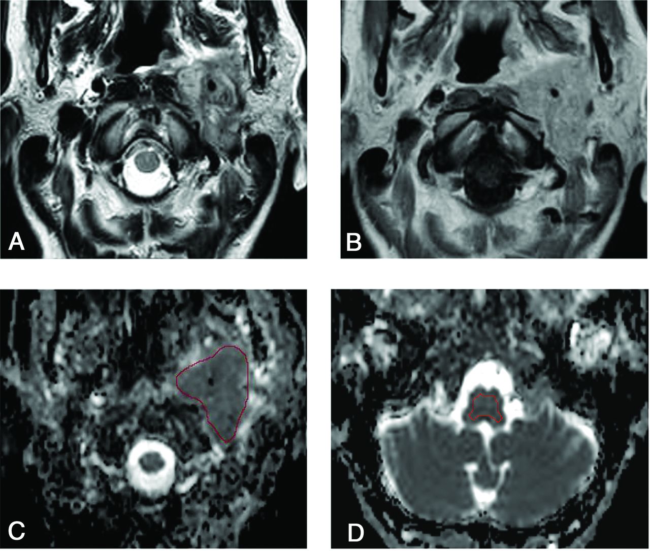
- FIG 1.
A 48-year-old woman positive for the SDHC mutation with a paraganglioma in the left jugular foramen. A and B, Axial T2-weighted and contrast-enhanced T1-weighted images demonstrate a heterogeneously enhancing, irregularly shaped tumor with flow voids in the left jugular foramen. C, An ROI is placed on the lesion on the ADC map. The mean ADC, maximum ADC, and minimum ADC values of reader 1 are 1.06, 1.47, and 0.53 × 10⁻³ mm2/s, respectively. D, Another ROI for an internal standard is placed on the medulla as an internal control (mean ADC, 0.75 × 10⁻³ mm2/s). The mean nADC, maximum nADC, and minimum nADC are 1.41, 1.96, and 0.71 × 10⁻³ mm2/s, respectively.
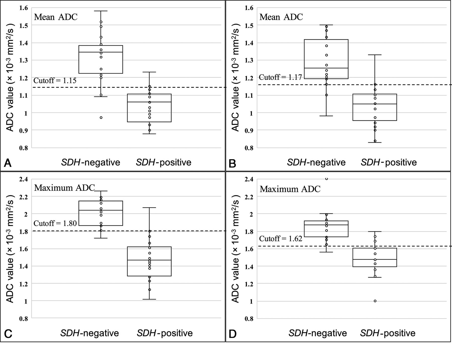
- FIG 2.
Comparison of mean and maximum ADC values between the SDH mutation–negative group and the SDH mutation–positive group (A and C, result of reader 1; B and D, result of reader 2).
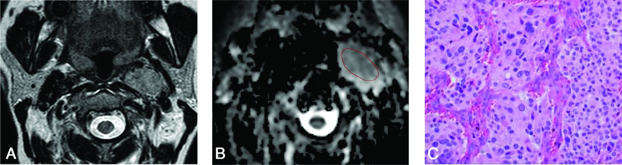
- FIG 3.
A 62-year-old woman negative for the SDH mutation with a paraganglioma in the right carotid space. A, Axial T2-weighted image demonstrates a heterogeneous well-defined tumor with flow voids in the right carotid space. B, The freehand ROI is placed on the lesion on the ADC map. Mean ADC, maximum ADC, and minimum ADC values of reader 1 are 1.31, 1.61, and 0.71 × 10⁻³ mm2/s, respectively. C, The resection specimen shows chief cells forming variable-size clusters in the zellballen pattern (H&E, ×40).
- FIG 4.
A 52-year-old woman positive for the SDHD mutation with a paraganglioma in the left carotid space. A, Axial T2-weighted image demonstrates a heterogeneous tumor with flow voids. B, The freehand ROI is placed on this lesion on the ADC map. The mean ADC, maximum ADC, and minimum ADC of reader 1 are 1.13, 1.57, and 0.51 × 10⁻³ mm2/s, respectively. C, The resection specimen shows a large and irregular cell nest and prominent vascularity (H&E, ×40).


Tables
SDH Mutation–Positive SDH Mutation–Negative P Value No. of lesions 30 18 NA Sex (male/female) 7:16 2:15 .37 Age (mean) (yr) 43.9 (SD, 16.2) (23 patients) 56.9 (SD, 10.7) (17 patients) .007 Maximum diameter (median) (IQR) (mm) 26.5 (20.6–33.0) 24.4 (21.2–36.0) .68 Salt-and-pepper appearance 24/30 13/18 .72 Ratio of head/neck region 13:17 13:5 .07 Adjacent osseous erosive changes of head region 13:13 12:13 1 Necrotic or cystic changes 18/30 10/18 .77 Note:—NA indicates not applicable; IQR, interquartile range.
- Table 2:
Diagnostic performance of ADC values in differentiating groups positive for the SDH mutation from those negative for it (both readers’ results)
ADC Mean (× 10–3
mm2/s)ADC Maximum (× 10–3
mm2/s)nADC Mean nADC Maximum Cutoff 1.15/1.17 1.80/1.62 1.52/1.53 2.32/2.07 Sensitivity 0.90/0.95 0.93/0.79 0.90/0.95 0.93/0.68 Specificity 0.83/0.78 0.94/0.94 0.83/0.83 0.94/1.00 PPV 0.90/0.82 0.97/0.94 0.90/0.86 0.97/1.00 NPV 0.83/0.93 0.90/0.81 0.83/0.94 0.90/0.75 Accuracy 0.88/0.87 0.94 /0.87 0.88/0.89 0.94/0.84 AUC 0.87/0.91 0.94/0.94 0.87/0.91 0.94/0.94 Note:—PPV indicates positive predictive value; NPV, negative predictive value; AUC, area under the curve.


