Advertisement

Main menu

  • Home
  • Content
    • Current Issue
    • Accepted Manuscripts
    • Article Preview
    • Past Issue Archive
    • Video Articles
    • AJNR Case Collection
    • Case of the Week Archive
    • Case of the Month Archive
    • Classic Case Archive
  • Special Collections
    • AJNR Awards
    • Low-Field MRI
    • Alzheimer Disease
    • ASNR Foundation Special Collection
    • Photon-Counting CT
    • View All
  • Multimedia
    • AJNR Podcasts
    • AJNR SCANtastic
    • Trainee Corner
    • MRI Safety Corner
    • Imaging Protocols
  • For Authors
    • Submit a Manuscript
    • Submit a Video Article
    • Submit an eLetter to the Editor/Response
    • Manuscript Submission Guidelines
    • Statistical Tips
    • Fast Publishing of Accepted Manuscripts
    • Graphical Abstract Preparation
    • Imaging Protocol Submission
    • Author Policies
  • About Us
    • About AJNR
    • Editorial Board
    • Editorial Board Alumni
  • More
    • Become a Reviewer/Academy of Reviewers
    • Subscribers
    • Permissions
    • Alerts
    • Feedback
    • Advertisers
    • ASNR Home

User menu

  • Alerts
  • Log in

Search

  • Advanced search
American Journal of Neuroradiology

American Journal of Neuroradiology

ASHNR American Society of Functional Neuroradiology ASHNR American Society of Pediatric Neuroradiology ASSR
  • Alerts
  • Log in

Advanced Search

  • Home
  • Content
    • Current Issue
    • Accepted Manuscripts
    • Article Preview
    • Past Issue Archive
    • Video Articles
    • AJNR Case Collection
    • Case of the Week Archive
    • Case of the Month Archive
    • Classic Case Archive
  • Special Collections
    • AJNR Awards
    • Low-Field MRI
    • Alzheimer Disease
    • ASNR Foundation Special Collection
    • Photon-Counting CT
    • View All
  • Multimedia
    • AJNR Podcasts
    • AJNR SCANtastic
    • Trainee Corner
    • MRI Safety Corner
    • Imaging Protocols
  • For Authors
    • Submit a Manuscript
    • Submit a Video Article
    • Submit an eLetter to the Editor/Response
    • Manuscript Submission Guidelines
    • Statistical Tips
    • Fast Publishing of Accepted Manuscripts
    • Graphical Abstract Preparation
    • Imaging Protocol Submission
    • Author Policies
  • About Us
    • About AJNR
    • Editorial Board
    • Editorial Board Alumni
  • More
    • Become a Reviewer/Academy of Reviewers
    • Subscribers
    • Permissions
    • Alerts
    • Feedback
    • Advertisers
    • ASNR Home
  • Follow AJNR on Twitter
  • Visit AJNR on Facebook
  • Follow AJNR on Instagram
  • Join AJNR on LinkedIn
  • RSS Feeds

AJNR Awards, New Junior Editors, and more. Read the latest AJNR updates

Research ArticlePediatrics

Transient Hyperintensity of the Infant Thyroid Gland on T1-Weighted MR Imaging: Correlation with Postnatal Age, Gestational Age, and Signal Intensity of the Pituitary Gland

H. Maki, M. Nakagawa, R. Kagaya, S. Kumazawa, K. Matsumoto, M. Hatano, Y. Miyake, W. Sugihara and Y. Shibamoto
American Journal of Neuroradiology May 2021, 42 (5) 955-960; DOI: https://doi.org/10.3174/ajnr.A7024
H. Maki
aDepartment of Radiology (H.M., M.N., S.K., K.M., W.S., Y.S.), Nagoya City University Graduate School of Medical Sciences, Mizuho-ku, Nagoya, Aichi, Japan
  • Find this author on Google Scholar
  • Find this author on PubMed
  • Search for this author on this site
  • ORCID record for H. Maki
M. Nakagawa
aDepartment of Radiology (H.M., M.N., S.K., K.M., W.S., Y.S.), Nagoya City University Graduate School of Medical Sciences, Mizuho-ku, Nagoya, Aichi, Japan
  • Find this author on Google Scholar
  • Find this author on PubMed
  • Search for this author on this site
  • ORCID record for M. Nakagawa
R. Kagaya
bDepartment of Radiology, Kariya Toyota General Hospital (R.K.), Kariya-shi, Aichi, Japan
  • Find this author on Google Scholar
  • Find this author on PubMed
  • Search for this author on this site
  • ORCID record for R. Kagaya
S. Kumazawa
aDepartment of Radiology (H.M., M.N., S.K., K.M., W.S., Y.S.), Nagoya City University Graduate School of Medical Sciences, Mizuho-ku, Nagoya, Aichi, Japan
  • Find this author on Google Scholar
  • Find this author on PubMed
  • Search for this author on this site
  • ORCID record for S. Kumazawa
K. Matsumoto
aDepartment of Radiology (H.M., M.N., S.K., K.M., W.S., Y.S.), Nagoya City University Graduate School of Medical Sciences, Mizuho-ku, Nagoya, Aichi, Japan
  • Find this author on Google Scholar
  • Find this author on PubMed
  • Search for this author on this site
  • ORCID record for K. Matsumoto
M. Hatano
cDepartment of Radiology, Nagoya City West Medical Center (M.H.), Nagoya, Japan
  • Find this author on Google Scholar
  • Find this author on PubMed
  • Search for this author on this site
  • ORCID record for M. Hatano
Y. Miyake
dDepartment of Radiology (Y.M.), Nagoya Daini Red Cross Hospital, Nagoya-shi, Aichi, Japan
  • Find this author on Google Scholar
  • Find this author on PubMed
  • Search for this author on this site
  • ORCID record for Y. Miyake
W. Sugihara
aDepartment of Radiology (H.M., M.N., S.K., K.M., W.S., Y.S.), Nagoya City University Graduate School of Medical Sciences, Mizuho-ku, Nagoya, Aichi, Japan
  • Find this author on Google Scholar
  • Find this author on PubMed
  • Search for this author on this site
  • ORCID record for W. Sugihara
Y. Shibamoto
aDepartment of Radiology (H.M., M.N., S.K., K.M., W.S., Y.S.), Nagoya City University Graduate School of Medical Sciences, Mizuho-ku, Nagoya, Aichi, Japan
  • Find this author on Google Scholar
  • Find this author on PubMed
  • Search for this author on this site
  • ORCID record for Y. Shibamoto
  • Article
  • Figures & Data
  • Info & Metrics
  • Responses
  • References
  • PDF
Loading

Article Figures & Data

Figures

  • Tables
  • FIG 1.
    • Download figure
    • Open in new tab
    • Download powerpoint
    FIG 1.

    Axial 3D GRE T1-weighted MR images of an infant at postnatal day 6 show the thyroid (A) and pituitary (B). For measurement of the signal intensity, ROIs are placed on the thyroid (black circle, A), sternocleidomastoid muscle (white circle, A), and anterior pituitary (black circle, B). The posterior pituitary is shown (arrow, B).
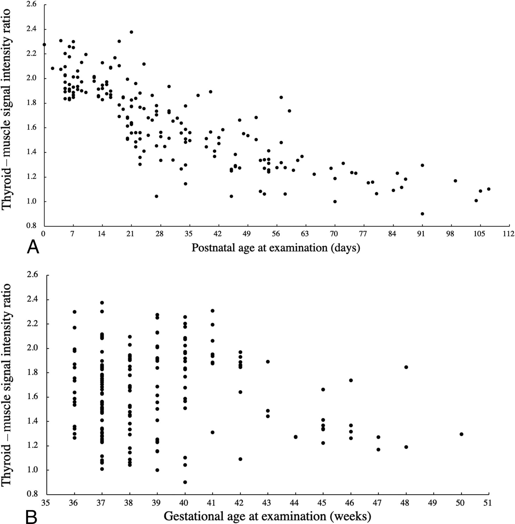
  • FIG 2.
    • Download figure
    • Open in new tab
    • Download powerpoint
    FIG 2.

    Scatterplots of the thyroid–muscle signal intensity ratio against the corresponding postnatal age at examination (A) and gestational age at examination (B). There is a negative correlation between the thyroid–muscle signal intensity ratio and postnatal age at examination (A). Conversely, there is no distinct relationship between the thyroid–muscle signal intensity ratio and gestational age at examination (B).
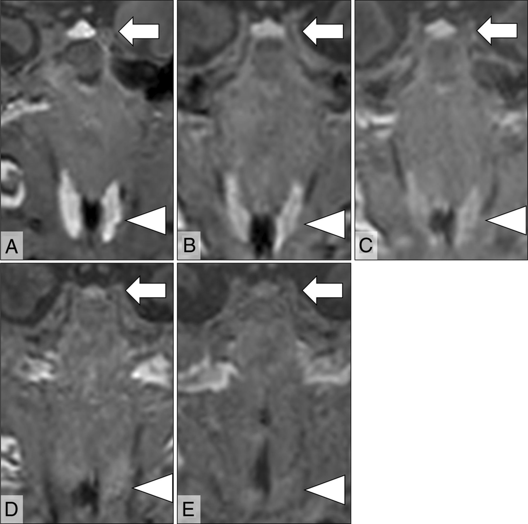
  • FIG 3.
    • Download figure
    • Open in new tab
    • Download powerpoint
    FIG 3.

    Five different cases showing the thyroid (arrowheads) and anterior pituitary (arrows) on coronal 3D GRE T1-weighted MR images examined at different postnatal ages: 4 days (A), 21 days (B), 33 days (C), 46 days (D), and 70 days (E). These patients' gestational ages at birth are 41, 34, 31, 31, and 29 weeks, respectively. At 4 days of postnatal age, the thyroid and anterior pituitary show extremely high signal intensity (A), and the signal intensity of those glands decreased proportionally to postnatal age (B–E). The signal intensities of the thyroid glands are similar to those of the anterior pituitary glands regardless of postnatal age, and the thyroid–anterior pituitary signal intensity ratios are almost 1 (A = 0.91, B = 1.04, C = 0.83, D = 0.81, E = 0.92).

Tables

  • Figures
  • Means of thyroid–muscle, anterior pituitary–muscle, and thyroid–anterior pituitary signal intensity ratios against the corresponding postnatal age at examination grouped according to week

    Postnatal Age at ExaminationSignal Intensity Ratio
    Thyroid–MuscleAnterior Pituitary–MuscleThyroid–Anterior Pituitary
    DayNoMean (SD)Mean (SD)Mean (SD)
    0–6202.03 (0.15)2.01 (0.30)1.03 (0.16)
    7–13221.99 (0.13)2.00 (0.23)1.01 (0.12)
    14–20261.88 (0.19)1.78 (0.29)1.07 (0.13)
    21–27311.67 (0.25)1.65 (0.17)1.02 (0.14)
    28–34201.54 (0.19)1.50 (0.19)1.03 (0.11)
    35–4191.57 (0.18)1.60 (0.27)1.00 (0.17)
    42–48121.38 (0.17)1.43 (0.20)0.98 (0.11)
    49–55131.34 (0.17)1.36 (0.21)1.00 (0.14)
    56–62101.40 (0.22)1.36 (0.18)1.03 (0.12)
    63–6921.25 (0.02)1.26 (0.18)1.02 (0.17)
    70–7651.19 (0.10)1.16 (0.10)1.03 (0.10)
    77–8331.13 (0.04)1.06 (0.02)1.06 (0.06)
    84–9041.16 (0.05)1.12 (0.04)1.03 (0.06)
    91–9721.10 (0.20)1.13 (0.03)0.97 (0.15)
    98–10421.09 (0.08)1.14 (0.12)0.96 (0.03)
    105–11121.10 (0.01)1.14 (0.11)0.97 (0.10)
    Total183
PreviousNext
Back to top

In this issue

American Journal of Neuroradiology: 42 (5)
American Journal of Neuroradiology
Vol. 42, Issue 5
1 May 2021
  • Table of Contents
  • Index by author
  • Complete Issue (PDF)
Advertisement
Print
Download PDF
Email Article

Thank you for your interest in spreading the word on American Journal of Neuroradiology.

NOTE: We only request your email address so that the person you are recommending the page to knows that you wanted them to see it, and that it is not junk mail. We do not capture any email address.

Enter multiple addresses on separate lines or separate them with commas.
Transient Hyperintensity of the Infant Thyroid Gland on T1-Weighted MR Imaging: Correlation with Postnatal Age, Gestational Age, and Signal Intensity of the Pituitary Gland
(Your Name) has sent you a message from American Journal of Neuroradiology
(Your Name) thought you would like to see the American Journal of Neuroradiology web site.
CAPTCHA
This question is for testing whether or not you are a human visitor and to prevent automated spam submissions.
Cite this article
H. Maki, M. Nakagawa, R. Kagaya, S. Kumazawa, K. Matsumoto, M. Hatano, Y. Miyake, W. Sugihara, Y. Shibamoto
Transient Hyperintensity of the Infant Thyroid Gland on T1-Weighted MR Imaging: Correlation with Postnatal Age, Gestational Age, and Signal Intensity of the Pituitary Gland
American Journal of Neuroradiology May 2021, 42 (5) 955-960; DOI: 10.3174/ajnr.A7024

Citation Manager Formats

  • BibTeX
  • Bookends
  • EasyBib
  • EndNote (tagged)
  • EndNote 8 (xml)
  • Medlars
  • Mendeley
  • Papers
  • RefWorks Tagged
  • Ref Manager
  • RIS
  • Zotero
0 Responses
Respond to this article
Share
Bookmark this article
Transient Hyperintensity of the Infant Thyroid Gland on T1-Weighted MR Imaging: Correlation with Postnatal Age, Gestational Age, and Signal Intensity of the Pituitary Gland
H. Maki, M. Nakagawa, R. Kagaya, S. Kumazawa, K. Matsumoto, M. Hatano, Y. Miyake, W. Sugihara, Y. Shibamoto
American Journal of Neuroradiology May 2021, 42 (5) 955-960; DOI: 10.3174/ajnr.A7024
del.icio.us logo Twitter logo Facebook logo Mendeley logo
  • Tweet Widget
  • Facebook Like
  • Google Plus One
Purchase

Jump to section

  • Article
    • Abstract
    • ABBREVIATIONS:
    • MATERIALS AND METHODS
    • RESULTS
    • DISCUSSION
    • CONCLUSIONS
    • References
  • Figures & Data
  • Info & Metrics
  • Responses
  • References
  • PDF

Related Articles

  • No related articles found.
  • PubMed
  • Google Scholar

Cited By...

  • No citing articles found.
  • Crossref
  • Google Scholar

This article has not yet been cited by articles in journals that are participating in Crossref Cited-by Linking.

More in this TOC Section

  • SyMRI & MR Fingerprinting in Brainstem Myelination
  • Comparison of Image Quality and Radiation Dose in Pediatric Temporal Bone CT Using Photon-Counting Detector CT and Energy-Integrating Detector CT
  • Dual-Layer Detector CT for PEDS Image Quality
Show more Pediatrics

Similar Articles

Advertisement

Indexed Content

  • Current Issue
  • Accepted Manuscripts
  • Article Preview
  • Past Issues
  • Editorials
  • Editor's Choice
  • Fellows' Journal Club
  • Letters to the Editor
  • Video Articles

Cases

  • Case Collection
  • Archive - Case of the Week
  • Archive - Case of the Month
  • Archive - Classic Case

More from AJNR

  • Trainee Corner
  • Imaging Protocols
  • MRI Safety Corner
  • Book Reviews

Multimedia

  • AJNR Podcasts
  • AJNR Scantastics

Resources

  • Turnaround Time
  • Submit a Manuscript
  • Submit a Video Article
  • Submit an eLetter to the Editor/Response
  • Manuscript Submission Guidelines
  • Statistical Tips
  • Fast Publishing of Accepted Manuscripts
  • Graphical Abstract Preparation
  • Imaging Protocol Submission
  • Evidence-Based Medicine Level Guide
  • Publishing Checklists
  • Author Policies
  • Become a Reviewer/Academy of Reviewers
  • News and Updates

About Us

  • About AJNR
  • Editorial Board
  • Editorial Board Alumni
  • Alerts
  • Permissions
  • Not an AJNR Subscriber? Join Now
  • Advertise with Us
  • Librarian Resources
  • Feedback
  • Terms and Conditions
  • AJNR Editorial Board Alumni

American Society of Neuroradiology

  • Not an ASNR Member? Join Now

© 2025 by the American Society of Neuroradiology All rights, including for text and data mining, AI training, and similar technologies, are reserved.
Print ISSN: 0195-6108 Online ISSN: 1936-959X

Powered by HighWire