Advertisement

Main menu

  • Home
  • Content
    • Current Issue
    • Accepted Manuscripts
    • Article Preview
    • Past Issue Archive
    • Video Articles
    • AJNR Case Collection
    • Case of the Week Archive
    • Case of the Month Archive
    • Classic Case Archive
  • Special Collections
    • AJNR Awards
    • Low-Field MRI
    • Alzheimer Disease
    • ASNR Foundation Special Collection
    • Photon-Counting CT
    • View All
  • Multimedia
    • AJNR Podcasts
    • AJNR SCANtastic
    • Trainee Corner
    • MRI Safety Corner
    • Imaging Protocols
  • For Authors
    • Submit a Manuscript
    • Submit a Video Article
    • Submit an eLetter to the Editor/Response
    • Manuscript Submission Guidelines
    • Statistical Tips
    • Fast Publishing of Accepted Manuscripts
    • Graphical Abstract Preparation
    • Imaging Protocol Submission
    • Author Policies
  • About Us
    • About AJNR
    • Editorial Board
    • Editorial Board Alumni
  • More
    • Become a Reviewer/Academy of Reviewers
    • Subscribers
    • Permissions
    • Alerts
    • Feedback
    • Advertisers
    • ASNR Home

User menu

  • Alerts
  • Log in

Search

  • Advanced search
American Journal of Neuroradiology

American Journal of Neuroradiology

ASHNR American Society of Functional Neuroradiology ASHNR American Society of Pediatric Neuroradiology ASSR
  • Alerts
  • Log in

Advanced Search

  • Home
  • Content
    • Current Issue
    • Accepted Manuscripts
    • Article Preview
    • Past Issue Archive
    • Video Articles
    • AJNR Case Collection
    • Case of the Week Archive
    • Case of the Month Archive
    • Classic Case Archive
  • Special Collections
    • AJNR Awards
    • Low-Field MRI
    • Alzheimer Disease
    • ASNR Foundation Special Collection
    • Photon-Counting CT
    • View All
  • Multimedia
    • AJNR Podcasts
    • AJNR SCANtastic
    • Trainee Corner
    • MRI Safety Corner
    • Imaging Protocols
  • For Authors
    • Submit a Manuscript
    • Submit a Video Article
    • Submit an eLetter to the Editor/Response
    • Manuscript Submission Guidelines
    • Statistical Tips
    • Fast Publishing of Accepted Manuscripts
    • Graphical Abstract Preparation
    • Imaging Protocol Submission
    • Author Policies
  • About Us
    • About AJNR
    • Editorial Board
    • Editorial Board Alumni
  • More
    • Become a Reviewer/Academy of Reviewers
    • Subscribers
    • Permissions
    • Alerts
    • Feedback
    • Advertisers
    • ASNR Home
  • Follow AJNR on Twitter
  • Visit AJNR on Facebook
  • Follow AJNR on Instagram
  • Join AJNR on LinkedIn
  • RSS Feeds

AJNR Awards, New Junior Editors, and more. Read the latest AJNR updates

LetterLetter

Reply:

M. Shapiro, K. Srivatanakul, E. Raz, M. Litao, E. Nossek and P.K. Nelson
American Journal of Neuroradiology April 2021, 42 (4) E31-E32; DOI: https://doi.org/10.3174/ajnr.A7098
M. Shapiro
aDepartment of Radiology and NeurologyNYU Grossman School of MedicineNew York, New York
  • Find this author on Google Scholar
  • Find this author on PubMed
  • Search for this author on this site
  • ORCID record for M. Shapiro
K. Srivatanakul
bDepartment of NeurosurgeryTokai UniversityKanagawa, Japan
  • Find this author on Google Scholar
  • Find this author on PubMed
  • Search for this author on this site
  • ORCID record for K. Srivatanakul
E. Raz
cDepartment of RadiologyNYU Grossman School of MedicineNew York, New York
  • Find this author on Google Scholar
  • Find this author on PubMed
  • Search for this author on this site
  • ORCID record for E. Raz
M. Litao
dDepartment of RadiologyNYU Grossman School of MedicineNew York, New York
  • Find this author on Google Scholar
  • Find this author on PubMed
  • Search for this author on this site
  • ORCID record for M. Litao
E. Nossek
eDepartment of NeurosurgeryNYU Grossman School of MedicineNew York, New York
  • Find this author on Google Scholar
  • Find this author on PubMed
  • Search for this author on this site
  • ORCID record for E. Nossek
P.K. Nelson
fDepartment of NeurosurgeryNYU Grossman School of MedicineNew York, New York
  • Find this author on Google Scholar
  • Find this author on PubMed
  • Search for this author on this site
  • ORCID record for P.K. Nelson
  • Article
  • Figures & Data
  • Info & Metrics
  • Responses
  • References
  • PDF
Loading

We are extremely thankful to the Hannover group for forwarding to the Editor their deep insights and critique. We see the resulting exchange as an excellent opportunity for a substantive anatomic discussion and debate in the Journal.

It appears that we agree on most points. We agree that study of vascular dural anatomy requires gross pathology and histology material, as acknowledged in the Limitations section of our publication. We agree that the channels we both reported go by varied names, hindering a comprehensive literature search. We also agree that the distinction between dural venous channels and bridging veins traversing the dura is not semantic, and furthermore, we agree that from the perspective of surgical safety this distinction is immaterial. We have also observed, in a less structured setting than your group (during surgery), the appearance of bridging veins traversing the dura to join with the recipient dural sinus.

The question as to whether a venous structure, once inside the dura, retains anatomic characteristics of a vein or assumes those of a venous sinus is a histologic question and, in our opinion, cannot be answered for all structures in question by either imaging or gross examination. Where we disagree is that we believe, based on existing evidence, that the structures we illustrated are sinuses:

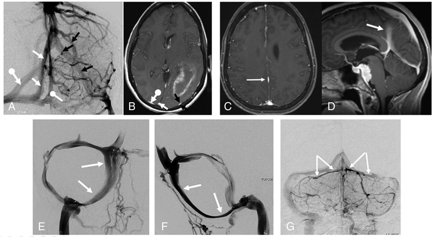
  1. They are lens-shaped, not round. This morphology closely resembles undisputed venous sinuses that traverse the dural sheets such as duplicated superior sagittal, falcine, and occipital sinuses (Fig 1A–F, Fig 2A, -B).

  2. We have observed what appear to be arachnoid granulations inside several of the channels we called “dural venous sinuses,” which are certainly not a structure expected in a bridging vein (Fig 2C,-D and Fig 6 in our original publication).

  3. Existence of tentorial venous sinuses is generally accepted, which is no proof of truth, but a strong supporting argument for the presence of similar channels in the supratentorial compartment (Fig 1G).

FIG 1.
  • Download figure
  • Open in new tab
  • Download powerpoint
FIG 1.

Examples of dural venous sinuses/channels similar to those described in our index article. Angiography (A) and MR imaging (B) of same patient, with multiple dural channels in addition to the usual superior sagittal sinus. The additional channels, marked by similar arrows on both angiogram and MR image, are flattened, lens-shaped structures, just like the dural venous channels we describe. C and D, Axial and sagittal MR images of patient with a falcine sinus variant—the same flattened morphology as in A and B. E and F, Morphology of the occipital sinus on a venous digital subtraction injection. G, Typical flattened appearance of tentorial venous sinuses.

FIG 2.
  • Download figure
  • Open in new tab
  • Download powerpoint
FIG 2.

A and B, Cinematic renderings, highlighting transition points (arrows) between round bridging vein and flattened dural venous channel in 2 different cases. A small fenestration versus arachnoid granulation is present in A (dashed arrow); neither structure would be expected in a bridging vein. Frontal (C) and lateral (D) views of another arachnoid granulation inside a dural venous channel.

We view the dura as a sort of arterial and venous “tabula rasa”—meaning any part of the dura can form a sinus when sufficient flow or other factors encourage it—both in utero and under pathologic circumstances such as a falcotentorial fistula. Indeed, we feel this view is entirely consistent with Padget’s investigations,1 repeatedly emphasizing the plexiform nature of dural venous structures.

Regarding the relationship between dural venous channels and dural fistulas, we feel that the varied opinions held by multiple groups as to their etiology and vascular pathoanatomy, given the relative paucity of data, are such that a productive discussion on the matter at this stage may be premature.

In conclusion, we believe that both bridging veins traversing the dura and supratenorial dural venous channels exist, and both are common. We continue to prefer the term “dural venous channel,” unless new data disprove this view. Perhaps “intradural venous channel” would be more of a semantic compromise. Regardless of whether any name at all will “catch,” we feel confident that both our efforts in this arena will continue.

ACKNOWLEDGMENTS

We thank Dr. Matthew G. Young (Department of Radiology, NYU Grossman School of Medicine, New York, New York) for generating the cinematic rendering images in Figure 1.

Reference

  1. 1.↵
    1. Padget DH
    . The cranial venous system in man in reference to development, adult configuration, and relation to the arteries. Am J Anat 1956;98:307–55 doi:10.1002/aja.1000980302 pmid:13362118
    CrossRefPubMed
  • © 2021 by American Journal of Neuroradiology
PreviousNext
Back to top

In this issue

American Journal of Neuroradiology: 42 (4)
American Journal of Neuroradiology
Vol. 42, Issue 4
1 Apr 2021
  • Table of Contents
  • Index by author
  • Complete Issue (PDF)
Advertisement
Print
Download PDF
Email Article

Thank you for your interest in spreading the word on American Journal of Neuroradiology.

NOTE: We only request your email address so that the person you are recommending the page to knows that you wanted them to see it, and that it is not junk mail. We do not capture any email address.

Enter multiple addresses on separate lines or separate them with commas.
Reply:
(Your Name) has sent you a message from American Journal of Neuroradiology
(Your Name) thought you would like to see the American Journal of Neuroradiology web site.
CAPTCHA
This question is for testing whether or not you are a human visitor and to prevent automated spam submissions.
Cite this article
M. Shapiro, K. Srivatanakul, E. Raz, M. Litao, E. Nossek, P.K. Nelson
Reply:
American Journal of Neuroradiology Apr 2021, 42 (4) E31-E32; DOI: 10.3174/ajnr.A7098

Citation Manager Formats

  • BibTeX
  • Bookends
  • EasyBib
  • EndNote (tagged)
  • EndNote 8 (xml)
  • Medlars
  • Mendeley
  • Papers
  • RefWorks Tagged
  • Ref Manager
  • RIS
  • Zotero
0 Responses
Respond to this article
Share
Bookmark this article
Reply:
M. Shapiro, K. Srivatanakul, E. Raz, M. Litao, E. Nossek, P.K. Nelson
American Journal of Neuroradiology Apr 2021, 42 (4) E31-E32; DOI: 10.3174/ajnr.A7098
del.icio.us logo Twitter logo Facebook logo Mendeley logo
  • Tweet Widget
  • Facebook Like
  • Google Plus One
Purchase

Jump to section

  • Article
    • ACKNOWLEDGMENTS
    • Reference
  • Figures & Data
  • Info & Metrics
  • Responses
  • References
  • PDF

Related Articles

  • No related articles found.
  • PubMed
  • Google Scholar

Cited By...

  • No citing articles found.
  • Crossref
  • Google Scholar

This article has not yet been cited by articles in journals that are participating in Crossref Cited-by Linking.

More in this TOC Section

  • Letter to the Editor regarding “Automated Volumetric Software in Dementia: Help or Hindrance to the Neuroradiologist?”
  • Reply:
  • Brain AVM’s Nidus: What if We Hadn’t Understood Anything?
Show more LETTERS

Similar Articles

Advertisement

Indexed Content

  • Current Issue
  • Accepted Manuscripts
  • Article Preview
  • Past Issues
  • Editorials
  • Editor's Choice
  • Fellows' Journal Club
  • Letters to the Editor
  • Video Articles

Cases

  • Case Collection
  • Archive - Case of the Week
  • Archive - Case of the Month
  • Archive - Classic Case

More from AJNR

  • Trainee Corner
  • Imaging Protocols
  • MRI Safety Corner
  • Book Reviews

Multimedia

  • AJNR Podcasts
  • AJNR Scantastics

Resources

  • Turnaround Time
  • Submit a Manuscript
  • Submit a Video Article
  • Submit an eLetter to the Editor/Response
  • Manuscript Submission Guidelines
  • Statistical Tips
  • Fast Publishing of Accepted Manuscripts
  • Graphical Abstract Preparation
  • Imaging Protocol Submission
  • Evidence-Based Medicine Level Guide
  • Publishing Checklists
  • Author Policies
  • Become a Reviewer/Academy of Reviewers
  • News and Updates

About Us

  • About AJNR
  • Editorial Board
  • Editorial Board Alumni
  • Alerts
  • Permissions
  • Not an AJNR Subscriber? Join Now
  • Advertise with Us
  • Librarian Resources
  • Feedback
  • Terms and Conditions
  • AJNR Editorial Board Alumni

American Society of Neuroradiology

  • Not an ASNR Member? Join Now

© 2025 by the American Society of Neuroradiology All rights, including for text and data mining, AI training, and similar technologies, are reserved.
Print ISSN: 0195-6108 Online ISSN: 1936-959X

Powered by HighWire