
Article Figures & Data
Figures
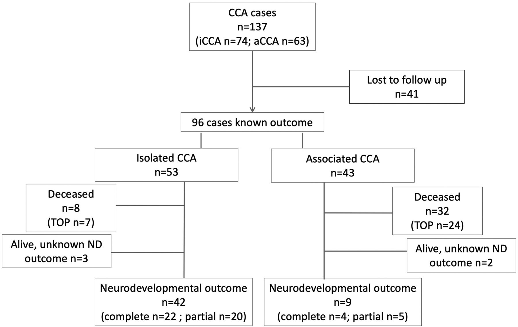
- FIG 1.
Flowchart detailing patient inclusion and exclusion criteria. ND indicates neurodevelopmental.
- FIG 2.
Comparison of mean MR imaging scores and 95% confidence intervals among the 4 CCA groups: isolated (complete and partial) and associated (complete and partial).
- FIG 3.
Example of associated partial CCA (white dashed arrow, D) in fetuses at 24 gestational weeks (A and B), 26 gestational weeks (C, D, F), and 31 gestational weeks (E). T2-weighted single-shot FSE images in the axial (A, B, E) and sagittal planes (C) and super-resolution 3D reconstruction of T2-weighted spin-echo FSE images through the coronal plane (D) and cortical surface (F). There is a malformation of cortical development with an abnormal “bump” in the insular region (dashed black arrow, A, C, F) and abnormal gyration in the posterior frontal cortex (dashed black arrow, E). There is a concurrent signal abnormality with low T2 signal intensity that follows the intermediate zone/subplate limit on the left (white arrow, A–D), also reaching the ventricular lining (black arrowhead, B), with slight ectasia of the homolateral posterior aspect of the lateral ventricle.


Tables
Summarized outcome by subgroup of corpus callosum agenesis
CCA Group Neurodevelopment Therapy School Normal Delay Phy Occ Spc Other Regular Assisted Not Age Mild Moderate Severe I C 15 5 1 1 9 2 1 12 9 P 16 2 1 1 11 11 7 A C 2 2 2 1 2 1 2 1 P 2 2 1 3 1 2 1 1 3 1 Note:—I indicates isolated; A, associated; C, complete; P, partial; Phy, physiotherapy; Occ, occupational therapy; Spc, speech therapy; Not Age, children not attending school, who were still under the local mandatory school age (5 years-old).


