Advertisement

Main menu

  • Home
  • Content
    • Current Issue
    • Accepted Manuscripts
    • Article Preview
    • Past Issue Archive
    • Video Articles
    • AJNR Case Collection
    • Case of the Week Archive
    • Case of the Month Archive
    • Classic Case Archive
  • Special Collections
    • AJNR Awards
    • Low-Field MRI
    • Alzheimer Disease
    • ASNR Foundation Special Collection
    • Photon-Counting CT
    • View All
  • Multimedia
    • AJNR Podcasts
    • AJNR SCANtastic
    • Trainee Corner
    • MRI Safety Corner
    • Imaging Protocols
  • For Authors
    • Submit a Manuscript
    • Submit a Video Article
    • Submit an eLetter to the Editor/Response
    • Manuscript Submission Guidelines
    • Statistical Tips
    • Fast Publishing of Accepted Manuscripts
    • Graphical Abstract Preparation
    • Imaging Protocol Submission
    • Author Policies
  • About Us
    • About AJNR
    • Editorial Board
    • Editorial Board Alumni
  • More
    • Become a Reviewer/Academy of Reviewers
    • Subscribers
    • Permissions
    • Alerts
    • Feedback
    • Advertisers
    • ASNR Home

User menu

  • Alerts
  • Log in

Search

  • Advanced search
American Journal of Neuroradiology

American Journal of Neuroradiology

ASHNR American Society of Functional Neuroradiology ASHNR American Society of Pediatric Neuroradiology ASSR
  • Alerts
  • Log in

Advanced Search

  • Home
  • Content
    • Current Issue
    • Accepted Manuscripts
    • Article Preview
    • Past Issue Archive
    • Video Articles
    • AJNR Case Collection
    • Case of the Week Archive
    • Case of the Month Archive
    • Classic Case Archive
  • Special Collections
    • AJNR Awards
    • Low-Field MRI
    • Alzheimer Disease
    • ASNR Foundation Special Collection
    • Photon-Counting CT
    • View All
  • Multimedia
    • AJNR Podcasts
    • AJNR SCANtastic
    • Trainee Corner
    • MRI Safety Corner
    • Imaging Protocols
  • For Authors
    • Submit a Manuscript
    • Submit a Video Article
    • Submit an eLetter to the Editor/Response
    • Manuscript Submission Guidelines
    • Statistical Tips
    • Fast Publishing of Accepted Manuscripts
    • Graphical Abstract Preparation
    • Imaging Protocol Submission
    • Author Policies
  • About Us
    • About AJNR
    • Editorial Board
    • Editorial Board Alumni
  • More
    • Become a Reviewer/Academy of Reviewers
    • Subscribers
    • Permissions
    • Alerts
    • Feedback
    • Advertisers
    • ASNR Home
  • Follow AJNR on Twitter
  • Visit AJNR on Facebook
  • Follow AJNR on Instagram
  • Join AJNR on LinkedIn
  • RSS Feeds

AJNR Awards, New Junior Editors, and more. Read the latest AJNR updates

Research ArticlePediatrics

Pediatric Head CT: Automated Quantitative Analysis with Quantile Regression

K.A. Cauley, Y. Hu and S.W. Fielden
American Journal of Neuroradiology February 2021, 42 (2) 382-388; DOI: https://doi.org/10.3174/ajnr.A6885
K.A. Cauley
aFrom the Departments of Radiology (K.A.C.)
  • Find this author on Google Scholar
  • Find this author on PubMed
  • Search for this author on this site
  • ORCID record for K.A. Cauley
Y. Hu
bBiomedical and Translational Informatics (Y.H.), Geisinger Medical Center, Danville, Pennsylvania
  • Find this author on Google Scholar
  • Find this author on PubMed
  • Search for this author on this site
  • ORCID record for Y. Hu
S.W. Fielden
cGeisinger Autism and Developmental Medicine Institute (S.W.F.), Lewisburg, Pennsylvania
  • Find this author on Google Scholar
  • Find this author on PubMed
  • Search for this author on this site
  • ORCID record for S.W. Fielden
  • Article
  • Figures & Data
  • Supplemental
  • Info & Metrics
  • Responses
  • References
  • PDF
Loading

Article Figures & Data

Figures

  • Tables
  • FIG 1.
    • Download figure
    • Open in new tab
    • Download powerpoint
    FIG 1.

    Products of automated head CT segmentation. Routine clinical head CT (A), brain-extracted image (B), and CSF eliminated (brain parenchyma) (C).
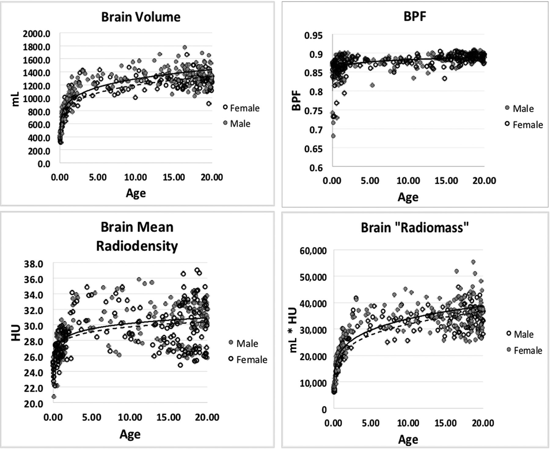
  • FIG 2.
    • Download figure
    • Open in new tab
    • Download powerpoint
    FIG 2.

    Scatterplots of computed brain metrics as a function of subject age: brain volume (A), BPF (B), mean brain radiodensity (C), and brain radiomass (D).

  • FIG 3.
    • Download figure
    • Open in new tab
    • Download powerpoint
    FIG 3.

    Box-and-whisker plots of BPF: 0–3 years (left) and 5–20 years (right). Tabular data below 0–3 years (Table 1). Data scatter is greater immediately after birth and decreases during the first 2 years, and the pattern is similar between the sexes. There is no significant difference between the mean BPF of the groupings. Table 2 shows statistically significant difference between the means of 5-year groupings.

  • FIG 4.
    • Download figure
    • Open in new tab
    • Download powerpoint
    FIG 4.

    Quantile regression of brain volume (upper graphs) and BPF (lower graphs), male on the left, female on the right. The 5%–95% and 10%–90% quantiles are shown.

Tables

  • Figures
    • View popup
    Table 1:

    Brain parenchymal fraction 0–3.0 years

    MaleFemale
    Age (yr)TotalMeanSDTotalMeanSD
    0–0.5200.8450.047170.840.062
    0.5–1.0150.860.018230.860.035
    1.0–1.5120.870.022130.870.024
    1.5–2.0100.860.03070.870.033
    2.0–2.540.870.00520.890.014
    2.5–3.020.890.00040.880.014
    • View popup
    Table 2:

    Brain parenchymal fraction 5–20 years

    Age (yr)TotalMean5–10 Years (P Value)10–15 Years (P Value)
    Male
     5–10180.872
     10–15200.879NS
     15–20720.891<.001.004
    Female
     5–10120.874
     10–15200.877NS
     15–20700.889.003.003
    • Note:—NS indicates not significant.

PreviousNext
Back to top

In this issue

American Journal of Neuroradiology: 42 (2)
American Journal of Neuroradiology
Vol. 42, Issue 2
1 Feb 2021
  • Table of Contents
  • Index by author
  • Complete Issue (PDF)
Advertisement
Print
Download PDF
Email Article

Thank you for your interest in spreading the word on American Journal of Neuroradiology.

NOTE: We only request your email address so that the person you are recommending the page to knows that you wanted them to see it, and that it is not junk mail. We do not capture any email address.

Enter multiple addresses on separate lines or separate them with commas.
Pediatric Head CT: Automated Quantitative Analysis with Quantile Regression
(Your Name) has sent you a message from American Journal of Neuroradiology
(Your Name) thought you would like to see the American Journal of Neuroradiology web site.
CAPTCHA
This question is for testing whether or not you are a human visitor and to prevent automated spam submissions.
Cite this article
K.A. Cauley, Y. Hu, S.W. Fielden
Pediatric Head CT: Automated Quantitative Analysis with Quantile Regression
American Journal of Neuroradiology Feb 2021, 42 (2) 382-388; DOI: 10.3174/ajnr.A6885

Citation Manager Formats

  • BibTeX
  • Bookends
  • EasyBib
  • EndNote (tagged)
  • EndNote 8 (xml)
  • Medlars
  • Mendeley
  • Papers
  • RefWorks Tagged
  • Ref Manager
  • RIS
  • Zotero
0 Responses
Respond to this article
Share
Bookmark this article
Pediatric Head CT: Automated Quantitative Analysis with Quantile Regression
K.A. Cauley, Y. Hu, S.W. Fielden
American Journal of Neuroradiology Feb 2021, 42 (2) 382-388; DOI: 10.3174/ajnr.A6885
del.icio.us logo Twitter logo Facebook logo Mendeley logo
  • Tweet Widget
  • Facebook Like
  • Google Plus One
Purchase

Jump to section

  • Article
    • Abstract
    • ABBREVIATION:
    • MATERIALS AND METHODS
    • RESULTS
    • DISCUSSION
    • CONCLUSIONS
    • Footnotes
    • References
  • Figures & Data
  • Supplemental
  • Info & Metrics
  • Responses
  • References
  • PDF

Related Articles

  • PubMed
  • Google Scholar

Cited By...

  • Head CT: Toward Making Full Use of the Information the X-Rays Give
  • Crossref (3)
  • Google Scholar

This article has been cited by the following articles in journals that are participating in Crossref Cited-by Linking.

  • Head CT: Toward Making Full Use of the Information the X-Rays Give
    K.A. Cauley, Y. Hu, S.W. Fielden
    American Journal of Neuroradiology 2021 42 8
  • Prediction of Shunt Malfunction Using Automated Ventricular Volume Analysis and Radiomics
    Ryan T. Kellogg, Jan Vargas, Matthew Nguyen, Anthony Nwanko, Sohil Patel, Kanchan Ghimire, Xue Feng
    Neurosurgery 2024
  • Importance of Neuroimaging in Infants with Microcephaly
    Akhil Maheshwari, Thierry AGM Huisman, Maroun Mhanna, Sabrina Rangwani, Gunes Orman
    Newborn 2023 2 2

More in this TOC Section

Pediatrics

  • SyMRI & MR Fingerprinting in Brainstem Myelination
  • Comparison of Image Quality and Radiation Dose in Pediatric Temporal Bone CT Using Photon-Counting Detector CT and Energy-Integrating Detector CT
  • Axenfeld-Rieger Syndrome: Neuroimaging Findings
Show more Pediatrics

Functional

  • Kurtosis and Epileptogenic Tubers: A Pilot Study
  • Glutaric Aciduria Type 1: DK vs. Conventional MRI
  • Predicting Outcomes in Tuberous Sclerosis Epilepsy
Show more Functional

Similar Articles

Advertisement

Indexed Content

  • Current Issue
  • Accepted Manuscripts
  • Article Preview
  • Past Issues
  • Editorials
  • Editor's Choice
  • Fellows' Journal Club
  • Letters to the Editor
  • Video Articles

Cases

  • Case Collection
  • Archive - Case of the Week
  • Archive - Case of the Month
  • Archive - Classic Case

More from AJNR

  • Trainee Corner
  • Imaging Protocols
  • MRI Safety Corner
  • Book Reviews

Multimedia

  • AJNR Podcasts
  • AJNR Scantastics

Resources

  • Turnaround Time
  • Submit a Manuscript
  • Submit a Video Article
  • Submit an eLetter to the Editor/Response
  • Manuscript Submission Guidelines
  • Statistical Tips
  • Fast Publishing of Accepted Manuscripts
  • Graphical Abstract Preparation
  • Imaging Protocol Submission
  • Evidence-Based Medicine Level Guide
  • Publishing Checklists
  • Author Policies
  • Become a Reviewer/Academy of Reviewers
  • News and Updates

About Us

  • About AJNR
  • Editorial Board
  • Editorial Board Alumni
  • Alerts
  • Permissions
  • Not an AJNR Subscriber? Join Now
  • Advertise with Us
  • Librarian Resources
  • Feedback
  • Terms and Conditions
  • AJNR Editorial Board Alumni

American Society of Neuroradiology

  • Not an ASNR Member? Join Now

© 2025 by the American Society of Neuroradiology All rights, including for text and data mining, AI training, and similar technologies, are reserved.
Print ISSN: 0195-6108 Online ISSN: 1936-959X

Powered by HighWire