
Article Figures & Data
Figures
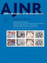
- FIGURE.
A 15-year-old boy presenting with migraines (A–C). Axial T2- and postcontrast T1-weighted images (A and B) show a cystic-appearing mass in the right lateral ventricle (arrows) without contrast enhancement. The mass appears solid on the CISS sequence (C). There is obstructive ventriculomegaly. Axial T2-weighted (D), FLAIR (E), and CISS (F) sequences in a 24-year-old asymptomatic woman. Images show a similar mass in the right lateral ventricle with a bright rim on FLAIR (E, arrowhead) and a solid texture on CISS (F).


