Advertisement

Main menu

  • Home
  • Content
    • Current Issue
    • Accepted Manuscripts
    • Article Preview
    • Past Issue Archive
    • Video Articles
    • AJNR Case Collection
    • Case of the Week Archive
    • Case of the Month Archive
    • Classic Case Archive
  • Special Collections
    • AJNR Awards
    • Low-Field MRI
    • Alzheimer Disease
    • ASNR Foundation Special Collection
    • Photon-Counting CT
    • View All
  • Multimedia
    • AJNR Podcasts
    • AJNR SCANtastic
    • Trainee Corner
    • MRI Safety Corner
    • Imaging Protocols
  • For Authors
    • Submit a Manuscript
    • Submit a Video Article
    • Submit an eLetter to the Editor/Response
    • Manuscript Submission Guidelines
    • Statistical Tips
    • Fast Publishing of Accepted Manuscripts
    • Graphical Abstract Preparation
    • Imaging Protocol Submission
    • Author Policies
  • About Us
    • About AJNR
    • Editorial Board
    • Editorial Board Alumni
  • More
    • Become a Reviewer/Academy of Reviewers
    • Subscribers
    • Permissions
    • Alerts
    • Feedback
    • Advertisers
    • ASNR Home

User menu

  • Alerts
  • Log in

Search

  • Advanced search
American Journal of Neuroradiology

American Journal of Neuroradiology

ASHNR American Society of Functional Neuroradiology ASHNR American Society of Pediatric Neuroradiology ASSR
  • Alerts
  • Log in

Advanced Search

  • Home
  • Content
    • Current Issue
    • Accepted Manuscripts
    • Article Preview
    • Past Issue Archive
    • Video Articles
    • AJNR Case Collection
    • Case of the Week Archive
    • Case of the Month Archive
    • Classic Case Archive
  • Special Collections
    • AJNR Awards
    • Low-Field MRI
    • Alzheimer Disease
    • ASNR Foundation Special Collection
    • Photon-Counting CT
    • View All
  • Multimedia
    • AJNR Podcasts
    • AJNR SCANtastic
    • Trainee Corner
    • MRI Safety Corner
    • Imaging Protocols
  • For Authors
    • Submit a Manuscript
    • Submit a Video Article
    • Submit an eLetter to the Editor/Response
    • Manuscript Submission Guidelines
    • Statistical Tips
    • Fast Publishing of Accepted Manuscripts
    • Graphical Abstract Preparation
    • Imaging Protocol Submission
    • Author Policies
  • About Us
    • About AJNR
    • Editorial Board
    • Editorial Board Alumni
  • More
    • Become a Reviewer/Academy of Reviewers
    • Subscribers
    • Permissions
    • Alerts
    • Feedback
    • Advertisers
    • ASNR Home
  • Follow AJNR on Twitter
  • Visit AJNR on Facebook
  • Follow AJNR on Instagram
  • Join AJNR on LinkedIn
  • RSS Feeds

AJNR Awards, New Junior Editors, and more. Read the latest AJNR updates

Research ArticlePediatrics
Open Access

DWI in Brains of Fetuses with Congenital Heart Disease: A Case-Control MR Imaging Study

J.-Y. Ren, H. Ji, M. Zhu and S.-Z. Dong
American Journal of Neuroradiology November 2021, 42 (11) 2040-2045; DOI: https://doi.org/10.3174/ajnr.A7267
J.-Y. Ren
aFrom the Department of Radiology, Shanghai Children’s Medical Center, School of Medicine, Shanghai Jiaotong University, Shanghai, P.R. China
  • Find this author on Google Scholar
  • Find this author on PubMed
  • Search for this author on this site
  • ORCID record for J.-Y. Ren
H. Ji
aFrom the Department of Radiology, Shanghai Children’s Medical Center, School of Medicine, Shanghai Jiaotong University, Shanghai, P.R. China
  • Find this author on Google Scholar
  • Find this author on PubMed
  • Search for this author on this site
  • ORCID record for H. Ji
M. Zhu
aFrom the Department of Radiology, Shanghai Children’s Medical Center, School of Medicine, Shanghai Jiaotong University, Shanghai, P.R. China
  • Find this author on Google Scholar
  • Find this author on PubMed
  • Search for this author on this site
  • ORCID record for M. Zhu
S.-Z. Dong
aFrom the Department of Radiology, Shanghai Children’s Medical Center, School of Medicine, Shanghai Jiaotong University, Shanghai, P.R. China
  • Find this author on Google Scholar
  • Find this author on PubMed
  • Search for this author on this site
  • ORCID record for S.-Z. Dong
  • Article
  • Figures & Data
  • Info & Metrics
  • Responses
  • References
  • PDF
Loading

Article Figures & Data

Figures

  • Tables
  • FIG 1.
    • Download figure
    • Open in new tab
    • Download powerpoint
    FIG 1.

    ADC map in a fetus at 26 weeks’ GA showing ROIs in the different regions. A and B, T2WI. C and D, The same GA, DWI. Pair-wise ADC values of the ROIs are manually delineated on each side of the frontal WM, periatrial WM, basal ganglia, thalamus, and cerebellar hemisphere, as well as a single measurement in the pons.
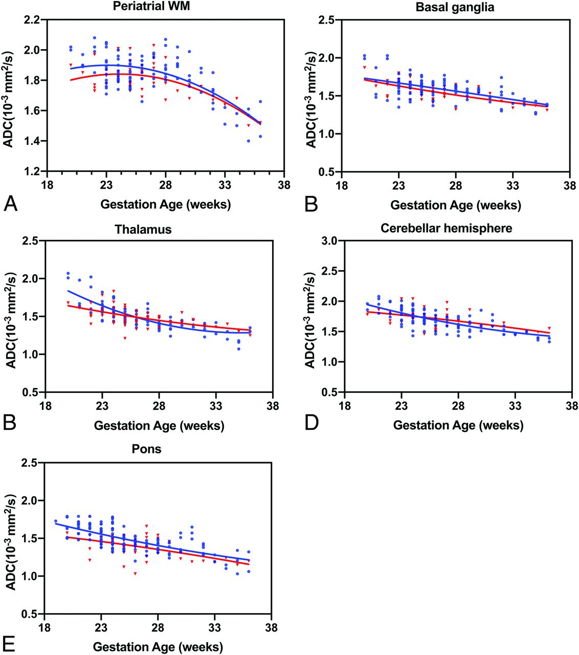
  • FIG 2.
    • Download figure
    • Open in new tab
    • Download powerpoint
    FIG 2.

    ADC values versus GA for all ROIs. Fetuses with CHD are referenced with red triangles, and blue circles indicate healthy controls. Periatrial WM (A), basal ganglia (B), thalamus (C), cerebellar hemisphere (D), pons (E).

  • FIG 3.
    • Download figure
    • Open in new tab
    • Download powerpoint
    FIG 3.

    Box-and-whisker plots representing the distribution of ADC measurements across the fetal brain anatomic structures and pregnancy trimesters. Asterisks indicate significant differences between the CHD (red) and control (blue) groups for the same structure. BG indicates basal ganglia; TH, thalamus; CH, cerebellar hemisphere; FWM, frontal WM; PWM, periatrial WM.

Tables

  • Figures
    • View popup
    Table 1:

    Clinical characteristics of our cohorta

    Control (n = 100)CHD (n = 50)P Valueb
    GA at MR imaging (mean) (wk)26.6 (SD, 3.9)26.1 (SD, 3.5).45
    Maternal age (mean) (yr)31.6 (SD, 5.0)33.1 (SD, 4.0).65
    Sex (No.) (%)
     Male46 (46.0%)26 (52.0%)
     Female54 (54.0%)24 (48.0%)
    Cardiac lesion
     TOFNA20 (40.0%)
     Double-outlet right ventricleNA10 (20.0%)
     TGANA15 (30.0%)
     HLHSNA5 (10.0%)
    • Note:—NA indicates not applicable.

    • ↵a Data are presented as number (%) unless otherwise noted.

    • ↵b By Wilcoxon rank sum test.

    • View popup
    Table 2:

    ADC values in the CHD and control groups in different brain areasa

    UnitControl (n = 100) (mean)CHD (n = 50) (mean)P Value
    GA <26 wk (No.)5025
     Frontal WM(10−3 mm2/s)1.87 (SD, 0.15)1.77 (SD, 0.14).01b
     Periatrial WM(10−3 mm2/s)1.88 (SD, 0.10)1.83 (SD, 0.08).04b
     Basal ganglia(10−3 mm2/s)1.65 (SD, 0.15)1.62 (SD, 0.11).47
     Thalamus(10−3 mm2/s)1.61 (SD, 0.16)1.55 (SD, 0.15).22
     CH(10−3 mm2/s)1.79 (SD, 0.15)1.77 (SD, 0.12).40
     Pons(10−3 mm2/s)1.53 (SD, 0.13)1.44 (SD, 0.13).03b
    GA ≥26 wk (No.)5025
     Frontal WM(10−3 mm2/s)1.94 (SD, 0.18)1.82 (SD, 0.15).006b
     Periatrial WM(10−3 mm2/s)1.80 (SD, 0.16)1.78 (SD, 0.13).41
     Basal ganglia(10−3 mm2/s)1.53 (SD, 0.13)1.50 (SD, 0.10).21
     Thalamus(10−3 mm2/s)1.39 (SD, 0.11)1.44 (SD, 0.08).04b
     CH(10−3 mm2/s)1.58 (SD, 0.13)1.65 (SD, 0.13).07
     Pons(10−3 mm2/s)1.37 (SD, 0.13)1.34 (SD, 0.15).38
    • Note:—CH indicates cerebellar hemisphere.

    • ↵a Data are presented as means.

    • ↵b P <.05.

PreviousNext
Back to top

In this issue

American Journal of Neuroradiology: 42 (11)
American Journal of Neuroradiology
Vol. 42, Issue 11
1 Nov 2021
  • Table of Contents
  • Index by author
  • Complete Issue (PDF)
Advertisement
Print
Download PDF
Email Article

Thank you for your interest in spreading the word on American Journal of Neuroradiology.

NOTE: We only request your email address so that the person you are recommending the page to knows that you wanted them to see it, and that it is not junk mail. We do not capture any email address.

Enter multiple addresses on separate lines or separate them with commas.
DWI in Brains of Fetuses with Congenital Heart Disease: A Case-Control MR Imaging Study
(Your Name) has sent you a message from American Journal of Neuroradiology
(Your Name) thought you would like to see the American Journal of Neuroradiology web site.
CAPTCHA
This question is for testing whether or not you are a human visitor and to prevent automated spam submissions.
Cite this article
J.-Y. Ren, H. Ji, M. Zhu, S.-Z. Dong
DWI in Brains of Fetuses with Congenital Heart Disease: A Case-Control MR Imaging Study
American Journal of Neuroradiology Nov 2021, 42 (11) 2040-2045; DOI: 10.3174/ajnr.A7267

Citation Manager Formats

  • BibTeX
  • Bookends
  • EasyBib
  • EndNote (tagged)
  • EndNote 8 (xml)
  • Medlars
  • Mendeley
  • Papers
  • RefWorks Tagged
  • Ref Manager
  • RIS
  • Zotero
0 Responses
Respond to this article
Share
Bookmark this article
DWI in Brains of Fetuses with Congenital Heart Disease: A Case-Control MR Imaging Study
J.-Y. Ren, H. Ji, M. Zhu, S.-Z. Dong
American Journal of Neuroradiology Nov 2021, 42 (11) 2040-2045; DOI: 10.3174/ajnr.A7267
del.icio.us logo Twitter logo Facebook logo Mendeley logo
  • Tweet Widget
  • Facebook Like
  • Google Plus One
Purchase

Jump to section

  • Article
    • Abstract
    • ABBREVIATIONS:
    • MATERIALS AND METHODS
    • RESULTS
    • DISCUSSION
    • CONCLUSIONS
    • Footnotes
    • References
  • Figures & Data
  • Info & Metrics
  • Responses
  • References
  • PDF

Related Articles

  • No related articles found.
  • PubMed
  • Google Scholar

Cited By...

  • No citing articles found.
  • Crossref (10)
  • Google Scholar

This article has been cited by the following articles in journals that are participating in Crossref Cited-by Linking.

  • Fetal MRI at 3 T: Principles to Optimize Success
    Fedel Machado-Rivas, Maria Camila Cortes-Albornoz, Onur Afacan, Maria Alejandra Bedoya, Camilo Calixto, Jungwhan John Choi, Matthew Ruggiero, Ali Gholipour, Camilo Jaimes
    RadioGraphics 2023 43 4
  • Harnessing the Power of Advanced Fetal Neuroimaging to Understand In Utero Footprints for Later Neuropsychiatric Disorders
    Josepheen De Asis-Cruz, Catherine Limperopoulos
    Biological Psychiatry 2023 93 10
  • Altered white matter connectivity in children with congenital heart disease with single ventricle physiology
    Brady J. Williamson, Maria E. Barnes-Davis, Jennifer Vannest, Julia S. Anixt, Haleh C. Heydarian, Lisa Kuan, Cameron S. Laue, Jayant Pratap, Mark Schapiro, Stephanie Y. Tseng, Darren S. Kadis
    Scientific Reports 2023 13 1
  • Structural Covariance Networks in the Fetal Brain Reveal Altered Neurodevelopment for Specific Subtypes of Congenital Heart Disease
    Siân Wilson, Daniel Cromb, Alexandra F. Bonthrone, Alena Uus, Anthony Price, Alexia Egloff, Milou P. M. Van Poppel, Johannes K. Steinweg, Kuberan Pushparajah, John Simpson, David F. A. Lloyd, Reza Razavi, Jonathan O'Muircheartaigh, A. David Edwards, Joseph V. Hajnal, Mary Rutherford, Serena J. Counsell
    Journal of the American Heart Association 2024
  • Advanced magnetic resonance imaging in human placenta: insights into fetal growth restriction and congenital heart disease
    Eric Sadiku, Liqun Sun, Christopher K. Macgowan, Mike Seed, Janna L. Morrison
    Frontiers in Cardiovascular Medicine 2024 11
  • Children with Congenital Heart Diseases Exhibit Altered Deep Gray Matter Structures
    Nils D. Forkert, Sarah J. MacEachern, Allison K. Duh, Peter Moon, Sarah Lee, Kristen W. Yeom
    Clinical Neuroradiology 2024 34 4
  • Regional changes in brain apparent diffusion coefficient in fetuses with complex congenital heart disease and normal pregnancy assessed using diffusion-weighted imaging
    Jia-Guang Song, Cong Sun, Mei Zhu, Jin-Xia Zhu, Nan Zhang, Guang-Bin Wang, Bin Zhao
    Frontiers in Neurology 2023 14
  • Sex differences in normal fetal regional brain apparent diffusion coefficient changes assessed by in utero DWI
    Jing-Ya Ren, Ming Zhu, Su-Zhen Dong
    Frontiers in Pediatrics 2024 12
  • Veränderte Diffusivität im fetalen Hirn bei angeborenen Herzfehlern
    Neuroradiologie Scan 2022 12 04
  • Volpe&#039;s Neurology of the Newborn
    Dorothy L. Bulas, Gilbert Vezina, Catherine Limperopoulos, Jamie L. Fraser, Adré J. du Plessis, Joseph J. Volpe
    2025

More in this TOC Section

  • SyMRI & MR Fingerprinting in Brainstem Myelination
  • Comparison of Image Quality and Radiation Dose in Pediatric Temporal Bone CT Using Photon-Counting Detector CT and Energy-Integrating Detector CT
  • Axenfeld-Rieger Syndrome: Neuroimaging Findings
Show more Pediatrics

Similar Articles

Advertisement

Indexed Content

  • Current Issue
  • Accepted Manuscripts
  • Article Preview
  • Past Issues
  • Editorials
  • Editor's Choice
  • Fellows' Journal Club
  • Letters to the Editor
  • Video Articles

Cases

  • Case Collection
  • Archive - Case of the Week
  • Archive - Case of the Month
  • Archive - Classic Case

More from AJNR

  • Trainee Corner
  • Imaging Protocols
  • MRI Safety Corner
  • Book Reviews

Multimedia

  • AJNR Podcasts
  • AJNR Scantastics

Resources

  • Turnaround Time
  • Submit a Manuscript
  • Submit a Video Article
  • Submit an eLetter to the Editor/Response
  • Manuscript Submission Guidelines
  • Statistical Tips
  • Fast Publishing of Accepted Manuscripts
  • Graphical Abstract Preparation
  • Imaging Protocol Submission
  • Evidence-Based Medicine Level Guide
  • Publishing Checklists
  • Author Policies
  • Become a Reviewer/Academy of Reviewers
  • News and Updates

About Us

  • About AJNR
  • Editorial Board
  • Editorial Board Alumni
  • Alerts
  • Permissions
  • Not an AJNR Subscriber? Join Now
  • Advertise with Us
  • Librarian Resources
  • Feedback
  • Terms and Conditions
  • AJNR Editorial Board Alumni

American Society of Neuroradiology

  • Not an ASNR Member? Join Now

© 2025 by the American Society of Neuroradiology All rights, including for text and data mining, AI training, and similar technologies, are reserved.
Print ISSN: 0195-6108 Online ISSN: 1936-959X

Powered by HighWire