
Article Figures & Data
Figures
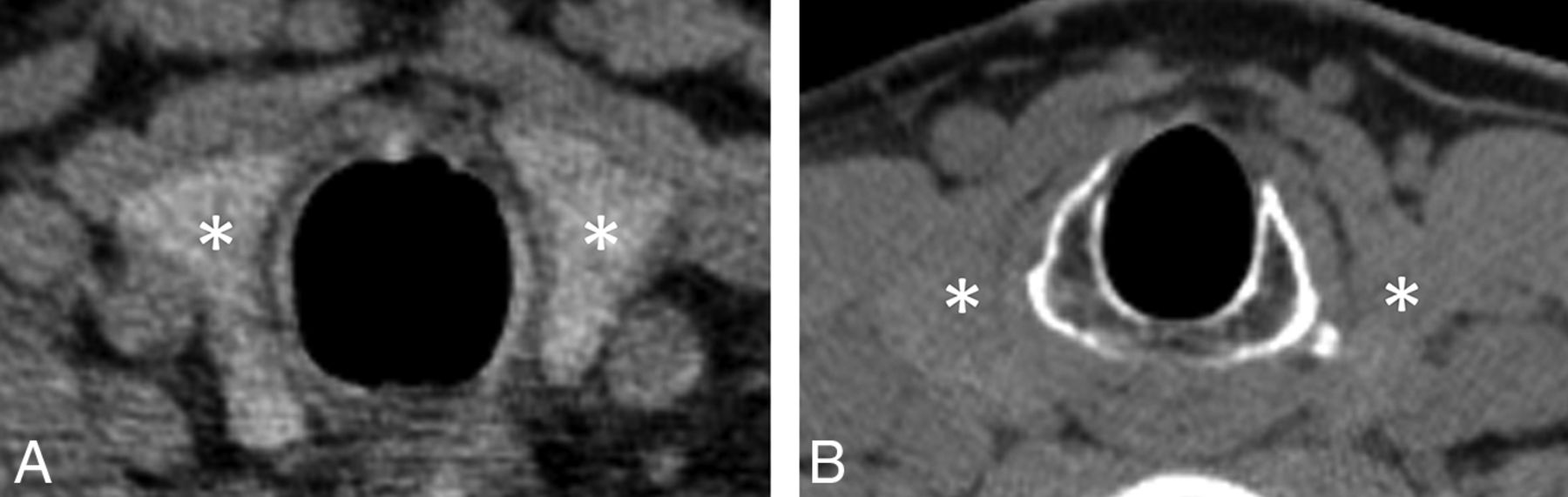
- FIG 1.
Axial, noncontrast, single-energy CT image (A) obtained in a patient with normal thyroid function demonstrates the normal hyperattenuating appearance of the thyroid gland (asterisks, A) relative to adjacent soft tissue. When present, this normal hyperattenuating appearance enables differentiation of parathyroid lesions adjacent to the thyroid gland from exophytic thyroid tissue. In contrast, the axial noncontrast single-energy CT image (B) obtained in a patient with long-standing Hashimoto disease demonstrates an iodine-deficient thyroid gland (asterisks, B), appearing nearly isodense to muscle, which renders differentiation of parathyroid lesions from exophytic thyroid tissue more difficult.
- FIG 2.
Virtual monoenergetic spectral curves (A) demonstrate noncontrast Hounsfield unit attenuation as a function of kiloelectron volts for the thyroid gland (red), sternocleidomastoid muscle (pink), and pathologically-proved parathyroid adenoma (blue) generated from ROIs placed on an axial noncontrast 70-keV virtual monoenergetic image (B) in a 56-year-old woman with primary hyperparathyroidism. The noncontrast Hounsfield unit attenuation difference between thyroid and the other tissues of interest is maximal at 40 keV. Corresponding axial arterial phase CT image (C) is also provided for comparison. T indicates thyroid; S, sternocleidomastoid; P, parathyroid.
- FIG 3.
Dot plot demonstrates the absolute difference in Hounsfield unit attenuation between 40 keV and 70 keV for thyroid gland (triangle), parathyroid lesions (X), and sternocleidomastoid muscles (square) in each of the 20 study participants.
- FIG 4.
Dot plot demonstrates contrast-to-noise ratios between the thyroid gland and pathologically-proved parathyroid lesions at 40 keV (circle) and 70 keV (line) for each of the 20 study participants.
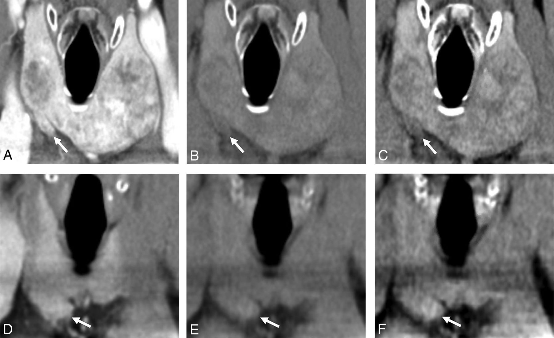
- FIG 5.
Coronal arterial phase (A), noncontrast 70-keV (B), and noncontrast 40-keV (C) images demonstrate a pathologically-proved right inferior parathyroid adenoma (arrows). Because the parathyroid adenoma appears isodense to the adjacent thyroid gland on the arterial phase image, it is uncertain whether the finding represents a parathyroid lesion or exophytic thyroid tissue. The parathyroid adenoma appears slightly hypoattenuating to the thyroid parenchyma on the standard (70-keV) noncontrast image; however, this attenuation difference is accentuated on the 40-keV image, indicating that the finding represents a parathyroid lesion rather than exophytic thyroid tissue. In contrast, coronal arterial phase (D), noncontrast 70-keV (E), and noncontrast 40-keV (F) images from a different patient demonstrate exophytic thyroid tissue (arrows) arising from the lower pole of the right thyroid lobe. On the arterial phase image alone, it is uncertain whether the finding represents exophytic thyroid tissue or a right inferior parathyroid lesion. Although the finding is isodense relative to the thyroid gland on the 70-keV noncontrast image, some uncertainty persists because of the nearly isoattenuating appearance of the thyroid gland relative to adjacent muscle, suggesting decreased iodine content from chronic thyroid disease. The 40-keV noncontrast image demonstrates substantially increased attenuation of the finding comparable with the increased attenuation of the remainder of the thyroid gland, indicating that the finding of interest represents exophytic thyroid tissue rather than a parathyroid lesion. In this patient, a biochemical cure was achieved with removal of a pathologically-proved parathyroid adenoma identified elsewhere in the neck (not shown), confirming that the finding depicted in images D, E, and F is indeed not a parathyroid lesion. Section thickness (2 mm), window level (40 HU), and window width (400 HU) are identical for all 6 images.


Tables
Characteristics Age (median) (range) (yr) 63 (33–81) Sex Male 9 Female 11 Operative findings Single-gland disease 16 Multigland disease 4 Concurrent hypothyroidism Yes 3 No 17 - Table 2:
Summary of differences in Hounsfield unit attenuation, contrast-to-noise, and image noise at 70 and 40 keV
40 keV (Median) (IQR) 70 keV (Median) (IQR) Difference (40-70 keV)
(Median) (IQR)P Comparison (P) Hounsfield unit attenuation Thyroid Absolute (HU) 158 (133–264) 84 (74–113) +67 (55–142) <.001 Par <.001 SCM % Change +89% (66–123) <.001 Par <.001 SCM Parathyroid Absolute (HU) 42 (29–59) 32 (22–41) +9 (4–17) <.001 Thy .22 SCM % Change +29% (13–58) <.001 Thy .09 SCM Sternocleidomastoid Absolute (HU) 67 (63–75) 57 (54–59) +11 (10–16) <.001 Thy .22 Par % Change +22% (17–28) <.001 Thy .09 Par Contrast-to-noise Thy/Par 4.7 (3.3–6.0) 3.8 (2.4–4.8) +0.8 (0.2–1.2) <.001 Thy/SCM 3.6 (1.7–5.0) 2.3 (0.9–3.1) +1.3 (0.7–1.9) <.001 Image noise (HU) 32 (23–37) 16 (12–19) +14 (11–20) <.001 Note:—Thy indicates thyroid; Par, parathyroid; SCM, sternocleidomastoid.


