
Article Figures & Data
Figures
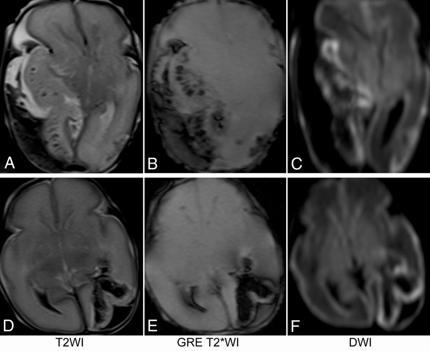
- FIG 1.
MR imaging of 2 preterm neonates who did not survive to discharge from the neonatal unit. The first patient (A–C) was born at 27 weeks 3 days’ gestation and had a large subpial hemorrhage and a large underlying hemorrhagic infarct. The second patient (D–F) was born at 28 weeks 1 day of gestation. She had a relatively small subpial hemorrhage and underlying hemorrhagic infarct but had engorged deep medullary veins in the bilateral cerebral hemispheres that may have had small thrombosis (not shown).
- FIG 2.
MR images of 3 term neonates. The subpial hemorrhage consistently shows hyperintense T1 signal, hypointense T2 signal, no restricted diffusion, and hypointense signal on GRE T2*WI. In the first patient (A–D), the underlying cerebral cortex and white matter have no hemorrhage. In the second patient (E–H), a mild fan-shaped hemorrhage is seen in the underlying white matter, resulting in a hypointense signal on T2WI, DWI, and T2*WI, while the cerebral cortex remains hyperintense on T2WI, DWI, and T2*WI. In the third patient (I–L), more severe hemorrhage is seen in the underlying white matter, leading to an obscured cerebral cortex on T2*WI and a partially obscured cortex on DWI.
- FIG 3.
Yin-yang sign. T2WI and DWI of 3 term neonates with subpial hemorrhage are shown. In the brain parenchyma underlying the subpial bleed, no hemorrhage is seen in the first patient (A and B); mild hemorrhage, in the second patient (C and D); and severe hemorrhage, in the third patient (E and F). Irrespective of the presence or degree of intraparenchymal hemorrhage, the combination of a dark subpial fluid collection and a bright underlying cerebral cortex forms a consistent, distinct image pattern (circled areas in A–F), resembling the yin-yang symbol in Chinese philosophy.
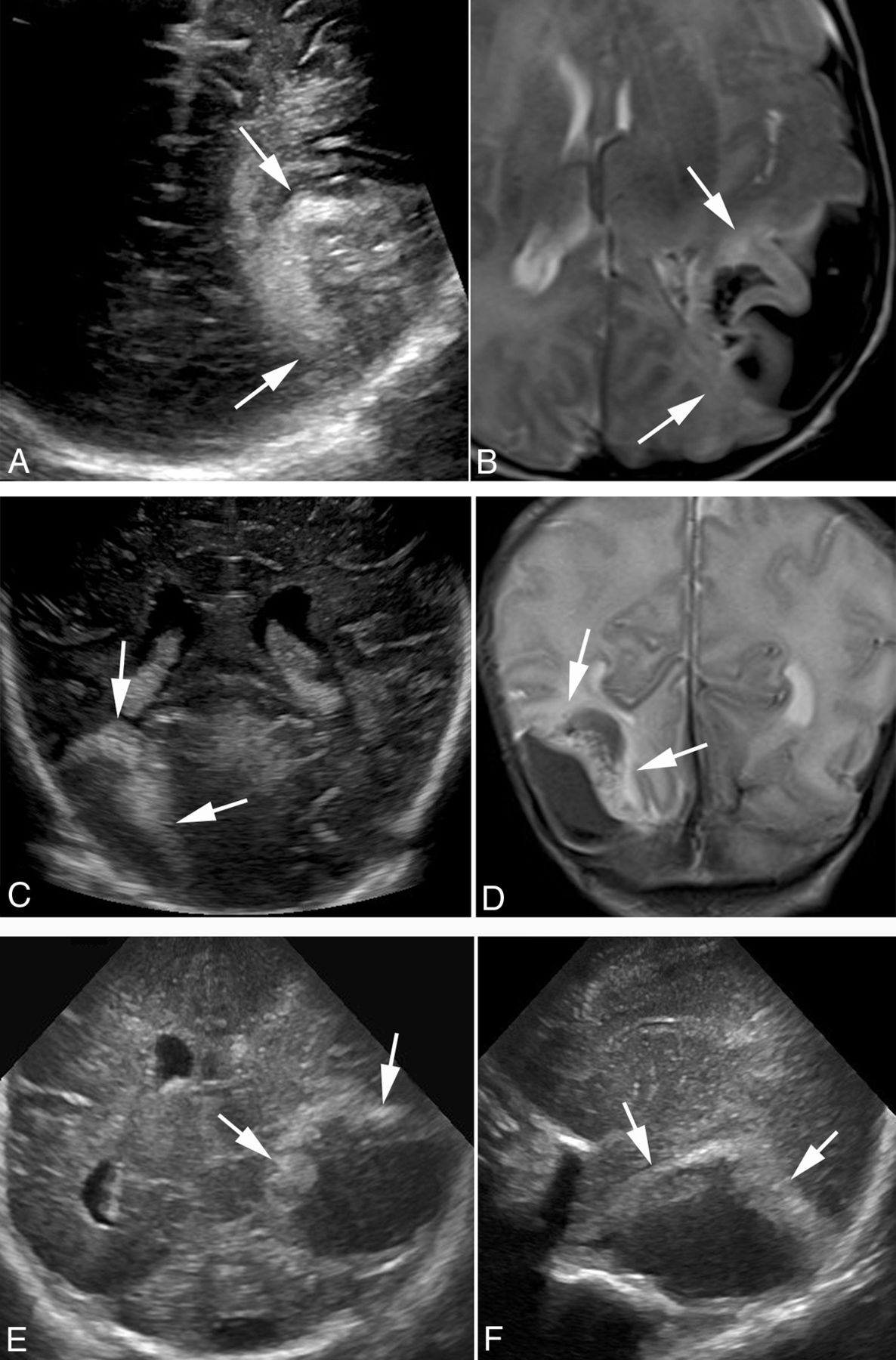
- FIG 4.
Ultrasound images of subpial hemorrhage with underlying cerebral infarct compared with MR images. The ultrasound images (A and C) were obtained <24 hours before the MR images (B and D) of a late-preterm neonate (A and B) and a term neonate (C and D). Ultrasound is able to detect both the subpial hemorrhage and underlying cerebral infarct in both patients (arrows). The subpial collection of bleeds is mildly echogenic in the first patient (A and B) and hypoechoic in the second patient (C and D), even though they are all hypointense on T2-weighed MR images. Ultrasound is unable to differentiate the infarcted cerebral cortex from the underlying white matter with hemorrhagic infarct. The ultrasound images in the lower panel are from a very preterm neonate who did not have brain MR imaging performed before succumbing to disease.


Tables
Clinical Parameter Subgroups Value Maternal age Mean age in years 33.5 (27–39) Gestational age at birth (weeks) Term (≥37) 11/16 Moderate-to-late preterm (32–37) 2/16 Very preterm (28–32) 2/16 Extremely preterm (≤ 28) 1/16 Sex Male 5/16 Female 11/16 Mode of delivery Vaginal 13/16 Cesarean delivery 3/16 Birth weight <2.5 kg 5/16 >2.5 kg 11/16 Mean 2.2 kg Birth assistance in vaginal delivery No assistance 10/13 Vacuum assistance 3/13 Clinical presentation Apnea 10/16 Seizure 9/16 Encephalopathy 3/16 APGAR score at 1 min 6.3 (range, 1–9) APGAR score at 5 min 8.4 (range, 4–9) Age at onset of symptoms 2.1 days (range, 1–6 days of life) Age at most recent follow-up 18.8 months (range, 1–90 months) Abnormal coagulation/hematology profile Maternal 0/16 Neonatal 1/16 (elevated hematocrit) Outcome Death 3/16 (1 extremely preterm, 2 very preterm) With neurologic deficits 2/16 No neurologic deficits 11/16 No. Percentage Imaging data MR imaging and US 7 MR imaging only 8 US only 1 Age at imaging (range) (day) MR imaging 3.2 (1–9) US 2.7 (1–7) MR imaging field strength (1.5T:3T) 12:3 MR imaging sequences Core sequencesa 15/15 GRE T2*WI 11/15 SWI 4/15 MRV performed On initial MRI 10/15 On repeat MRI within 4 days 3/15 MRA performed On initial MRI 9/15 On repeat MRI within 6 weeks 3/15 Lesion laterality (right:left) 10:6 62%:38% Lesion location Temporal 11/16 69% Frontal 3/16 19% Parietal 1/16 6% Occipital 1/16 6% Parenchymal diffusion restriction 15/15 100% Parenchymal hemorrhage 11/15 50% Intraventricular hemorrhage 3/15 18% Midline shift 8/15 50% Yin-yang sign on MRI 15/15 100% Yin-yang sign on US 6/6 100% Note:—US indicates ultrasound.
↵a The core MR images include sagittal and axial T1WI, axial and coronal FSE T2WI, and axial and coronal DWI.
- Table 3:
Appearance of subpial hemorrhage and underlying brain parenchyma on initial MR imaging and US
Subpial Bleed, Parenchymal Lesion MRI US T1WI T2WI DWI T2*WI ↑ ↓ ↓ ↓ ↓ or ↑ Cortical infarct with no WM hemorrhage (4/15) Cortex ↓ ↑ ↑ ↔ ↑ WM ↓ ↑ ↑ ↔ Cortical infarct with small WM hemorrhage (6/15) Cortex ↓ ↑ ↑ ↔ ↑ WM Mixed Mixed ↓ ↓ Cortical infarct with large WM hemorrhage (5/15) Cortex ↓ ↑ ↑ ↓ ↑ WM Mixed Mixed ↓ ↓ Note:—↔ indicates isointense or isoechoic; ↑, hyperintense or hyperechoic; ↓, hypointense or hypoechoic; US, ultrasound.
- Table 4:
Appearances of subpial hemorrhage and underlying brain parenchyma on follow-up MR imaging
Time Interval between Initial and Follow-Up MR Imaging No. of Scans Follow-Up MR (Appearances Compared with Initial MRI) Subpial Hemorrhage Underlying Cerebral Infarct 1–6 days 8 Similar size and shape Similar size and shape 7–13 weeks 4 Evolved into fluid collection with less mass effect Decreased size 13–28 weeks 6 Evolved into fluid collection with no mass effect Decreased size


