Advertisement

Main menu

  • Home
  • Content
    • Current Issue
    • Accepted Manuscripts
    • Article Preview
    • Past Issue Archive
    • Video Articles
    • AJNR Case Collection
    • Case of the Week Archive
    • Case of the Month Archive
    • Classic Case Archive
  • Special Collections
    • AJNR Awards
    • Low-Field MRI
    • Alzheimer Disease
    • ASNR Foundation Special Collection
    • Photon-Counting CT
    • View All
  • Multimedia
    • AJNR Podcasts
    • AJNR SCANtastic
    • Trainee Corner
    • MRI Safety Corner
    • Imaging Protocols
  • For Authors
    • Submit a Manuscript
    • Submit a Video Article
    • Submit an eLetter to the Editor/Response
    • Manuscript Submission Guidelines
    • Statistical Tips
    • Fast Publishing of Accepted Manuscripts
    • Graphical Abstract Preparation
    • Imaging Protocol Submission
    • Author Policies
  • About Us
    • About AJNR
    • Editorial Board
    • Editorial Board Alumni
  • More
    • Become a Reviewer/Academy of Reviewers
    • Subscribers
    • Permissions
    • Alerts
    • Feedback
    • Advertisers
    • ASNR Home

User menu

  • Alerts
  • Log in

Search

  • Advanced search
American Journal of Neuroradiology

American Journal of Neuroradiology

ASHNR American Society of Functional Neuroradiology ASHNR American Society of Pediatric Neuroradiology ASSR
  • Alerts
  • Log in

Advanced Search

  • Home
  • Content
    • Current Issue
    • Accepted Manuscripts
    • Article Preview
    • Past Issue Archive
    • Video Articles
    • AJNR Case Collection
    • Case of the Week Archive
    • Case of the Month Archive
    • Classic Case Archive
  • Special Collections
    • AJNR Awards
    • Low-Field MRI
    • Alzheimer Disease
    • ASNR Foundation Special Collection
    • Photon-Counting CT
    • View All
  • Multimedia
    • AJNR Podcasts
    • AJNR SCANtastic
    • Trainee Corner
    • MRI Safety Corner
    • Imaging Protocols
  • For Authors
    • Submit a Manuscript
    • Submit a Video Article
    • Submit an eLetter to the Editor/Response
    • Manuscript Submission Guidelines
    • Statistical Tips
    • Fast Publishing of Accepted Manuscripts
    • Graphical Abstract Preparation
    • Imaging Protocol Submission
    • Author Policies
  • About Us
    • About AJNR
    • Editorial Board
    • Editorial Board Alumni
  • More
    • Become a Reviewer/Academy of Reviewers
    • Subscribers
    • Permissions
    • Alerts
    • Feedback
    • Advertisers
    • ASNR Home
  • Follow AJNR on Twitter
  • Visit AJNR on Facebook
  • Follow AJNR on Instagram
  • Join AJNR on LinkedIn
  • RSS Feeds

AJNR Awards, New Junior Editors, and more. Read the latest AJNR updates

LetterLetter
Open Access

Possible Acute Disseminated Encephalomyelitis Related to Severe Acute Respiratory Syndrome Coronavirus 2 Infection

P.S. Utukuri, A. Bautista, A. Lignelli and G. Moonis
American Journal of Neuroradiology September 2020, 41 (9) E82-E83; DOI: https://doi.org/10.3174/ajnr.A6714
P.S. Utukuri
aDepartment of Neuroradiology
  • Find this author on Google Scholar
  • Find this author on PubMed
  • Search for this author on this site
  • ORCID record for P.S. Utukuri
A. Bautista
bDepartment of Neurology
  • Find this author on Google Scholar
  • Find this author on PubMed
  • Search for this author on this site
  • ORCID record for A. Bautista
A. Lignelli
aDepartment of Neuroradiology
  • Find this author on Google Scholar
  • Find this author on PubMed
  • Search for this author on this site
  • ORCID record for A. Lignelli
G. Moonis
aDepartment of Neuroradiology
  • Find this author on Google Scholar
  • Find this author on PubMed
  • Search for this author on this site
  • ORCID record for G. Moonis
  • Article
  • Figures & Data
  • Info & Metrics
  • Responses
  • References
  • PDF
Loading

As of June 1, 2020, there were 1.84 million confirmed cases of coronavirus disease 2019 (COVID-19) in the United States with more than 372,000 of the cases in New York City. Most of the clinical manifestations of the infection are related to the respiratory and gastrointestinal systems. Several neurologic manifestations of COVID-19 infection have also been documented, predominantly presenting as olfactory or gustatory dysfunction, encephalopathy, and cerebrovascular accidents. We present a less common neurologic symptom presumed to be acute disseminated encephalomyelitis (ADEM), presenting as transverse myelitis in an adult male patient with no preceding upper respiratory symptoms.

The patient is a 44-year-old healthy man who initially presented with urinary retention for 2 days with additional symptoms of bilateral lower extremity weakness and numbness and an inability to walk. There was no history of back trauma, only a history of back pain for which he had taken nonsteroidal anti-inflammatory drugs. There were no upper respiratory symptoms or fever. There was no known recent viral illness or vaccinations.

Initial examination was remarkable only for lethargy, dysarthria, bilateral arm ataxia, urinary retention, and weakness of both legs that was barely antigravity. Initial imaging work-up included a head CT and MR imaging of the lumbar spine. The head CT findings were normal.

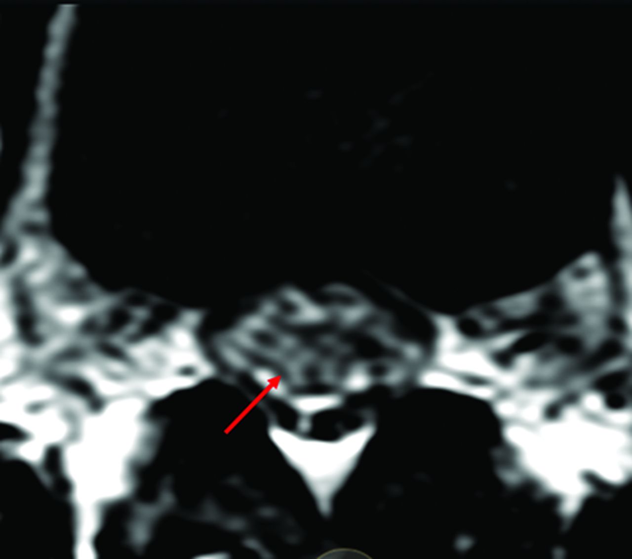
The lumbar spine MR imaging demonstrated slight expansion of the conus medullaris with mild T2 hyperintensity and minimal foci of enhancement (Fig 1). The patient was admitted with an impression of transverse myelitis of unclear etiology.

FIG 1.
  • Download figure
  • Open in new tab
  • Download powerpoint
FIG 1.

Axial T2 image through the conus demonstrating abnormally increased T2 signal (arrow) with slight expansion of the conus.

Per hospital policy, every admitted patient was tested for Severe Acute Respiratory Syndrome coronavirus 2 (SARS-CoV-2) via a nasopharyngeal swab using a real-time reverse transcription polymerase chain reaction (RT-PCR) test, which resulted positively 1 day after admission.

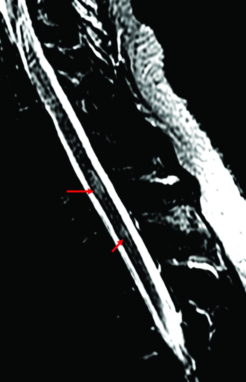
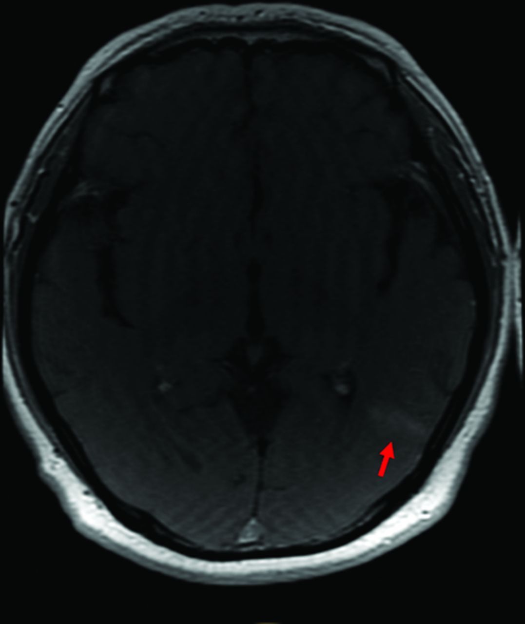
Further imaging of the remainder of the brain and spinal axis demonstrated nonenhancing T2 hyperintense lesions throughout the cervical and thoracic spinal cord (Fig 2) and several periventricular and juxtacortical lesions within the brain (Fig 3). The inferior cervical lesion demonstrated slight associated cord expansion. There was homogeneous brisk enhancement associated with the dominant left parietal lobe juxtacortical/cortical lesion (Fig 4). A lumbar puncture was eventually performed because of ongoing and progressing symptoms.

FIG 2.
  • Download figure
  • Open in new tab
  • Download powerpoint
FIG 2.

Sagittal inversion recovery sequence demonstrating abnormal T2 hyperintense lesions within the lower cervical and upper thoracic spinal cord (arrows).

FIG 3.
  • Download figure
  • Open in new tab
  • Download powerpoint
FIG 3.

Axial FLAIR images of the brain demonstrating FLAIR hyperintense lesions (arrows) within the left posterior parietal lobe (A) and periventricular region (B).

FIG 4.
  • Download figure
  • Open in new tab
  • Download powerpoint
FIG 4.

Axial T1 postcontrast image demonstrating brisk homogeneous enhancement of the dominant lesion (arrow).

The initial serum tests showed normal complete blood cell count, metabolic panel, urine studies, and inflammatory markers (sedimentation rate, C-reactive protein level) and were negative for HIV. D-dimer was 2.8 μg/mL FEU (reference range, <0.8 μg/mL FEU). Serum cardiolipin antibody immunoglobulin M was mildly elevated at 39 MPL (reference range, 0–12 mpl). Findings of other rheumatologic work-ups were unrevealing. There were no deficiencies in vitamin B12, vitamin E, or copper. Serum protein electrophoresis, angiotensin-converting enzyme levels, and syphilis screen findings were normal.

The CSF profile was largely unremarkable, with white blood cells counts of 6/μL (0–5/μL), of which lymphocytes were 92%; protein 36. CSF bacterial and viral PCR studies were negative. Cytology and flow cytometry were also unrevealing. Oligoclonal bands were absent. The immunoglobulin G (IgG) index was normal. CSF PCR analysis for SARS-CoV-2 was initially questionably positive; however, it was eventually negative to date.

In this patient with unexplained acute encephalopathy and multifocal neurologic signs and symptoms, the constellation of imaging findings in this case is suggestive of ADEM, which may have been triggered by a SARS-CoV-2 infection. The brain lesions are within the periventricular or juxtacortical white matter, predominantly favoring a primary demyelinating process. None of the FLAIR hyperintense lesions in the brain showed hemorrhage as is commonly seen in cases of encephalitis; in addition, the results of the CSF bacterial and viral PCR tests were also negative, making the diagnosis of infectious encephalitis less likely. No T1 hypointense lesions were present. The diagnosis of a first multiple sclerosis attack was thought less likely because of the absence of oligoclonal bands, a normal IgG index, cortical gray matter involvement, and clinical features of encephalopathy.

ADEM is presumed to be a post- or parainfectious process. Typically, ADEM presents in children or adolescents, usually younger than 15 years of age, though cases have been reported in all ages. Approximately 75% of patients have a history of a recent upper respiratory infection or vaccination. Despite reports of the possible association between infection and ADEM, there is no clear understanding of the relationship between the infectious agent and the onset of demyelination. There is experimental evidence in mice for a relationship between coronavirus and CNS demyelination. The literature did report detection of coronavirus in the CNS of a 15-year-old adolescent, and most recently, there has been a report of acute myelitis in an older adult patient with known COVID-19 from Wuhan, China.1⇓-3 The pathophysiology remains uncertain. The patient was initially treated with IV methylprednisolone and subsequently IV immunoglobulin with modest improvement. At the time of this writing, the patient has been discharged to an acute rehabilitation facility.

Footnotes

  • Disclosures: Gul Moonis—UNRELATED: Royalties: Wolters Kluwer, approximately $150 for Neuroradiology: A Core Review.

Indicates open access to non-subscribers at www.ajnr.org

References

  1. 1.↵
    1. Ann Yeh E,
    2. Collins A,
    3. Cohen ME, et al
    . Detection of coronavirus in the central nervous system of a child with acute disseminated encephalomyelitis. Pediatrics 2004;113(1 Pt 1):e73–76 doi:10.1542/peds.113.1.e73 pmid:14702500
    Abstract/FREE Full Text
  2. 2.↵
    1. Zhao K,
    2. Huang J,
    3. Dai D, et al
    . Acute myelitis after SARS-CoV2 infection: a case report. https://doi.org/10.1101/2020.03.16.20035105. Accessed June 1, 2020
  3. 3.↵
    Acute disseminated encephalomyelitis (ADEM). Radiopaedia.org. https://radiopaedia.org/cases/acute-disseminated-encephalomyelitis-adem. Accessed June 1, 2020
  • © 2020 by American Journal of Neuroradiology
PreviousNext
Back to top

In this issue

American Journal of Neuroradiology: 41 (9)
American Journal of Neuroradiology
Vol. 41, Issue 9
1 Sep 2020
  • Table of Contents
  • Index by author
  • Complete Issue (PDF)
Advertisement
Print
Download PDF
Email Article

Thank you for your interest in spreading the word on American Journal of Neuroradiology.

NOTE: We only request your email address so that the person you are recommending the page to knows that you wanted them to see it, and that it is not junk mail. We do not capture any email address.

Enter multiple addresses on separate lines or separate them with commas.
Possible Acute Disseminated Encephalomyelitis Related to Severe Acute Respiratory Syndrome Coronavirus 2 Infection
(Your Name) has sent you a message from American Journal of Neuroradiology
(Your Name) thought you would like to see the American Journal of Neuroradiology web site.
CAPTCHA
This question is for testing whether or not you are a human visitor and to prevent automated spam submissions.
Cite this article
P.S. Utukuri, A. Bautista, A. Lignelli, G. Moonis
Possible Acute Disseminated Encephalomyelitis Related to Severe Acute Respiratory Syndrome Coronavirus 2 Infection
American Journal of Neuroradiology Sep 2020, 41 (9) E82-E83; DOI: 10.3174/ajnr.A6714

Citation Manager Formats

  • BibTeX
  • Bookends
  • EasyBib
  • EndNote (tagged)
  • EndNote 8 (xml)
  • Medlars
  • Mendeley
  • Papers
  • RefWorks Tagged
  • Ref Manager
  • RIS
  • Zotero
0 Responses
Respond to this article
Share
Bookmark this article
Possible Acute Disseminated Encephalomyelitis Related to Severe Acute Respiratory Syndrome Coronavirus 2 Infection
P.S. Utukuri, A. Bautista, A. Lignelli, G. Moonis
American Journal of Neuroradiology Sep 2020, 41 (9) E82-E83; DOI: 10.3174/ajnr.A6714
del.icio.us logo Twitter logo Facebook logo Mendeley logo
  • Tweet Widget
  • Facebook Like
  • Google Plus One
Purchase

Jump to section

  • Article
    • Footnotes
    • References
  • Figures & Data
  • Info & Metrics
  • Responses
  • References
  • PDF

Related Articles

  • No related articles found.
  • PubMed
  • Google Scholar

Cited By...

  • Unravelling the clinical complexity of myelin oligodendrocyte glycoprotein antibody-associated acute disseminated encephalomyelitis in children: a comprehensive analysis
  • Acute Ascending Necrotizing Myelitis After COVID-19 Infection: A Clinicopathologic Report
  • Spectrum of spinal cord involvement in COVID-19: A systematic review
  • Crossref (19)
  • Google Scholar

This article has been cited by the following articles in journals that are participating in Crossref Cited-by Linking.

  • Acute Transverse Myelitis (ATM):Clinical Review of 43 Patients With COVID-19-Associated ATM and 3 Post-Vaccination ATM Serious Adverse Events With the ChAdOx1 nCoV-19 Vaccine (AZD1222)
    Gustavo C. Román, Fernando Gracia, Antonio Torres, Alexis Palacios, Karla Gracia, Diógenes Harris
    Frontiers in Immunology 2021 12
  • Central neuroinflammation in Covid-19: a systematic review of 182 cases with encephalitis, acute disseminated encephalomyelitis, and necrotizing encephalopathies
    Raha Zamani, Rozhina Pouremamali, Nima Rezaei
    Reviews in the Neurosciences 2022 33 4
  • Spinal cord involvement in COVID-19: A review
    Ravindra Kumar Garg, Vimal Kumar Paliwal, Ankit Gupta
    The Journal of Spinal Cord Medicine 2023 46 3
  • Clinical and Pathophysiologic Spectrum of Neuro-COVID
    Josef Finsterer, Fulvio A Scorza
    Molecular Neurobiology 2021 58 8
  • Acute disseminated encephalomyelitis and COVID-19: A systematic synthesis of worldwide cases
    Laura Zelada-Ríos, Kevin Pacheco-Barrios, Milagros Galecio-Castillo, Cesar Yamunaqué-Chunga, Kelvin Álvarez-Toledo, Ricardo Otiniano-Sifuentes
    Journal of Neuroimmunology 2021 359
  • Emerging potential mechanisms and predispositions to the neurological manifestations of COVID-19
    Aaron Jesuthasan, Flavia Massey, Hadi Manji, Michael S. Zandi, Sarah Wiethoff
    Journal of the Neurological Sciences 2021 428
  • Neuroradiologic manifestations of COVID-19: what the emergency radiologist needs to know
    Michael F. Goldberg, Morton F. Goldberg
    Emergency Radiology 2020 27 6
  • Neurological and Head/Eyes/Ears/Nose/Throat Manifestations of COVID-19: A Systematic Review and Meta-Analysis
    Aravind Ganesh, Isabella R. Reis, Malavika Varma, David G. Patry, Lara J. Cooke
    Canadian Journal of Neurological Sciences / Journal Canadien des Sciences Neurologiques 2022 49 4
  • Severe Acute Respiratory Syndrome Coronavirus 2-Induced Neurological Complications
    Shijia Yu, Mingjun Yu
    Frontiers in Cell and Developmental Biology 2020 8
  • Encephalopathy as a prognostic factor in adults with acute disseminated encephalomyelitis following COVID-19
    Stefano Gelibter, Gabriele Bellavia, Carla Arbasino, Natale Arnò, Margaret Glorioso, Sara Mazza, Rosanna Murelli, Massimo Sciarretta, Carlo Dallocchio
    Journal of Neurology 2022 269 5

More in this TOC Section

  • Brain AVM’s Nidus: What if We Hadn’t Understood Anything?
  • Letter to the Editor regarding “Automated Volumetric Software in Dementia: Help or Hindrance to the Neuroradiologist?”
  • Reply:
Show more LETTERS

Similar Articles

Advertisement

Indexed Content

  • Current Issue
  • Accepted Manuscripts
  • Article Preview
  • Past Issues
  • Editorials
  • Editor's Choice
  • Fellows' Journal Club
  • Letters to the Editor
  • Video Articles

Cases

  • Case Collection
  • Archive - Case of the Week
  • Archive - Case of the Month
  • Archive - Classic Case

More from AJNR

  • Trainee Corner
  • Imaging Protocols
  • MRI Safety Corner
  • Book Reviews

Multimedia

  • AJNR Podcasts
  • AJNR Scantastics

Resources

  • Turnaround Time
  • Submit a Manuscript
  • Submit a Video Article
  • Submit an eLetter to the Editor/Response
  • Manuscript Submission Guidelines
  • Statistical Tips
  • Fast Publishing of Accepted Manuscripts
  • Graphical Abstract Preparation
  • Imaging Protocol Submission
  • Evidence-Based Medicine Level Guide
  • Publishing Checklists
  • Author Policies
  • Become a Reviewer/Academy of Reviewers
  • News and Updates

About Us

  • About AJNR
  • Editorial Board
  • Editorial Board Alumni
  • Alerts
  • Permissions
  • Not an AJNR Subscriber? Join Now
  • Advertise with Us
  • Librarian Resources
  • Feedback
  • Terms and Conditions
  • AJNR Editorial Board Alumni

American Society of Neuroradiology

  • Not an ASNR Member? Join Now

© 2025 by the American Society of Neuroradiology All rights, including for text and data mining, AI training, and similar technologies, are reserved.
Print ISSN: 0195-6108 Online ISSN: 1936-959X

Powered by HighWire