
Article Figures & Data
Figures
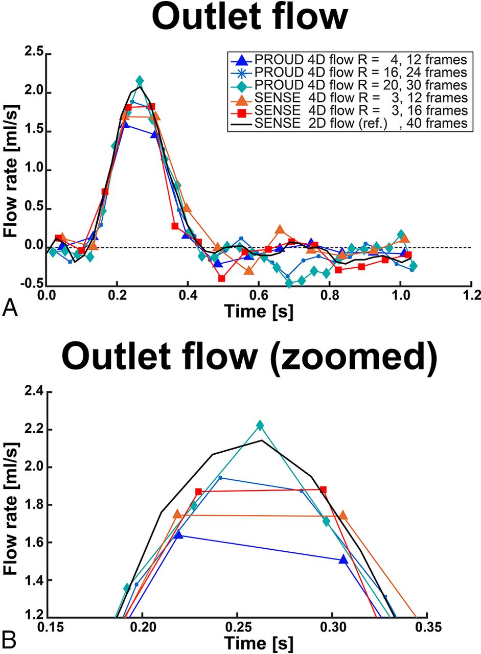
- FIG 1.
A, Flow rates of the outlet ROI of exemplary low (12 frames), moderate (16–24 frames), and high temporal resolution (30–40 frames). B, Zoomed view into the peak flow rate of the outlet ROI.
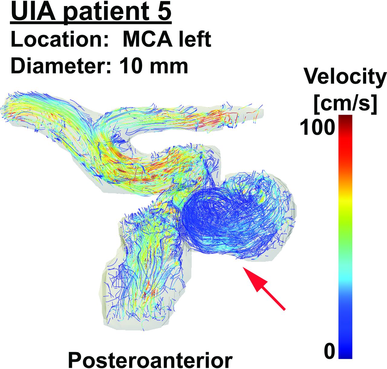
- FIG 2.
Streamlines of patient 5 with an UIA to visualize blood flow patterns in the aneurysm (red arrow) of 10-mm diameter located at the left MCA. A video of streamlines of all five patients with UIAs over the cardiac cycle can be seen in the On-line Video. MCA indicates middle cerebral artery.
- FIG 3.
Example WSS and OSI analysis of patient 5. Time-averaged WSS on the left, peak-systolic WSS in the middle, and OSI on the right for datasets with either 10 (upper row) or 30 (lower row) cardiac frames. MCA indicates middle cerebral artery; a.u., arbitrary units.
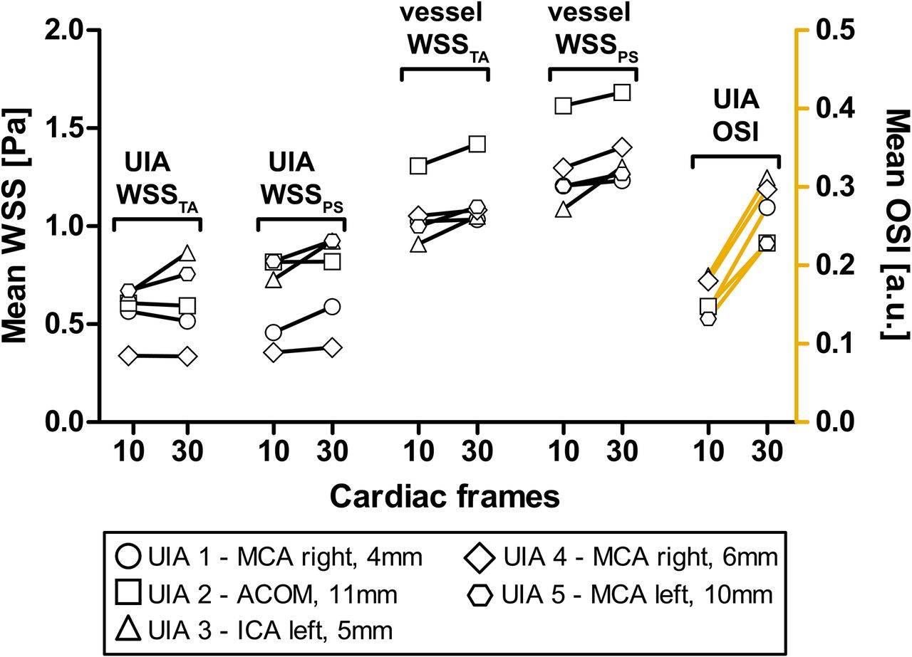
- FIG 4.
The changes in time-averaged WSS, peak systolic WSS, and OSI in the aneurysm and in the surrounding vessel when increasing the temporal resolution from 10 to 30 cardiac frames, or from 95 to 32 ms, respectively. ACOM indicates anterior communicating artery; ICA, internal carotid artery; MCA, middle cerebral artery; a.u., arbitrary units.


Tables
- Table 1:
Results of the flow phantom analysis in terms of peak flow rate difference, stroke volume difference, and the SD of the diastolic flow
Sequence Acceleration Factor Cardiac Frames Mean Peak Flow Rate Difference (mL/s) (%) Mean Stroke Volume Difference (mL) (%) Mean Diastolic Flow Rate SE (mL/s) 4D PROUD-1 32 12 −0.65 (−46.7%) −0.036 (−15.3%) 0.028 4D PROUD-2 24 12 −0.52 (−35.0%) −0.024 (−5.3%) 0.018 4D PROUD-3 16 12 −0.47 (−32.9%) −0.040 (−14.3%) 0.033 4D PROUD-5 8 12 −0.45 (−31.6%) −0.031 (−9.4%) 0.035 4D PROUD-4 12 12 −0.36 (−25.5%) −0.024 (−7.4%) 0.021 4D PROUD-6 4 12 −0.34 (−23.9%) −0.027 (−10.4%) 0.027 4D SENSE-1 3 12 −0.31 (−22.5%) −0.007 (−5.2%) 0.047 4D SENSE-2 3 16 −0.27 (−21.5%) −0.027 (−11.5%) 0.031 4D PROUD-5 16 24 −0.19 (−15.0%) −0.002 (−3.2%) 0.029 4D PROUD-3 32 24 −0.18 (−13.5%) −0.013 (−3.7%) 0.029 4D PROUD-6 8 24 −0.17 (−10.4%) −0.028 (−10.6%) 0.024 4D PROUD-6 24 24 −0.13 (−8.0%) −0.008 (−0.8%) 0.028 4D PROUD-5 26 40 −0.08 (−8.8%) −0.011 (−2.0%) 0.027 4D PROUD-4 30 30 −0.06 (−7.8%) 0.005 (−3.3%) 0.022 4D PROUD-6 10 30 0.05 (1.9%) −0.029 (−12.4%) 0.027 4D PROUD-6 13 40 −0.04 (−2.4%) −0.031 (−11.4%) 0.024 4D PROUD-5 20 30 −0.03 (−4.3%) 0.003 (−4.9%) 0.034 2D reference-2 2 40 −0.05 (−3.7%) −0.006 (−3.0%) 0.010 2D reference-1a 2 40 0.00 (0.0%) 0.000 (−0.0%) 0.011 Note:—SE indicates standard error.
↵a Used as a comparison baseline.
- Table 2:
Results of the healthy subject analysis in terms of peak flow rate difference, peak flow rate repeatability, stroke volume difference, stroke volume repeatability, and pulsatility index difference
Comparison Pairs Bland-Altman Orthogonal Regression A (Frames) B (Frames) Bias LoA P Equation ρ Peak flow rate difference (mL/s) 2D reference-1 (30) 4D PROUD-1 (30) 0.31 ± 0.13 1.41 .22 y = 1.14x – 0.80 0.67 2D reference-1 (30) 4D PROUD-1 (15) 0.51 ± 0.13 1.44 .03 y = 1.15x – 1.04 0.66 2D reference-1 (30) 4D PROUD-1 (7) 0.76 ± 0.13 1.44 <.01 y = 0.90x – 0.42 0.60 2D reference-1 (30) 4D SENSE (7) 0.79 ± 0.17 1.81 .01 y = 0.83x – 0.19 0.36 Peak flow rate repeatability (mL/s) 4D PROUD-1 (30) 4D PROUD-2 (30) 0.14 ± 0.08 0.90 .61 y = 0.87x + 0.28 0.87 4D PROUD-1 (15) 4D PROUD-2 (15) 0.12 ± 0.08 0.87 .90 y = 0.84x + 0.37 0.88 4D PROUD-1 (7) 4D PROUD-2 (7) 0.09 ± 0.07 0.71 .77 y = 0.90x + 0.17 0.89 2D reference-1 (30) 2D reference-2 (30) 0.19 ± 0.08 0.86 .38 y = 1.01x – 0.23 0.86 Stroke volume difference (mL) 2D reference-1 (30) 4D PROUD-1 (30) 0.11 ± 0.09 0.94 .53 y = 0.99x – 0.08 0.64 2D reference-1 (30) 4D PROUD-1 (15) 0.11 ± 0.09 0.94 .45 y = 0.97x – 0.04 0.64 2D reference-1 (30) 4D PROUD-1 (7) 0.14 ± 0.09 0.93 .36 y = 0.95x – 0.03 0.64 2D reference-1 (30) 4D SENSE (7) 0.15 ± 0.11 1.18 .36 y = 1.06x – 0.29 0.46 Stroke volume repeatability (mL) 4D PROUD-1 (30) 4D PROUD-2 (30) 0.08 ± 0.04 0.43 .57 y = 0.98x – 0.05 0.93 4D PROUD-1 (15) 4D PROUD-2 (15) 0.08 ± 0.04 0.42 .60 y = 0.98x – 0.04 0.92 4D PROUD-1 (7) 4D PROUD-2 (7) 0.07 ± 0.04 0.44 .64 y = 1.00x – 0.08 0.92 2D reference-1 (30) 2D reference-2 (30) 0.06 ± 0.04 0.47 .80 y = 1.10x – 0.28 0.92 Pulsatility index difference (a.u.) 2D reference-1 (30) 4D PROUD-1 (30) 0.01 ± 0.04 0.39 .41 y = 1.97x – 0.82 0.15 2D reference-1 (30) 4D PROUD-1 (15) 0.18 ± 0.04 0.39 <.01 y = 1.51x – 0.60 0.10 2D reference-1 (30) 4D PROUD-1 (7) 0.31 ± 0.03 0.33 <.01 y = 0.01x + 0.51 0.01 2D reference-1 (30) 4D SENSE (7) 0.31 ± 0.03 0.35 <.01 y = 0.03x + 0.50 0.02 Note:—LoA indicates limits of agreement; a.u., arbitrary units.


Jump to section
Related Articles
Cited By...
- Report from the society of magnetic resonance angiography: clinical applications of 7T neurovascular MR in the assessment of intracranial vascular disease
- Cerebrovascular super-resolution 4D Flow MRI - using deep learning to non-invasively quantify velocity, flow, and relative pressure
- A realistic way to investigate the design, and mechanical properties of flow diverter stents