
Article Figures & Data
Figures
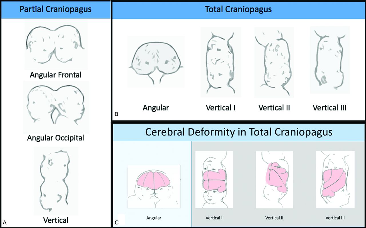
- Fig 1.
Stone and Goodrich7 classification for CPT. Partial CPT lack substantial shared dural venous sinuses. Total CPT share a large portion of dural venous sinuses and present with pronounced brain compression, leading to distortion within the cranium. The 2 main subtypes are based on the long-axis angle between twins: angular and vertical. Shared calvaria causes deformity of each twin’s brain. Reproduced with permission from Stone and Goodrich, 2006.7
- Fig 2.
A, T1-weighted MR imaging does not clearly show conjoined brain tissue in this total CPT. However, no clear dura between cerebral hemispheres and interdigitating of gyri (black arrow) is seen. B, Intraoperative photograph from the same patient demonstrating conjoined brain tissue (white arrow) along the axis defined by the flanking neurosurgical sponges (yellow arrows).
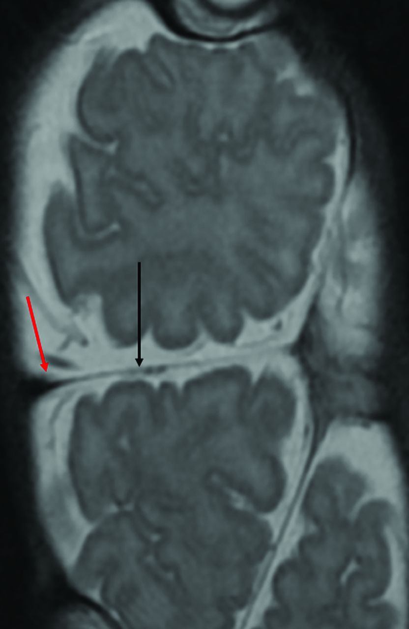
- Fig 3.
Coronal T2-weighted image demonstrates cerebral tissue separated by a single-layer transverse dural septum (black arrow). The CVS courses in the periphery of the septum (red arrow).
- Fig 4.
A, Surgical-separation strategy may entail sequential division of venous sinus branches from the nondominant twin (dashed line), allowing the anatomically predisposed dominant twin to keep the CVS (black arrow) and associated dura. In the nondominant twin, subsequent reversal of venous drainage to collateral basal channels may be induced (Reproduced, with permission, from Stone and Goodrich,7 2006). B, Postcontrast MR venography demonstrates the CVS (white arrow).
- Fig 5.
Coronal CT (A) of TA CPT shows shared brain parenchyma, including a diencephalic bridge (arrow). MRA of the brain (B) from the same patients demonstrates a shared MCA (arrows), with an appearance similar to that of the anterior cerebral artery. This set of twins cannot be separated.
- Fig 6.
A, 2D sonogram obtained in the axial plane through the skull of the fetal CPT shows the hyperechoic joined calvaria (green arrows). The point of skull union between the twins is also clearly seen (yellow arrow). B, Fetal MR imaging in the coronal oblique plane through both brains of the CPT. The CVS is seen along the lateral margin of the inner calvaria at the point of bony union (yellow arrow), with the associated dural shelf separating the brains (green arrow). It is likely that the brain is fused where brain surfaces touch, and no dural or CSF cleft is seen (orange arrow). The torso and spine of 1 twin is also seen in this plane (white arrow).
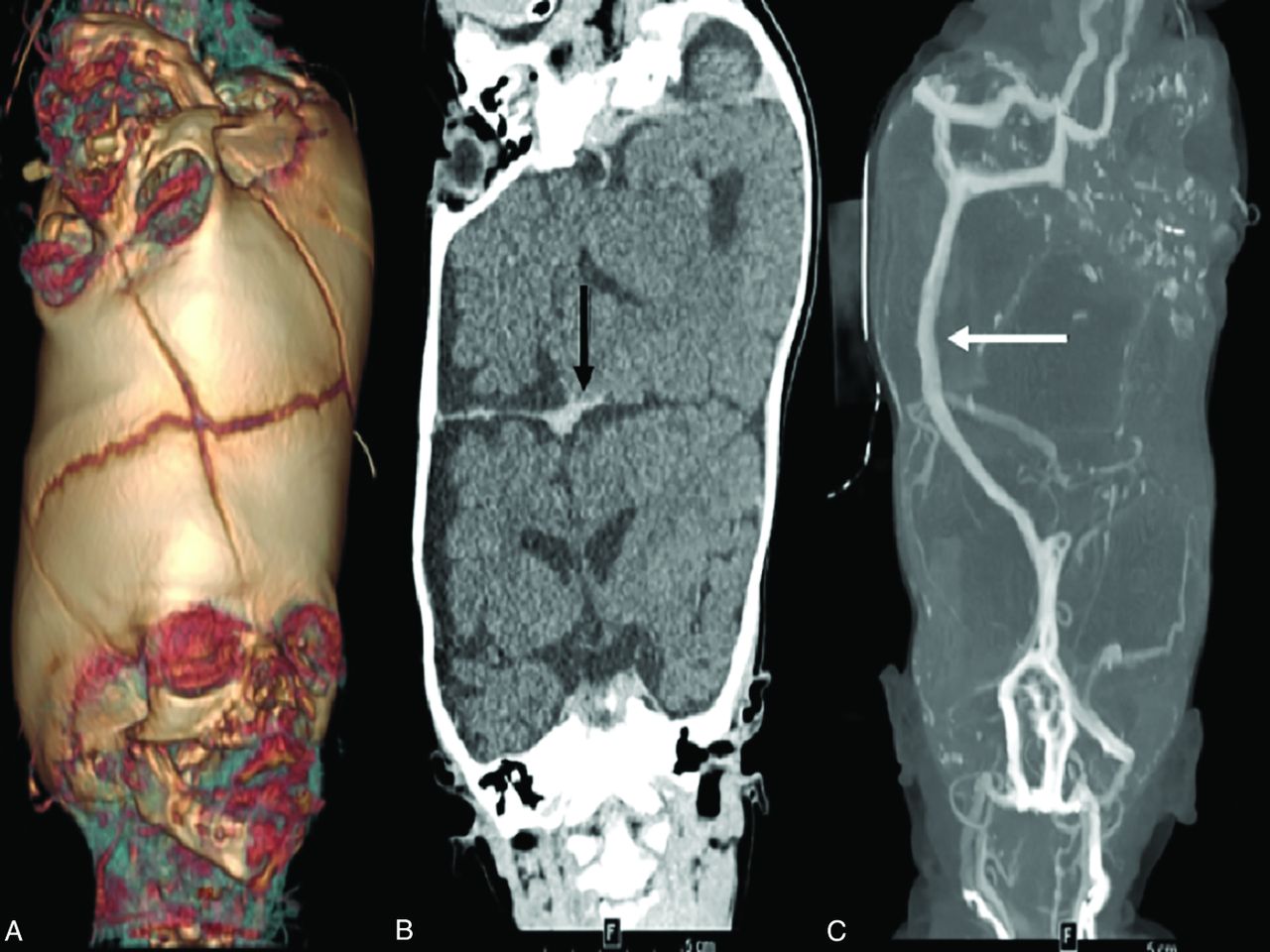
- Fig 7.
3D surface rendering from CT (A) demonstrates suture patterns in the fused calvaria. A coronal contrast-enhanced CT venogram (B) demonstrates the orientation of the cerebral hemispheres and the presence of a dural septum (arrow). Maximum intensity projection of a CT venogram (C) demonstrates a shared superior sagittal sinus (arrow).
- Fig 8.
T2-weighted coronal image (A) is concerning for parenchymal bridging between the parietal lobes (arrow). Coronal postcontrast enhanced T1-weighted image (B) and dynamic MR angiography and venography (C–F) demonstrate a circumferential sinus communicating with both superior sagittal sinuses and dominant occipital sinuses without evidence of arterial anastomosis. Tractography (G) can demonstrate contiguity of white matter tracts in the cephalocaudal direction depicted in blue (arrow).
- Fig 9.
Multiple venous phase left ICA injections show how an occlusion balloon is used to define venous flow between twins (A and B). A coil (C) is then placed in a venous sinus to help promote venous collateral formation before surgery.


