Advertisement

Main menu

  • Home
  • Content
    • Current Issue
    • Accepted Manuscripts
    • Article Preview
    • Past Issue Archive
    • Video Articles
    • AJNR Case Collection
    • Case of the Week Archive
    • Case of the Month Archive
    • Classic Case Archive
  • Special Collections
    • Advancing NeuroMRI with High-Relaxivity Contrast Agents
    • AJNR Awards
    • Low-Field MRI
    • Alzheimer Disease
    • ASNR Foundation Special Collection
    • Photon-Counting CT
    • View All
  • Multimedia
    • AJNR Podcasts
    • AJNR SCANtastic
    • Trainee Corner
    • MRI Safety Corner
    • Imaging Protocols
  • For Authors
    • Submit a Manuscript
    • Submit a Video Article
    • Submit an eLetter to the Editor/Response
    • Manuscript Submission Guidelines
    • Statistical Tips
    • Fast Publishing of Accepted Manuscripts
    • Graphical Abstract Preparation
    • Imaging Protocol Submission
    • Author Policies
  • About Us
    • About AJNR
    • Editorial Board
    • Editorial Board Alumni
  • More
    • Become a Reviewer/Academy of Reviewers
    • Subscribers
    • Permissions
    • Alerts
    • Feedback
    • Advertisers
    • ASNR Home

User menu

  • Alerts
  • Log in

Search

  • Advanced search
American Journal of Neuroradiology

American Journal of Neuroradiology

ASHNR American Society of Functional Neuroradiology ASHNR American Society of Pediatric Neuroradiology ASSR
  • Alerts
  • Log in

Advanced Search

  • Home
  • Content
    • Current Issue
    • Accepted Manuscripts
    • Article Preview
    • Past Issue Archive
    • Video Articles
    • AJNR Case Collection
    • Case of the Week Archive
    • Case of the Month Archive
    • Classic Case Archive
  • Special Collections
    • Advancing NeuroMRI with High-Relaxivity Contrast Agents
    • AJNR Awards
    • Low-Field MRI
    • Alzheimer Disease
    • ASNR Foundation Special Collection
    • Photon-Counting CT
    • View All
  • Multimedia
    • AJNR Podcasts
    • AJNR SCANtastic
    • Trainee Corner
    • MRI Safety Corner
    • Imaging Protocols
  • For Authors
    • Submit a Manuscript
    • Submit a Video Article
    • Submit an eLetter to the Editor/Response
    • Manuscript Submission Guidelines
    • Statistical Tips
    • Fast Publishing of Accepted Manuscripts
    • Graphical Abstract Preparation
    • Imaging Protocol Submission
    • Author Policies
  • About Us
    • About AJNR
    • Editorial Board
    • Editorial Board Alumni
  • More
    • Become a Reviewer/Academy of Reviewers
    • Subscribers
    • Permissions
    • Alerts
    • Feedback
    • Advertisers
    • ASNR Home
  • Follow AJNR on Twitter
  • Visit AJNR on Facebook
  • Follow AJNR on Instagram
  • Join AJNR on LinkedIn
  • RSS Feeds

AJNR Awards, New Junior Editors, and more. Read the latest AJNR updates


Improved Turnaround Times | Median time to first decision: 12 days

Research ArticlePediatric Neuroimaging

Fetal and Neonatal MRI Predictors of Aggressive Early Clinical Course in Vein of Galen Malformation

L. Arko, M. Lambrych, A. Montaser, D Zurakowski and D.B. Orbach
American Journal of Neuroradiology June 2020, 41 (6) 1105-1111; DOI: https://doi.org/10.3174/ajnr.A6585
L. Arko
aFrom the Departments of Neurosurgery (L.A., M.L., A.M., D.B.O.)
  • Find this author on Google Scholar
  • Find this author on PubMed
  • Search for this author on this site
  • ORCID record for L. Arko
M. Lambrych
aFrom the Departments of Neurosurgery (L.A., M.L., A.M., D.B.O.)
  • Find this author on Google Scholar
  • Find this author on PubMed
  • Search for this author on this site
  • ORCID record for M. Lambrych
A. Montaser
aFrom the Departments of Neurosurgery (L.A., M.L., A.M., D.B.O.)
  • Find this author on Google Scholar
  • Find this author on PubMed
  • Search for this author on this site
  • ORCID record for A. Montaser
D Zurakowski
bAnesthesiology and Surgery (D.Z.)
  • Find this author on Google Scholar
  • Find this author on PubMed
  • Search for this author on this site
  • ORCID record for D Zurakowski
D.B. Orbach
aFrom the Departments of Neurosurgery (L.A., M.L., A.M., D.B.O.)
cNeurointerventional Radiology (D.B.O.), Boston Children’s Hospital, Harvard Medical School, Boston, Massachusetts.
  • Find this author on Google Scholar
  • Find this author on PubMed
  • Search for this author on this site
  • ORCID record for D.B. Orbach
  • Article
  • Figures & Data
  • Supplemental
  • Info & Metrics
  • Responses
  • References
  • PDF
Loading

Article Figures & Data

Figures

  • Tables
  • Fig 1.
    • Download figure
    • Open in new tab
    • Download powerpoint
    Fig 1.

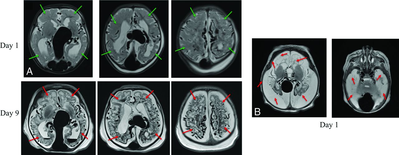
    Aggressive early evolution of a vein of Galen malformation (NAR cohort) (A, left panels). After med-flighting this neonate with VOGM to our center for urgent treatment, the MR imaging obtained on day-of-life 9 showed diffuse bihemispheric brain injuries (red arrows) not seen on the initial on day-of-life 1 MR imaging (green arrows showing analogous regions). Increased soft-tissue edema in the neck and scalp on day 0 is secondary to overwhelming heart failure (B, right panels). Another patient, a one-day-old neonate with VOGM presents with complete liquefactive gliosis of both cerebral hemispheres (white brain signal resembles white CSF signal on T2-weighted imaging).

  • Fig 2.
    • Download figure
    • Open in new tab
    • Download powerpoint
    Fig 2.

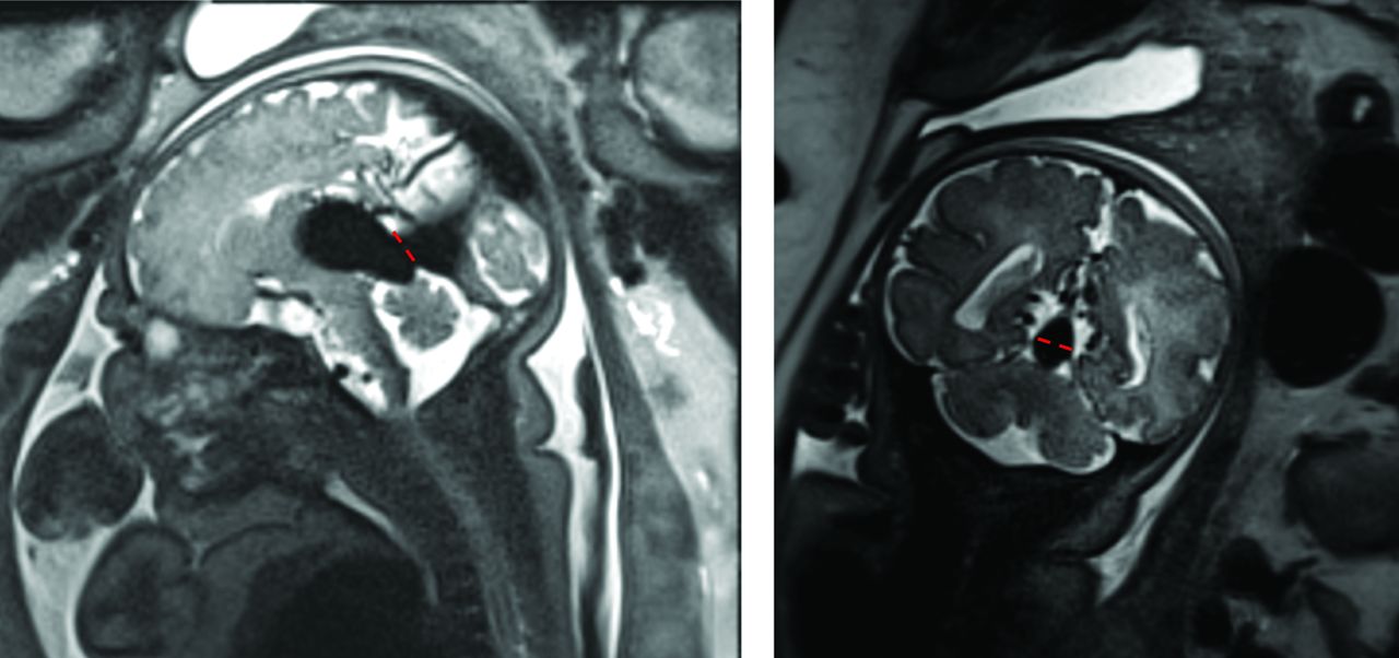
    Measurement of the mediolateral diameter of the falcine sinus at its shortest point. The left image shows a sagittal view of a fetal MR imaging, with the dashed red line at the section of shortest height of the falcine sinus. The right image shows a coronal view with the mediolateral diameter of the sinus demonstrated with red dashes, measured at this same shortest section. This diameter efficiently differentiated the IT from the NAR cohort, as did the cross-sectional area of the sinus, measured at this same section.

  • Fig 3.
    • Download figure
    • Open in new tab
    • Download powerpoint
    Fig 3.

    ROC curves showing measurements of the straight or falcine sinus area (SS-A) and the straight or falcine sinus maximal mediolateral diameter (SS-MD) at the shortest point of the sinus. Measurements from neonatal MR imaging (A) and from fetal MR imaging (B).

Tables

  • Figures
    • View popup
    Table 1:

    Fetal MR imaging scan univariate data

    RegionReader 1Reader 2ICC
    NARInfant TreatmentP ValueNARInfant TreatmentP Value
    No.MedianIQRNo.MedianIQRNo.MedianIQRNo.MedianIQR
    Varix ML (mm)101813–2241911–251.00101813–2452012–231.000.98
    Varix AP (mm)103028–3341912–25<.0193021–3452016–27.150.75
    Varix CC (mm)102018–214168–25.64101812–2151811–23.770.89
    Varix V (CM3)1052–5431–5.73952–6552–61.000.93
    SS-MD (mm)10108–12464–6<.011085–11533–5<.0010.95
    SS-P (mm)94339–4942822–34<.001103928–4152017–31<.010.87
    SS-A (mm2)108556–11252018–45<.01
    BA-MD (mm)533–4332–0.57
    R ICA-MD (mm)543–5332-0.57
    L ICA-MD (mm)533–5332-0.39
    R Sig-MD (mm)8109–12464–8<.0011076–8554–6<.0010.72
    L Sig-MD (mm)8117–12465–7.051097–9564–6<.010.94
    • Note:—AP indicates anterior-posterior diameter; IQR, interquartile range; L, left; ML, mediolateral diameter; R, right; V, volume; ICC, intraclass correlation coefficient. Statistical significance was set using a conservative criterion of P < .01 to account for multiple testing.

    • View popup
    Table 2:

    Early MR imaging scan data for neonatal at-risk and infant treatment cohorts

    RegionReader 1Reader 2ICC
    NARInfant TreatmentPNARInfant TreatmentP
    No.MedIQRNo.MedIQRNo.MedIQRNo.MedIQR
    Varix ML (mm)212418–27112119–25.46212317–27112118–26.970.921
    Varix AP (mm)213019–35112020–27.22213025–35112220–29.050.633
    Varix CC (mm)202420–27111916–24.16212116–29111815–24.140.875
    Varix V (CM3)2164–101143–8.242094–131143–8.150.961
    SS-MD (mm)17108–141165–8.002197–111132–6<.0010.983
    SS-A (mm2)2164–81133–4.00208565–123112818–55<.001 
    SS-P (mm)174036–48112921–37.00203532–43112721–35.030.969
    BA-MD (mm)2143–51143–4.032143–41132–3<.010.877
    R ICA-MD (mm)2144–51132–4.002143–41132–3<.0010.851
    L ICA-MD (mm)2143–51133–3.002143–41132–3<.010.655
    R Sig-MD (mm)2197–101174–9.042165–81152–6.020.642
    L Sig-MD (mm)2188–111176–9.102176–81143–6<.0010.775
    • Note:—Med indicates median; AP, anterior-posterior diameter; IQR, interquartile range; L, left; ML, mediolateral diameter; R, right; V, volume; ICC, intraclass correlation coefficient. Statistical significance was set using a conservative criterion of P < .01 to account for mutliple testing.

    • View popup
    Table 3:

    Presence of VOGM morphology in early MR imaging scan data for neonatal at-risk and infant treatment cohorts

    VOGM MorphologyChoroidalMuralTotal
    Reviewer 1
     Cohort
      NAR19221
      IT9211
      Total28432
    χ²(1, n = 32) = 0.495, P = .48
    Reviewer 2
     Cohort
      NAR21021
      IT4711
      Total25732
    χ²(1, n = 32) = 17.105, P < .001
    κ = 0.48, P = .05
    • View popup
    Table 4:

    Presence of occipital sinus morphology in early MR imaging scan data for neonatal at-risk and infant treatment cohorts

    Presence of Occipital SinusNoYesTotal
    Reviewer 1
     Cohort
      NAR02121
      IT6511
      Total62632
    χ²(1, n = 32) = 14.1, P < .001
    Reviewer 2
     Cohort
      NAR02121
      IT4711
      Total42832
    χ²(1, n = 32) = 8.3, P < .01
    κ = 0.819, P < .001
    • View popup
    Table 5:

    Cutoff values for NAR on fetal and neonatal MR imaginga

    AUC95% CIP ValueCutoff ValueSensitivitySpecificity
    Neonatal
     SS-MD0.8660.736–0.996<.001>6.2 mm86%82%
     SS-A0.8360.690–0.983.002>58 mm280%82%
    Fetal scans
     SS-MD0.9800.919–1.000.003>5.2 mm90%100%
     SS-A0.9410.823–1.000.007>33 mm290%80%
    • ↵a Probabilities determined by logistic regression, where straight or falcine sinus diameter on MR imaging was identified as a multivariable predictor of clinical outcome (AUC = 0.89, P < .001).

    • View popup
    Table 6:

    Likelihood of clinical evolution to NAR as a function of the mediolateral diameter of the straight or falcine sinus at its shortest pointa

    Straight or Falcine Sinus Diameter (mm)Probability of NAR95% CI
    04%1%–30%
    18%2%–38%
    214%3%–44%
    324%8%–52%
    438%18%–60%
    553%33%–72%
    668%49%–83%
    780%60%–92%
    888%69%–96%
    993%75%–98%
    1096%80%–99%
    1198%84%–99%
    1299%87%–99%
    13100%90%–100%
    • ↵a Probabilities determined by logistic regression, where straight or falcine sinus diameter on MR imaging was identified as a multivariable predictor of clinical outcome (AUC = 0.89, P < .001).

PreviousNext
Back to top

In this issue

American Journal of Neuroradiology: 41 (6)
American Journal of Neuroradiology
Vol. 41, Issue 6
1 Jun 2020
  • Table of Contents
  • Index by author
  • Complete Issue (PDF)
Advertisement
Print
Download PDF
Email Article

Thank you for your interest in spreading the word on American Journal of Neuroradiology.

NOTE: We only request your email address so that the person you are recommending the page to knows that you wanted them to see it, and that it is not junk mail. We do not capture any email address.

Enter multiple addresses on separate lines or separate them with commas.
Fetal and Neonatal MRI Predictors of Aggressive Early Clinical Course in Vein of Galen Malformation
(Your Name) has sent you a message from American Journal of Neuroradiology
(Your Name) thought you would like to see the American Journal of Neuroradiology web site.
CAPTCHA
This question is for testing whether or not you are a human visitor and to prevent automated spam submissions.
Cite this article
L. Arko, M. Lambrych, A. Montaser, D Zurakowski, D.B. Orbach
Fetal and Neonatal MRI Predictors of Aggressive Early Clinical Course in Vein of Galen Malformation
American Journal of Neuroradiology Jun 2020, 41 (6) 1105-1111; DOI: 10.3174/ajnr.A6585

Citation Manager Formats

  • BibTeX
  • Bookends
  • EasyBib
  • EndNote (tagged)
  • EndNote 8 (xml)
  • Medlars
  • Mendeley
  • Papers
  • RefWorks Tagged
  • Ref Manager
  • RIS
  • Zotero
0 Responses
Respond to this article
Share
Bookmark this article
Fetal and Neonatal MRI Predictors of Aggressive Early Clinical Course in Vein of Galen Malformation
L. Arko, M. Lambrych, A. Montaser, D Zurakowski, D.B. Orbach
American Journal of Neuroradiology Jun 2020, 41 (6) 1105-1111; DOI: 10.3174/ajnr.A6585
del.icio.us logo Twitter logo Facebook logo Mendeley logo
  • Tweet Widget
  • Facebook Like
  • Google Plus One
Purchase

Jump to section

  • Article
    • Abstract
    • ABBREVIATIONS:
    • MATERIALS AND METHODS
    • RESULTS
    • DISCUSSION
    • CONCLUSIONS
    • Footnotes
    • References
  • Figures & Data
  • Supplemental
  • Info & Metrics
  • Responses
  • References
  • PDF

Related Articles

  • PubMed
  • Google Scholar

Cited By...

  • Cerebrovascular Anomalies in the Fetus
  • Comparing Vascular Morphology and Hemodynamics in Patients with Vein of Galen Malformations Using Intracranial 4D Flow MRI
  • Brain Injury in Fetuses with Vein of Galen Malformation and Nongalenic Arteriovenous Fistulas: Static Snapshot or a Portent of More?
  • Percutaneous transuterine fetal cerebral embolisation to treat vein of Galen malformations at risk of urgent neonatal decompensation: study protocol for a clinical trial of safety and feasibility
  • Crossref
  • Google Scholar

This article has not yet been cited by articles in journals that are participating in Crossref Cited-by Linking.

More in this TOC Section

  • Pediatric Glioma:Imaging Clusters,Molecular Traits
  • Clinical SVR of Fetal Brain MRI
  • FRACTURE MR in Congenital Vertebral Anomalies
Show more PEDIATRIC NEUROIMAGING

Similar Articles

Advertisement

Indexed Content

  • Current Issue
  • Accepted Manuscripts
  • Article Preview
  • Past Issues
  • Editorials
  • Editor's Choice
  • Fellows' Journal Club
  • Letters to the Editor
  • Video Articles

Cases

  • Case Collection
  • Archive - Case of the Week
  • Archive - Case of the Month
  • Archive - Classic Case

More from AJNR

  • Trainee Corner
  • Imaging Protocols
  • MRI Safety Corner
  • Book Reviews

Multimedia

  • AJNR Podcasts
  • AJNR Scantastics

Resources

  • Turnaround Time
  • Submit a Manuscript
  • Submit a Video Article
  • Submit an eLetter to the Editor/Response
  • Manuscript Submission Guidelines
  • Statistical Tips
  • Fast Publishing of Accepted Manuscripts
  • Graphical Abstract Preparation
  • Imaging Protocol Submission
  • Evidence-Based Medicine Level Guide
  • Publishing Checklists
  • Author Policies
  • Become a Reviewer/Academy of Reviewers
  • News and Updates

About Us

  • About AJNR
  • Editorial Board
  • Editorial Board Alumni
  • Alerts
  • Permissions
  • Not an AJNR Subscriber? Join Now
  • Advertise with Us
  • Librarian Resources
  • Feedback
  • Terms and Conditions
  • AJNR Editorial Board Alumni

American Society of Neuroradiology

  • Not an ASNR Member? Join Now

© 2025 by the American Society of Neuroradiology All rights, including for text and data mining, AI training, and similar technologies, are reserved.
Print ISSN: 0195-6108 Online ISSN: 1936-959X

Powered by HighWire