Advertisement

Main menu

  • Home
  • Content
    • Current Issue
    • Accepted Manuscripts
    • Article Preview
    • Past Issue Archive
    • Video Articles
    • AJNR Case Collection
    • Case of the Week Archive
    • Case of the Month Archive
    • Classic Case Archive
  • Special Collections
    • AJNR Awards
    • Low-Field MRI
    • Alzheimer Disease
    • ASNR Foundation Special Collection
    • Photon-Counting CT
    • View All
  • Multimedia
    • AJNR Podcasts
    • AJNR SCANtastic
    • Trainee Corner
    • MRI Safety Corner
    • Imaging Protocols
  • For Authors
    • Submit a Manuscript
    • Submit a Video Article
    • Submit an eLetter to the Editor/Response
    • Manuscript Submission Guidelines
    • Statistical Tips
    • Fast Publishing of Accepted Manuscripts
    • Graphical Abstract Preparation
    • Imaging Protocol Submission
    • Author Policies
  • About Us
    • About AJNR
    • Editorial Board
    • Editorial Board Alumni
  • More
    • Become a Reviewer/Academy of Reviewers
    • Subscribers
    • Permissions
    • Alerts
    • Feedback
    • Advertisers
    • ASNR Home

User menu

  • Alerts
  • Log in

Search

  • Advanced search
American Journal of Neuroradiology

American Journal of Neuroradiology

ASHNR American Society of Functional Neuroradiology ASHNR American Society of Pediatric Neuroradiology ASSR
  • Alerts
  • Log in

Advanced Search

  • Home
  • Content
    • Current Issue
    • Accepted Manuscripts
    • Article Preview
    • Past Issue Archive
    • Video Articles
    • AJNR Case Collection
    • Case of the Week Archive
    • Case of the Month Archive
    • Classic Case Archive
  • Special Collections
    • AJNR Awards
    • Low-Field MRI
    • Alzheimer Disease
    • ASNR Foundation Special Collection
    • Photon-Counting CT
    • View All
  • Multimedia
    • AJNR Podcasts
    • AJNR SCANtastic
    • Trainee Corner
    • MRI Safety Corner
    • Imaging Protocols
  • For Authors
    • Submit a Manuscript
    • Submit a Video Article
    • Submit an eLetter to the Editor/Response
    • Manuscript Submission Guidelines
    • Statistical Tips
    • Fast Publishing of Accepted Manuscripts
    • Graphical Abstract Preparation
    • Imaging Protocol Submission
    • Author Policies
  • About Us
    • About AJNR
    • Editorial Board
    • Editorial Board Alumni
  • More
    • Become a Reviewer/Academy of Reviewers
    • Subscribers
    • Permissions
    • Alerts
    • Feedback
    • Advertisers
    • ASNR Home
  • Follow AJNR on Twitter
  • Visit AJNR on Facebook
  • Follow AJNR on Instagram
  • Join AJNR on LinkedIn
  • RSS Feeds

AJNR Awards, New Junior Editors, and more. Read the latest AJNR updates

Brief ReportAdult Brain

Vessel Wall MRI Enhancement in Noninflammatory Cerebral Amyloid Angiopathy

Q. Hao, N.M. Tsankova, H. Shoirah, C.P. Kellner and K. Nael
American Journal of Neuroradiology March 2020, 41 (3) 446-448; DOI: https://doi.org/10.3174/ajnr.A6445
Q. Hao
aFrom the Departments of Neurology (Q.H.)
  • Find this author on Google Scholar
  • Find this author on PubMed
  • Search for this author on this site
  • ORCID record for Q. Hao
N.M. Tsankova
bPathology (N.M.T.)
  • Find this author on Google Scholar
  • Find this author on PubMed
  • Search for this author on this site
  • ORCID record for N.M. Tsankova
H. Shoirah
cNeurosurgery (H.S., C.P.K.)
  • Find this author on Google Scholar
  • Find this author on PubMed
  • Search for this author on this site
  • ORCID record for H. Shoirah
C.P. Kellner
dand Radiology (K.N.), Icahn School of Medicine at Mount Sinai, New York, New York
  • Find this author on Google Scholar
  • Find this author on PubMed
  • Search for this author on this site
  • ORCID record for C.P. Kellner
K. Nael
eDepartment of Radiological Sciences (K.N.), David Geffen School of Medicine, University of California at Los Angeles, Los Angeles, California.
  • Find this author on Google Scholar
  • Find this author on PubMed
  • Search for this author on this site
  • ORCID record for K. Nael
  • Article
  • Figures & Data
  • Supplemental
  • Info & Metrics
  • Responses
  • References
  • PDF
Loading

Abstract

SUMMARY: Cerebral amyloid angiopathy is characterized by deposition of amyloid-β fibrils in the walls of small-to-medium-sized blood vessels. In this retrospective review of 5 patients with histologically confirmed noninflammatory cerebral amyloid angiopathy, high-resolution vessel wall MRI showed arterial wall enhancement in 2 patients (40%). Despite common consensus of equating vessel wall enhancement with inflammation, this report demonstrates that β-amyloid accumulation alone without inflammation can be associated with arterial wall enhancement in a subset of patients.

ABBREVIATIONS:

CAA
cerebral amyloid angiopathy
VWMRI
vessel wall MR imaging

Cerebral amyloid angiopathy (CAA) is characterized by deposition of amyloid-β fibrils in the vessel walls of leptomeningeal and cortical arteries, arterioles, and, less often, capillaries and veins of the central nervous system.1 Histologically, Puchtler alkaline Congo Red stain remains the standard diagnostic method for staining amyloid deposition, which can be seen in the adventitia, tunica media, or all layers of vessel wall.

Along the spectrum of amyloid-deposition-related angiopathies, there are 3 entities: 1) CAA (generally there is an absence of wall inflammation); 2) inflammatory CAA (varying amounts of perivascular inflammation are present, including multinucleated giant cells); and 3) amyloid-β–related angiitis (in which there is destructive vasculitis).2 There is often overlap in the clinical presentations of these entities, which may include focal neurologic deficits, cognitive impairment, or seizure related to hemorrhagic or ischemic changes. The clinical diagnosis of CAA relies on clinical history and imaging evidence of microbleeds or lobar hemorrhage that are not explained by other etiologies.1

High-resolution vessel wall MR imaging (VWMRI) is an emerging technique that is used to assess various vasculopathies, such as atherosclerotic disease, vasculitis, Moyamoya disease, reversible cerebral vasoconstriction syndrome, and dissection.3 While vessel wall enhancement using VWMRI has been reported in the inflammatory variety of amyloid-related angiopathies,2,3 very little is known about the VWMRI findings in patients with noninflammatory CAA. We report the VWMRI findings in a patient with histologically confirmed CAA.

Brief Report

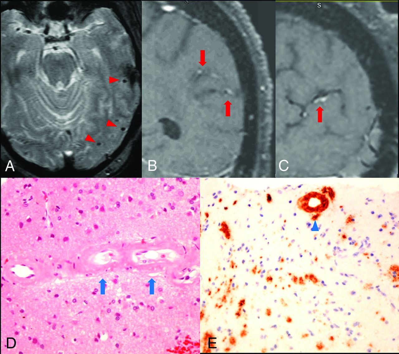
A 75-year-old patient with a history of hypertension, diabetes, progressive memory loss, and behavior changes during 6 months presented with worsening of mental status followed by seizures and status epilepticus. The initial neurologic examination re-vealed impaired attention and language; however, cranial nerves, motor strength, and gait were unremarkable. The patient had fluctuating mental status during the first 3 days and developed a clinical seizure on the fourth day with subsequent nonconvulsive status epilepticus, which was revealed on video-electroencephalography and required multiple antiepileptic agents. MR imaging showed no acute infarction but a few chronic microhemorrhages (Fig 1A) and some white matter changes. Due to the presence of chronic microhemorrhages, the differential diagnosis of vasculitis or amyloid angiopathy was entertained. Conventional angiography showed normal intracranial arteries. VWMRI (3D T1-weighted fast-spin-echo sequence with variable refocusing flip angle acquisition with inherent black-blood effect [sampling perfection with application-optimized contrasts by using different flip angle evolution, SPACE sequence; Siemens, Erlangen, Germany]: FOV, 230 mm; TR/TE, 700/18 ms; matrix, 256 mm2; 144 sections × 0.9-mm thick; voxels, 0.9 × 0.9 × 0.9 mm3) on the eighth day of admission showed arterial wall enhancement along the cortical branches of the left middle cerebral artery in the left parietal and temporal lobes (Fig 1B, -C). Findings of clinical and laboratory work-ups were negative for CNS infection, systemic vasculitis, or metabolic abnormalities except for elevated blood glucose (340 mg/dL on admission). Biopsy revealed β-amyloid accumulation in the vessel wall without associated inflammation (Fig 1D, -E).

Fig 1.
  • Download figure
  • Open in new tab
  • Download powerpoint
Fig 1.

Arterial wall enhancement is associated with amyloid accumulation within the vessel wall. A, Gradient-echo image shows microhemorrhages in the left temporal lobe (red arrowheads). B and C, coronal and sagittal views of postcontrast T1-weighted VWMRI show enhancement in the wall of cortical branches of the left middle cerebral artery in the parietal and temporal lobes (red arrows). D, Hematoxylin-eosin stain of a left temporal lobe sample shows thickened, hyalinized blood vessels containing amorphous eosinophilic material (blue arrows) in small- and medium-sized arteries within the leptomeninges and superficial cortical gray matter. E, Immunostain for β-amyloid shows amyloid accumulation within the vessel wall (blue arrowhead). No inflammatory cells were observed surrounding the vessels.

Following institutional review board approval, we subsequently reviewed digital charts of patients with a histologically proved diagnosis of noninflammatory CAA between August 2010 and February 2019. From a total of 10 patients identified, only 4 patients had adequate imaging for inclusion in this study (no MR imaging, n = 2; inadequate MR imaging image quality to assess vessel wall enhancement, n = 4). Of these 4 patients, 1 demonstrated arterial wall enhancement similar to that in our report. Clinical and demographic data, imaging findings including VWMRI and the number of microhemorrhages, and histopathologic findings are summarized in the On-line Table.

DISCUSSION

Intracranial vessel wall enhancement detected by high-resolution MR imaging has been considered a sign of inflammation and/or vasa vasorum in-growth in patients with atherosclerosis or vasculitis.3,4 Although vessel wall enhancement can be expected with vascular inflammatory processes such as primary angiitis of the central nervous system or an inflammatory type of amyloid such as amyloid β–related angiitis, in our report, we show that vessel wall enhancement can also be seen in a subset of patients with noninflammatory CAA (40%). This suggests that β-amyloid accumulation by itself and without inflammation can also result in vessel wall enhancement. This report, to our knowledge, is the first on VWMRI findings in patients with histopathologically proved noninflammatory CAA. An earlier study by De Havenon et al5 showed vessel wall enhancement in about 50% of patients with CAA; however, in this study, there was no discrimination between inflammatory and noninflammatory CAA because the patients did not have histopathology to confirm the diagnosis.

One hypothesis or explanation for our findings is potential change and disruption of the blood-brain barrier in CAA. CAA-positive vessels often exhibit morphologic changes such as loss of smooth-muscle cells and vessel wall thickening, which, in turn, can result in increased leakiness and an increased incidence of microbleeds.6,7 It is likely that this increased permeability is responsible for wall enhancement in a subset of CAA (despite the absence of inflammation). Most interesting, our patients who exhibited vessel wall enhancement also had numerous microhemorrhages, while those without vessel wall enhancement had none or just a few.

There are several limitations to our study. First, our sample size is very small, precluding any meaningful statistical analysis. Second, there is potential selection bias in this retrospectively collected series. Third, due to the retrospective nature of our study, there may have been variability in our VWMRI sequence parameters. Also, we did not use delay alternating with nutation for tailored excitation (DANTE) preparation to refine our blood suppression. However, we used the best option available to us from our vendors to perform these clinical studies using FDA-approved sequences in a clinical environment. Future prospective radiologic-histologic studies using dedicated VWMRI are needed to better characterize the arterial wall changes in CAA with and without inflammation, and their role in CAA-related cerebral pathologies.

Footnotes

  • Disclosures: Kambiz Nael—UNRELATED: Board Membership: Olea Medical, Comments: Medical Advisory Board. Qing Hao—RELATED: Support for Travel to Meetings for the Study or Other Purposes: Icahn School of Medicine at Mount Sinai, Comments: conference support from departmental research fund.

  • Paper previously presented, in part, at: Annual Meeting of American Society of Neuroradiology, May 18–23, 2019; Boston, Massachusetts.

References

  1. 1.↵
    1. Biffi A,
    2. Greenberg SM
    . Cerebral amyloid angiopathy: a systematic review. J Clin Neurol 2011;7:1–9 doi:10.3988/jcn.2011.7.1.1 pmid:21519520
    CrossRefPubMed
  2. 2.↵
    1. Moussaddy A,
    2. Levy A,
    3. Strbian D, et al
    . Inflammatory cerebral amyloid angiopathy, amyloid-beta-related angiitis, and primary angiitis of the central nervous system: similarities and differences. Stroke 2015;46:e210–13 doi:10.1161/STROKEAHA.115.010024 pmid:26185185
    FREE Full Text
  3. 3.↵
    1. Alexander MD,
    2. Yuan C,
    3. Rutman A, et al
    . High-resolution intracranial vessel wall imaging: imaging beyond the lumen. J Neurol Neurosurg Psychiatry 2016;87:589–97 doi:10.1136/jnnp-2015-312020 pmid:26746187
    Abstract/FREE Full Text
  4. 4.↵
    1. Lehman VT,
    2. Brinjikji W,
    3. Kallmes DF, et al
    . Clinical interpretation of high-resolution vessel wall MRI of intracranial arterial diseases. Br J Radiol 2016;89:20160496 doi:10.1259/bjr.20160496 pmid:27585640
    CrossRefPubMed
  5. 5.↵
    1. De Havenon A,
    2. Lobo R,
    3. Eisenmenger L, et al
    . MRI detection of vessel wall inflammation and contrast leakage in cerebral amyloid angiopathy. In: Proceedings of the International Stroke Conference, Houston, TX, Feb 22–24, 2017: WP138
  6. 6.↵
    1. Freeze WM,
    2. Bacskai BJ,
    3. Frosch MP, et al
    . Blood-brain barrier leakage and microvascular lesions in cerebral amyloid angiopathy. Stroke 2019;50:328–35 doi:10.1161/STROKEAHA.118.023788 pmid:30661497
    CrossRefPubMed
  7. 7.↵
    1. Charidimou A,
    2. Boulouis G,
    3. Gurol ME, et al
    . Emerging concepts in sporadic cerebral amyloid angiopathy. Brain 2017;140:1829–50 doi:10.1093/brain/awx047 pmid:28334869
    CrossRefPubMed
  • Received October 17, 2019.
  • Accepted after revision January 9, 2020.
  • © 2020 by American Journal of Neuroradiology
View Abstract
PreviousNext
Back to top

In this issue

American Journal of Neuroradiology: 41 (3)
American Journal of Neuroradiology
Vol. 41, Issue 3
1 Mar 2020
  • Table of Contents
  • Index by author
  • Complete Issue (PDF)
Advertisement
Print
Download PDF
Email Article

Thank you for your interest in spreading the word on American Journal of Neuroradiology.

NOTE: We only request your email address so that the person you are recommending the page to knows that you wanted them to see it, and that it is not junk mail. We do not capture any email address.

Enter multiple addresses on separate lines or separate them with commas.
Vessel Wall MRI Enhancement in Noninflammatory Cerebral Amyloid Angiopathy
(Your Name) has sent you a message from American Journal of Neuroradiology
(Your Name) thought you would like to see the American Journal of Neuroradiology web site.
CAPTCHA
This question is for testing whether or not you are a human visitor and to prevent automated spam submissions.
Cite this article
Q. Hao, N.M. Tsankova, H. Shoirah, C.P. Kellner, K. Nael
Vessel Wall MRI Enhancement in Noninflammatory Cerebral Amyloid Angiopathy
American Journal of Neuroradiology Mar 2020, 41 (3) 446-448; DOI: 10.3174/ajnr.A6445

Citation Manager Formats

  • BibTeX
  • Bookends
  • EasyBib
  • EndNote (tagged)
  • EndNote 8 (xml)
  • Medlars
  • Mendeley
  • Papers
  • RefWorks Tagged
  • Ref Manager
  • RIS
  • Zotero
0 Responses
Respond to this article
Share
Bookmark this article
Vessel Wall MRI Enhancement in Noninflammatory Cerebral Amyloid Angiopathy
Q. Hao, N.M. Tsankova, H. Shoirah, C.P. Kellner, K. Nael
American Journal of Neuroradiology Mar 2020, 41 (3) 446-448; DOI: 10.3174/ajnr.A6445
del.icio.us logo Twitter logo Facebook logo Mendeley logo
  • Tweet Widget
  • Facebook Like
  • Google Plus One
Purchase

Jump to section

  • Article
    • Abstract
    • ABBREVIATIONS:
    • DISCUSSION
    • Footnotes
    • References
  • Figures & Data
  • Supplemental
  • Info & Metrics
  • Responses
  • References
  • PDF

Related Articles

  • PubMed
  • Google Scholar

Cited By...

  • Occult Amyloid-{beta}-Related Angiitis: Neuroimaging Findings at 1.5T, 3T, and 7T MRI
  • Vessel Wall Enhancement on Black-Blood MRI Predicts Acute and Future Stroke in Cerebral Amyloid Angiopathy
  • Crossref (9)
  • Google Scholar

This article has been cited by the following articles in journals that are participating in Crossref Cited-by Linking.

  • Current Clinical Applications of Intracranial Vessel Wall MR Imaging
    Raghav R. Mattay, Jose F. Saucedo, Vance T. Lehman, Jiayu Xiao, Emmanuel C. Obusez, Scott B. Raymond, Zhaoyang Fan, Jae W. Song
    Seminars in Ultrasound, CT and MRI 2021 42 5
  • Vessel Wall Enhancement on Black-Blood MRI Predicts Acute and Future Stroke in Cerebral Amyloid Angiopathy
    J.S. McNally, A. Sakata, M.D. Alexander, L.D. Dewitt, J.A. Sonnen, S.T. Menacho, G.J. Stoddard, S.-E. Kim, A.H. de Havenon
    American Journal of Neuroradiology 2021 42 6
  • Diagnosing Cerebral Amyloid Angiopathy–Related Inflammation
    Andreas Charidimou
    Neurology 2024 103 2
  • Imaging of Amyloid-beta-related Arteritis
    Aaron Bangad, Mehdi Abbasi, Sam Payabvash, Adam de Havenon
    Neuroimaging Clinics of North America 2024 34 1
  • Inflammation in Cerebral Amyloid Angiopathy‐Related Transient Focal Neurological Episodes
    Amina Sellimi, Larysa Panteleienko, Dermot Mallon, Simon Fandler‐Höfler, Rupert Oliver, Victoria Harvey, Michael S. Zandi, Gargi Banerjee, David J. Werring
    Annals of Neurology 2025 97 3
  • Occult Amyloid-β-Related Angiitis: Neuroimaging Findings at 1.5T, 3T, and 7T MRI
    Can Özütemiz, Haitham M. Hussein, Salman Ikramuddin, H. Brent Clark, Andreas Charidimou, Christopher Streib
    American Journal of Neuroradiology 2024 45 8
  • Radiopaedia.org
    Frank Gaillard, Rohit Sharma, Chesterlee Ocampo
    2014
  • Imaging of intracranial arterial disease: a comparison between MRI and unenhanced CT
    Carlo Lucci, Ina Rissanen, Richard A. P. Takx, Anja G. van der Kolk, Anita A. Harteveld, Jan W. Dankbaar, Mirjam I. Geerlings, Pim A. de Jong, Jeroen Hendrikse
    Frontiers in Radiology 2024 4
  • The Diagnostic Value of Contrast‐Enhanced Vessel Wall MRI for Diagnosing Neuropsychiatric Systemic Lupus Erythematosus
    Satoru Ide, Yuya Fujita, Yu Murakami, Koichiro Futatsuya, Yuta Yoshimatsu, Jun Tsukamoto, Haruka Oku, Toshihiro Sakamoto, Yoshiya Tanaka, Takatoshi Aoki
    Journal of Magnetic Resonance Imaging 2025

More in this TOC Section

  • Diagnostic Neuroradiology of Monoclonal Antibodies
  • NCCT vs. MRI for Brain Atrophy in Acute Stroke
  • Clinical Outcomes After Chiari I Decompression
Show more ADULT BRAIN

Similar Articles

Advertisement

Indexed Content

  • Current Issue
  • Accepted Manuscripts
  • Article Preview
  • Past Issues
  • Editorials
  • Editor's Choice
  • Fellows' Journal Club
  • Letters to the Editor
  • Video Articles

Cases

  • Case Collection
  • Archive - Case of the Week
  • Archive - Case of the Month
  • Archive - Classic Case

More from AJNR

  • Trainee Corner
  • Imaging Protocols
  • MRI Safety Corner

Multimedia

  • AJNR Podcasts
  • AJNR Scantastics

Resources

  • Turnaround Time
  • Submit a Manuscript
  • Submit a Video Article
  • Submit an eLetter to the Editor/Response
  • Manuscript Submission Guidelines
  • Statistical Tips
  • Fast Publishing of Accepted Manuscripts
  • Graphical Abstract Preparation
  • Imaging Protocol Submission
  • Evidence-Based Medicine Level Guide
  • Publishing Checklists
  • Author Policies
  • Become a Reviewer/Academy of Reviewers
  • News and Updates

About Us

  • About AJNR
  • Editorial Board
  • Editorial Board Alumni
  • Alerts
  • Permissions
  • Not an AJNR Subscriber? Join Now
  • Advertise with Us
  • Librarian Resources
  • Feedback
  • Terms and Conditions
  • AJNR Editorial Board Alumni

American Society of Neuroradiology

  • Not an ASNR Member? Join Now

© 2025 by the American Society of Neuroradiology All rights, including for text and data mining, AI training, and similar technologies, are reserved.
Print ISSN: 0195-6108 Online ISSN: 1936-959X

Powered by HighWire