Advertisement

Main menu

  • Home
  • Content
    • Current Issue
    • Accepted Manuscripts
    • Article Preview
    • Past Issue Archive
    • Video Articles
    • AJNR Case Collection
    • Case of the Week Archive
    • Case of the Month Archive
    • Classic Case Archive
  • Special Collections
    • AJNR Awards
    • Low-Field MRI
    • Alzheimer Disease
    • ASNR Foundation Special Collection
    • Photon-Counting CT
    • View All
  • Multimedia
    • AJNR Podcasts
    • AJNR SCANtastic
    • Trainee Corner
    • MRI Safety Corner
    • Imaging Protocols
  • For Authors
    • Submit a Manuscript
    • Submit a Video Article
    • Submit an eLetter to the Editor/Response
    • Manuscript Submission Guidelines
    • Statistical Tips
    • Fast Publishing of Accepted Manuscripts
    • Graphical Abstract Preparation
    • Imaging Protocol Submission
    • Author Policies
  • About Us
    • About AJNR
    • Editorial Board
    • Editorial Board Alumni
  • More
    • Become a Reviewer/Academy of Reviewers
    • Subscribers
    • Permissions
    • Alerts
    • Feedback
    • Advertisers
    • ASNR Home

User menu

  • Alerts
  • Log in

Search

  • Advanced search
American Journal of Neuroradiology

American Journal of Neuroradiology

ASHNR American Society of Functional Neuroradiology ASHNR American Society of Pediatric Neuroradiology ASSR
  • Alerts
  • Log in

Advanced Search

  • Home
  • Content
    • Current Issue
    • Accepted Manuscripts
    • Article Preview
    • Past Issue Archive
    • Video Articles
    • AJNR Case Collection
    • Case of the Week Archive
    • Case of the Month Archive
    • Classic Case Archive
  • Special Collections
    • AJNR Awards
    • Low-Field MRI
    • Alzheimer Disease
    • ASNR Foundation Special Collection
    • Photon-Counting CT
    • View All
  • Multimedia
    • AJNR Podcasts
    • AJNR SCANtastic
    • Trainee Corner
    • MRI Safety Corner
    • Imaging Protocols
  • For Authors
    • Submit a Manuscript
    • Submit a Video Article
    • Submit an eLetter to the Editor/Response
    • Manuscript Submission Guidelines
    • Statistical Tips
    • Fast Publishing of Accepted Manuscripts
    • Graphical Abstract Preparation
    • Imaging Protocol Submission
    • Author Policies
  • About Us
    • About AJNR
    • Editorial Board
    • Editorial Board Alumni
  • More
    • Become a Reviewer/Academy of Reviewers
    • Subscribers
    • Permissions
    • Alerts
    • Feedback
    • Advertisers
    • ASNR Home
  • Follow AJNR on Twitter
  • Visit AJNR on Facebook
  • Follow AJNR on Instagram
  • Join AJNR on LinkedIn
  • RSS Feeds

AJNR Awards, New Junior Editors, and more. Read the latest AJNR updates

Research ArticleClinical Report

Macrocerebellum in Achondroplasia: A Further CNS Manifestation of FGFR3 Mutations?

H.M. Pascoe, J.Y.-M. Yang, J. Chen, A.M. Fink and S. Kumbla
American Journal of Neuroradiology February 2020, 41 (2) 338-342; DOI: https://doi.org/10.3174/ajnr.A6369
H.M. Pascoe
aFrom the Departments of Medical Imaging (H.M.P., A.M.F., S.K.)
  • Find this author on Google Scholar
  • Find this author on PubMed
  • Search for this author on this site
  • ORCID record for H.M. Pascoe
J.Y.-M. Yang
bNeurosurgery (J.Y.-M.Y.), The Royal Children’s Hospital, Parkville, Victoria, Australia
cNeuroscience Research (J.Y.-M.Y.)
dDevelopmental Imaging (J.Y.-M.Y., J.C.), Murdoch Children’s Research Institute, Parkville, Victoria, Australia
eDepartment of Paediatrics (J.Y.-M.Y.), University of Melbourne, Parkville, Victoria, Australia
  • Find this author on Google Scholar
  • Find this author on PubMed
  • Search for this author on this site
  • ORCID record for J.Y.-M. Yang
J. Chen
dDevelopmental Imaging (J.Y.-M.Y., J.C.), Murdoch Children’s Research Institute, Parkville, Victoria, Australia
  • Find this author on Google Scholar
  • Find this author on PubMed
  • Search for this author on this site
  • ORCID record for J. Chen
A.M. Fink
aFrom the Departments of Medical Imaging (H.M.P., A.M.F., S.K.)
fDepartment of Perinatal Medicine (A.M.F.), Mercy Hospital for Women, Heidelberg, Victoria, Australia
  • Find this author on Google Scholar
  • Find this author on PubMed
  • Search for this author on this site
  • ORCID record for A.M. Fink
S. Kumbla
aFrom the Departments of Medical Imaging (H.M.P., A.M.F., S.K.)
gDepartment of Diagnostic Imaging (S.K.), Monash Health, Clayton, Victoria, Australia.
  • Find this author on Google Scholar
  • Find this author on PubMed
  • Search for this author on this site
  • ORCID record for S. Kumbla
  • Article
  • Figures & Data
  • Info & Metrics
  • Responses
  • References
  • PDF
Loading

Abstract

SUMMARY: Achondroplasia is the result of a mutation in the fibroblast growth factor receptor 3 gene (FGFR3). Appearances suggestive of macrocerebellum have not been described in this patient group. We retrospectively reviewed MR imaging studies of the brain in 23 children with achondroplasia. A constellation of imaging findings that are recognized in macrocerebellum was observed, including cerebellar hemisphere enlargement (inferior and superior extension, wrapping around the brainstem); an effaced retro- and infravermian cerebellar subarachnoid CSF space; a shortened midbrain; distortion of the tectal plate; and mass effect on the brainstem. All MR imaging studies exhibited some of these findings. Quantitative analysis confirmed an increased cerebellar volume compared with age- and sex-matched controls. We hypothesized that this may be due to direct effects of the FGFR3 mutation on cerebellar morphogenesis.

ABBREVIATIONS:

PCF
posterior cranial fossa
PCFBV
posterior cranial fossa brain volume
STBV
supratentorial brain volume

Achondroplasia is the most common form of inherited disproportionate short stature affecting approximately 250,000 people worldwide. It is an autosomal dominant condition. The responsible mutation is almost always a glycine-to-arginine substitution at codon 380 in the transmembrane domain of the fibroblast growth factor receptor 3 (FGFR3) gene, which results in overactivation of the receptor. Neuroimaging is frequently performed in children with achondroplasia because there are known associated intracranial abnormalities.1⇓⇓-4

Macrocerebellum, an abnormally enlarged cerebellum, while rare, is associated with many syndromic conditions5⇓⇓⇓⇓⇓⇓-12 and has also been described as an isolated finding.13⇓-15 It has, however, never been reported in achondroplasia. We suspected an increased cerebellar volume in patients with achondroplasia based on observed qualitative MR imaging findings. However, given the high incidence of skull base deformities in achondroplasia, the possibility that these were contributing to the MR imaging appearances by causing crowding of the posterior cranial fossa (PCF) structures was considered. Skull base deformation occurs due to impaired endochondral ossification and premature fusion of the posterior fossa synchondroses.1,3 A recent quantitative study of the PCF in patients with achondroplasia, however, did not show a decreased bony PCF volume compared with controls but did confirm an increased cerebellar volume.3

We hypothesized that the appearances described in macrocerebellum are present to varying degrees in patients with achondroplasia and that these are due to a true increase in the cerebellar volume rather than secondary to skull base changes. In our case series, we describe the qualitative imaging features suggestive of macrocerebellum and use volumetric analysis to confirm our findings.

Case Series

A retrospective MR imaging data base search was conducted using the Radiology Information System at the Medical Imaging Department, The Royal Children’s Hospital, Melbourne, Australia. We identified all children (from birth to 18 years of age) who had a diagnosis of achondroplasia and an MR imaging study of the brain performed for any indication between January 2000 and November 2018. The study was approved by our institutional Human Research Ethics Committee as a clinical audit.

The inclusion criteria were a diagnosis of achondroplasia and the availability of a diagnostic-quality MR examination of the brain in the study period. If the patient had >1 MR imaging in the study period, the most recent MR imaging was used. Twenty-three patients were included (12 boys and 11 girls). Ages ranged from 3 months to 15 years. An age- and sex-matched healthy control was selected for each patient. The controls were identified from the MR imaging database at The Royal Children’s Hospital using the following criteria: 1) normal brain anatomy, and 2) absence of a neurologic disorder. Reasons for their MR imaging referral were either sensorineural hearing loss or headache. MR imaging studies were performed on a 1.5 or 3T scanner.

Both qualitative and quantitative analyses were performed.

For qualitative analysis, the volumetric T1WIs were retrospectively evaluated by a pediatric radiology fellow (H.M.P.) and 2 pediatric neuroradiologists (S.K. and A.M.F.), and the results were consensus-based. The presence of the following qualitative imaging features (Fig 1A–C) was assessed:

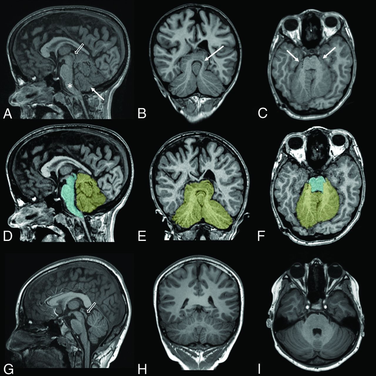
  1. Extension of the cerebellar hemispheres inferior to the vermis (on the midsagittal plane; Fig 1A).

  2. Extension of the superomedial portions of the cerebellar hemispheres above the tentorium cerebelli (on the coronal plane; Fig 1B).

  3. Wrapping of the cerebellar hemispheres around the brainstem with extension around the lateral aspects of the brainstem at the level of the middle cerebellar peduncles (on the axial plane; Fig 1C).

  4. Effacement of the retro- and infravermian subarachnoid CSF spaces (on the midsagittal plane; Fig 1A).

  5. Apparent shortening of the midbrain with or without distortion of the tectal plate (on the midsagittal plane; Fig 1A).

  6. Mass effect on the brainstem (on the midsagittal plane; Fig 1A). The features assessed were anterior bowing of the brainstem, narrowing of the prepontine cistern, and effacement of the fourth ventricle.

FIG 1.
  • Download figure
  • Open in new tab
  • Download powerpoint
FIG 1.

T1WIs of an 11-year-old child with achondroplasia (A–C) and the same T1WIs with superimposed cerebellum (yellow) and brainstem (cyan) segmentations (D–F). T1WIs from a healthy 11-year-old child (G–I) are provided for comparison. Extension of the cerebellar hemispheres inferior to the vermis is seen on the midsagittal plane (arrow in A); extension of the superomedial portions of the hemispheres above the tentorium cerebelli is seen on the coronal plane (arrow in B); and wrapping of the cerebellar hemispheres around the brainstem with extension around the lateral aspects of the brainstem at the level of the middle cerebellar peduncles is seen on the axial plane (arrows in C). Also seen on the midsagittal plane (A) are effacement of the retro- and infravermian subarachnoid CSF spaces, mass effect on the brainstem (which is anteriorly bowed [asterisk] with narrowing of the prepontine cistern and partial effacement of the fourth ventricle), and shortening of the midbrain with distortion of the tectal plate (open arrow). Note the normal morphology of the midbrain and tectal plate in G (open arrow).

Quantitative analysis of the cerebellar and brainstem volumes was performed in each patient and the age- and sex-matched healthy control using automated brain tissue segmentation. T2WIs were used for analyzing patients younger than 1 year of age, and T1WIs for patients older than 1 year of age. All T2WIs used for the younger subjects had 0.4-mm in-plane resolution and 4-mm slice thickness. All 3D T1WIs had similar (0.5–0.9 mm) in-plane resolution and 0.9-mm slice thickness. The imaging sequence used and the resolution were comparable between the patient and the paired control to avoid introducing bias in our quantitative analysis. Morphologically Adaptive Neonatal Tissue Segmentation (MANTIS) software (https://www.mantisoftware.com/)16 was used in the younger group, and a surface-based brain parcellation software, FreeSurfer Version 6.0 (http://surfer.nmr.mgh.harvard.edu),17 was used in the older group. Manual editing was subsequently performed to ensure anatomic accuracy. This was performed by the neurosurgery fellow (J.Y.-M.Y.) and reviewed by a pediatric radiology fellow (H.M.P.) and a pediatric neuroradiologist (S.K.). The cerebellar segmentation included both the cerebellar hemispheres and vermis. The brainstem segmentation included the midbrain, pons, and medulla. The posterior cranial fossa brain volume (PCFBV) was derived by summing the cerebellum and brainstem volumes.

The supratentorial brain volume (STBV) was also calculated for all cases. This was performed using the brain tissue classification tool from SPM12 (http://www.fil.ion.ucl.ac.uk/spm/software/spm12)18 to calculate the total brain volume, and the PCFBV was then subtracted from this.

Statistical analysis was performed using the R statistical and computing software and graphics (http://www.r-project.org). Group summary statistics were expressed in means and SDs. Comparison between the patients and their age- and sex-matched controls was analyzed using the paired Wilcoxon rank sum test. P values < .05 were considered statistically significant.

Results of the qualitative neuroimaging features of macrocerebellum are summarized in Table 1. Each of the MR imaging findings was recorded as present or not. Figure 1A–C demonstrates the qualitative MR imaging findings, with a healthy control for comparison shown in Fig 1G–I.

View this table:
  • View inline
  • View popup
Table 1:

Summary of the qualitative features of macrocerebellum in patients with achondroplasia

The results of quantitative analysis are summarized in Table 2. Figure 1D–F demonstrates the cerebellar and brainstem segmentations in standard orthogonal 2D MR imaging planes. Figure 2 demonstrates the cerebellar and brainstem segmentation as a 3D volume rendering.

FIG 2.
  • Download figure
  • Open in new tab
  • Download powerpoint
FIG 2.

Cerebellum (yellow) and brainstem (cyan) segmentations of the same patient with achondroplasia as in Fig 1, shown as 3D volume renders.

View this table:
  • View inline
  • View popup
Table 2:

Quantitative analysis of the cerebellum and brainstem

The achondroplasia patients had a significantly larger PCFBV compared with their age- and sex-matched healthy controls (P = .02). This volume difference was due to a larger cerebellum (P = .009). There was no brainstem volume difference between the 2 study groups (P = .92). The patients with achondroplasia also had significantly larger STBVs compared with their age- and sex-matched healthy controls (P < .001).

DISCUSSION

The prevalence of skull base changes in patients with achondroplasia is well-documented; however, the morphology of the PCF content has not been widely reported.3 In particular, appearances described in macrocerebellum,19 such as those we observed, have not been described in this patient group.

The cerebellar enlargement in macrocerebellum has been reported to mainly involve the cerebellar hemispheres13,15 and has been described as an isolated finding13⇓-15 as well as in many syndromic conditions.5⇓⇓⇓⇓⇓⇓-12 As a consequence of the cerebellar hemispheric enlargement, the hemispheres extend into available surrounding spaces, resulting in wrapping around the brainstem as well as upward and/or downward herniation.15 All of the patients with achondroplasia in our study demonstrated some of these qualitative signs of apparent cerebellar hemisphere enlargement. Ancillary changes of apparent macrocerebellum observed in the PCF were the following: effacement of the retro- and infravermian subarachnoid CSF spaces; apparent shortening of the midbrain, with associated distortion of the tectal plate in severe cases; and mass effect on the brainstem. Most of these features have been previously described in macrocerebellum.5,19

On the basis of our qualitative results, it remained possible that deformation of the bony PCF causing overcrowding could be responsible for the MR imaging appearance of an apparent macrocerebellum. However, in line with the findings by Calandrelli et al,3 we confirmed that compared with healthy controls, cerebellar volumes were increased in patients with achondroplasia, indicating true macrocerebellum. In contradistinction to Calandrelli et al, however, we found no difference in the brainstem volume between patients and controls. This may be due to the small number of cases in both series; thus, larger series would be needed to establish whether there is any brainstem involvement in achondroplasia.

Fibroblast growth factor receptors are transmembrane tyrosine kinases that are expressed in the developing brain.20 They are encoded by 4 genes (FGFR1–4),21 including the FGFR3 gene, which is mutated in achondroplasia. Fibroblast growth factors bind to the fibroblast growth factor receptors, and this signaling is an important regulator of cerebral cortex development.22 Studies have demonstrated malformations involving the cerebral cortex, such as expansion of the occipitotemporal cortex22 and oversulcation of the mesiotemporal lobes4 in the presence of activating FGFR3 mutations. These findings may also explain our finding of significantly higher suptratentorial brain volume in patients with achondroplasia compared with healthy controls.

Less well-known is that fibroblast growth factor signaling has also been shown in mice to have an important role in regulating the formation of the cerebellum from the early embryonic period into early postnatal life.23 Three neural layers are present in the adult cerebellar cortex (the molecular, Purkinje cell, and granular cells layers), and most of the cerebellar neurons migrate to the cortex from the ventricular zone during the early embryonic period under the influences of molecular signaling, including fibroblast growth factor signaling. In the mouse cerebellum, FGFR3 has been detected in the cerebellum as early as embryonic day 12.5. FGFR3 is then expressed at varying levels in different parts of the cerebellar nuclei and cortical layers throughout the remainder of the prenatal period extending to 21 days postnatally.23 Shazeeb et al24 reported no FGFR3 expression in the cerebellum of 3-week-old mice.

There is no current literature on the appearance of the cerebellum in achondroplasia. However, given our qualitative and quantitative findings, the known expression of FGFR3 in the cerebellum during early development in mice, and the known growth-promoting effects of FGFR3 mutations in the supratentorial compartment, we hypothesized that macrocerebellum in achondroplasia may be due to the direct effects of the FGFR3 mutation on cerebellar morphogenesis. Further genetic and pathologic studies are required to substantiate or refute this hypothesis.

A major limitation of our study is that it is retrospective, with a relatively small sample size. The prevalence of macrocerebellum in patients with achondroplasia can thus not be established from this study. Additionally, segmenting the cerebellar hemispheres and vermis separately was beyond the scope of the methodology of this study; thus, the relative contribution of each to the enlarged cerebellum cannot be determined.

CONCLUSIONS

Our study is the first to describe macrocerebellum with its typical appearance in children with achondroplasia. We confirmed an increase in the cerebellar volume quantitatively through volumetric analysis and hypothesized that macrocerebellum may be due to the direct effects of the FGFR3 mutation. Thus, macrocerebellum can be inferred when the qualitative imaging appearances we described are identified. These features should be sought when imaging patients with achondroplasia, alongside the other previously reported PCF and supratentorial features. Further pathologic and genetic studies will be required to gain a better understanding of our observations.

Acknowledgments

We thank the support from The Royal Children's Hospital Foundation, Murdoch Children's Research Institute, The University of Melbourne Department of Pediatrics, and the Victorian Government's Operational Infrastructure Support Program.

Footnotes

  • Dr Joseph Yuan-Mou Yang receives grant support from the Royal Children’s Hospital Foundation (RCH1000).

References

  1. 1.↵
    1. Kao SC,
    2. Waziri MH,
    3. Smith WL, et al
    . MR imaging of the craniovertebral junction, cranium, and brain in children with achondroplasia. AJR Am J Roentgenol 1989;153:565–69 doi:10.2214/ajr.153.3.565 pmid:2763957
    CrossRefPubMed
  2. 2.↵
    1. Pauli RM
    . Achondroplasia: a comprehensive clinical review. Orphanet J Rare Dis 2019;14:1 doi:10.1186/s13023-018-0972-6 pmid:30606190
    CrossRefPubMed
  3. 3.↵
    1. Calandrelli R,
    2. Panfili M,
    3. D’Apolito G, et al
    . Quantitative approach to the posterior cranial fossa and craniocervical junction in asymptomatic children with achondroplasia. Neuroradiology 2017;59:1031–41 doi:10.1007/s00234-017-1887-y pmid:28819680
    CrossRefPubMed
  4. 4.↵
    1. Manikkam SA,
    2. Chetcuti K,
    3. Howell KB, et al
    . Temporal lobe malformations in achondroplasia: expanding the brain imaging phenotype associated with FGFR3-related skeletal dysplasias. AJNR Am J Neuroradiol 2018;39:380–84 doi:10.3174/ajnr.A5468 pmid:29170271
    Abstract/FREE Full Text
  5. 5.↵
    1. Alqahtani E,
    2. Huisman TA,
    3. Boltshauser E, et al
    . Mucopolysaccharidoses type I and II: new neuroimaging findings in the cerebellum. Eur J Paediatr Neurol 2014;18:211–17 doi:10.1016/j.ejpn.2013.11.014 pmid:24423630
    CrossRefPubMed
  6. 6.↵
    1. Poretti A,
    2. Snow J,
    3. Summers AC, et al
    . Joubert syndrome: neuroimaging findings in 110 patients in correlation with cognitive function and genetic cause. J Med Genet 2017;54:521–29 doi:10.1136/jmedgenet-2016-104425 pmid:28087721
    Abstract/FREE Full Text
  7. 7.↵
    1. Shiohama T,
    2. Fujii K,
    3. Miyashita T, et al
    . Brain morphology in children with nevoid basal cell carcinoma syndrome. Am J Med Genet A 2017;173:946–52 doi:10.1002/ajmg.a.38115 pmid:28328116
    CrossRefPubMed
  8. 8.↵
    1. Schaefer GB,
    2. Bodensteiner JB,
    3. Buehler BA, et al
    . The neuroimaging findings in Sotos syndrome. Am J Med Genet 1997;68:462–65 doi:10.1002/(sici)1096-8628(19970211)68:4<462::aid-ajmg18>3.0.co;2-q pmid:9021022
    CrossRefPubMed
  9. 9.↵
    1. Gripp KW,
    2. Hopkins E,
    3. Doyle D, et al
    . High incidence of progressive postnatal cerebellar enlargement in Costello syndrome: brain overgrowth associated with HRAS mutations as the likely cause of structural brain and spinal cord abnormalities. Am J Med Genet A 2010;152:1161–68 doi:10.1002/ajmg.a.33391 pmid:20425820
    CrossRefPubMed
  10. 10.↵
    1. Jones W,
    2. Hesselink J,
    3. Courchesne E, et al
    . Cerebellar abnormalities in infants and toddlers with Williams syndrome. Dev Med Child Neurol 2002;44:688–94 doi:10.1017/s0012162201002766 pmid:12418794
    CrossRefPubMed
  11. 11.↵
    1. van der Knaap MS,
    2. Salomons GS,
    3. Li R, et al
    . Unusual variants of Alexander’s disease. Ann Neurol 2005;57:327–38 doi:10.1002/ana.20381 pmid:15732098
    CrossRefPubMed
  12. 12.↵
    1. Kau T,
    2. Karlo C,
    3. Güngör T, et al
    . Increased cerebellar volume in the early stage of fucosidosis: a case control study. Neuroradiology 2011;53:509–16 doi:10.1007/s00234-011-0855-1 pmid:21384124
    CrossRefPubMed
  13. 13.↵
    1. Bodensteiner JB,
    2. Schaefer GB,
    3. Keller GM, et al
    . Macrocerebellum: neuroimaging and clinical features of a newly recognized condition. J Child Neurol 1997;12:365–68 doi:10.1177/088307389701200605 pmid:9309519
    CrossRefPubMed
  14. 14.↵
    1. D’Arco F,
    2. Ugga L,
    3. Caranci F, et al
    . Isolated macrocerebellum: description of six cases and literature review. Quant Imaging Med Surg 2016;6:496–503 doi:10.21037/qims.2016.06.10 pmid:27942468
    CrossRefPubMed
  15. 15.↵
    1. Izbudak I,
    2. Acer N,
    3. Poretti A, et al
    . Macrocerebellum: volumetric and diffusion tensor imaging analysis. Turk Neurosurg 2015;25:948–53 pmid:26617148
    PubMed
  16. 16.↵
    1. Beare RJ,
    2. Chen J,
    3. Kelly CE, et al
    . Neonatal brain tissue classification with morphological adaptation and unified segmentation. Front Neuroinform 2016;10:12 doi:10.3389/fninf.2016.00012 pmid:27065840
    CrossRefPubMed
  17. 17.↵
    1. Fischl B
    . FreeSurfer. Neuroimage 2012;62:774–81 doi:10.1016/j.neuroimage.2012.01.021 pmid:22248573
    CrossRefPubMed
  18. 18.↵
    1. Ashburner J,
    2. Friston KJ
    . Unified segmentation. Neuroimage 2005;26:839–51 doi:10.1016/j.neuroimage.2005.02.018 pmid:15955494
    CrossRefPubMed
  19. 19.↵
    1. Poretti A,
    2. Mall V,
    3. Smitka M, et al
    . Macrocerebellum: significance and pathogenic considerations. Cerebellum 2012;11:1026–36 doi:10.1007/s12311-012-0379-1 pmid:22528964
    CrossRefPubMed
  20. 20.↵
    1. Hevner RF
    . The cerebral cortex malformation in thanatophoric dysplasia: neuropathology and pathogenesis. Acta Neuropathol 2005;110:208–21 doi:10.1007/s00401-005-1059-8 pmid:16133544
    CrossRefPubMed
  21. 21.↵
    1. Zhang X,
    2. Ibrahimi OA,
    3. Olsen SK, et al
    . Receptor specificity of the fibroblast growth factor family: the complete mammalian FGF family. J Biol Chem 2006;281:15694–700 doi:10.1074/jbc.M601252200 pmid:16597617
    Abstract/FREE Full Text
  22. 22.↵
    1. Thomson RE,
    2. Kind PC,
    3. Graham NA, et al
    . FGF receptor 3 activation promotes selective growth and expansion of occipitotemporal cortex. Neural Dev 2009;4:4 doi:10.1186/1749-8104-4-4 pmid:19192266
    CrossRefPubMed
  23. 23.↵
    1. Yaguchi Y,
    2. Yu T,
    3. Ahmed MU, et al
    . Fibroblast growth factor (FGF) gene expression in the developing cerebellum suggests multiple roles for FGF signaling during cerebellar morphogenesis and development. Dev Dyn 2009;238:2058–72 doi:10.1002/dvdy.22013 pmid:19544582
    CrossRefPubMed
  24. 24.↵
    1. Shazeeb MS,
    2. Cox MK,
    3. Gupta A, et al
    . Skeletal characterization of the FGFR3 mouse model of achondroplasia using micro-CT and MRI volumetric imaging. Sci Rep 2018;8:469 doi:10.1038/s41598-017-18801-0 pmid:29323153
    CrossRefPubMed
  • Received August 14, 2019.
  • Accepted after revision November 2, 2019.
  • © 2020 by American Journal of Neuroradiology
View Abstract
PreviousNext
Back to top

In this issue

American Journal of Neuroradiology: 41 (2)
American Journal of Neuroradiology
Vol. 41, Issue 2
1 Feb 2020
  • Table of Contents
  • Index by author
  • Complete Issue (PDF)
Advertisement
Print
Download PDF
Email Article

Thank you for your interest in spreading the word on American Journal of Neuroradiology.

NOTE: We only request your email address so that the person you are recommending the page to knows that you wanted them to see it, and that it is not junk mail. We do not capture any email address.

Enter multiple addresses on separate lines or separate them with commas.
Macrocerebellum in Achondroplasia: A Further CNS Manifestation of FGFR3 Mutations?
(Your Name) has sent you a message from American Journal of Neuroradiology
(Your Name) thought you would like to see the American Journal of Neuroradiology web site.
CAPTCHA
This question is for testing whether or not you are a human visitor and to prevent automated spam submissions.
Cite this article
H.M. Pascoe, J.Y.-M. Yang, J. Chen, A.M. Fink, S. Kumbla
Macrocerebellum in Achondroplasia: A Further CNS Manifestation of FGFR3 Mutations?
American Journal of Neuroradiology Feb 2020, 41 (2) 338-342; DOI: 10.3174/ajnr.A6369

Citation Manager Formats

  • BibTeX
  • Bookends
  • EasyBib
  • EndNote (tagged)
  • EndNote 8 (xml)
  • Medlars
  • Mendeley
  • Papers
  • RefWorks Tagged
  • Ref Manager
  • RIS
  • Zotero
0 Responses
Respond to this article
Share
Bookmark this article
Macrocerebellum in Achondroplasia: A Further CNS Manifestation of FGFR3 Mutations?
H.M. Pascoe, J.Y.-M. Yang, J. Chen, A.M. Fink, S. Kumbla
American Journal of Neuroradiology Feb 2020, 41 (2) 338-342; DOI: 10.3174/ajnr.A6369
del.icio.us logo Twitter logo Facebook logo Mendeley logo
  • Tweet Widget
  • Facebook Like
  • Google Plus One
Purchase

Jump to section

  • Article
    • Abstract
    • ABBREVIATIONS:
    • DISCUSSION
    • CONCLUSIONS
    • Acknowledgments
    • Footnotes
    • References
  • Figures & Data
  • Info & Metrics
  • Responses
  • References
  • PDF

Related Articles

  • No related articles found.
  • PubMed
  • Google Scholar

Cited By...

  • No citing articles found.
  • Crossref (4)
  • Google Scholar

This article has been cited by the following articles in journals that are participating in Crossref Cited-by Linking.

  • Impairment of motor skills in children with achondroplasia—usefulness of brain and cranio-cervical junction evaluation by quantitative magnetic resonance imaging: a case-control study
    Rosalinda Calandrelli, Fabio Pilato, Luca Massimi, Roberta Onesimo, Gabriella D’Apolito, Lorenzo Tenore, Domenico Romeo, Chiara Leoni, Giuseppe Zampino, Cesare Colosimo
    Acta Radiologica 2022 63 12
  • Embryonic cranial cartilage defects in the Fgfr3Y367C/+ mouse model of achondroplasia
    Susan M. Motch Perrine, Nishchal Sapkota, Kazuhiko Kawasaki, Yejia Zhang, Danny Z. Chen, Mizuho Kawasaki, Emily L. Durham, Yann Heuzé, Laurence Legeai‐Mallet, Joan T. Richtsmeier
    The Anatomical Record 2025 308 7
  • Anatomical Characteristics of Cervicomedullary Compression on MRI Scans in Children with Achondroplasia
    Isabella Trautwein, Daniel Behme, Philip Kunkel, Jasper Gerdes, Klaus Mohnike
    Journal of Imaging 2024 10 11
  • Cognitive and Behavioral Profile in a Child with Macrocerebellum—A Rare Congenital Brain Malformation
    Betsy Baby, Gowthami Nair, Maniyan Lathikakumari Arya, Madhusoodanan Uranguladi, Ajina Khan, Soumya Sundaram
    Journal of Pediatric Neurology 2024

More in this TOC Section

Clinical Report

  • Clinical and Imaging Features of Primary Intradural Extramedullary Ewing Sarcoma
  • A Practical Guide to Microtia Scoring: Step-by-Step
  • Revisiting a Rare Anomaly Described 25 Years Ago in the AJNR: A Journey from Pediatric Hemifacial Microsomia and Middle Cranial Fossa Aplasia to CSF-Lymphatic Fistula and Spontaneous Intracranial Hypotension as an Adult
Show more Clinical Report

Pediatrics

  • Clinical and Imaging Features of Primary Intradural Extramedullary Ewing Sarcoma
  • A Practical Guide to Microtia Scoring: Step-by-Step
  • Revisiting a Rare Anomaly Described 25 Years Ago in the AJNR: A Journey from Pediatric Hemifacial Microsomia and Middle Cranial Fossa Aplasia to CSF-Lymphatic Fistula and Spontaneous Intracranial Hypotension as an Adult
Show more Pediatrics

Similar Articles

Advertisement

Indexed Content

  • Current Issue
  • Accepted Manuscripts
  • Article Preview
  • Past Issues
  • Editorials
  • Editor's Choice
  • Fellows' Journal Club
  • Letters to the Editor
  • Video Articles

Cases

  • Case Collection
  • Archive - Case of the Week
  • Archive - Case of the Month
  • Archive - Classic Case

More from AJNR

  • Trainee Corner
  • Imaging Protocols
  • MRI Safety Corner
  • Book Reviews

Multimedia

  • AJNR Podcasts
  • AJNR Scantastics

Resources

  • Turnaround Time
  • Submit a Manuscript
  • Submit a Video Article
  • Submit an eLetter to the Editor/Response
  • Manuscript Submission Guidelines
  • Statistical Tips
  • Fast Publishing of Accepted Manuscripts
  • Graphical Abstract Preparation
  • Imaging Protocol Submission
  • Evidence-Based Medicine Level Guide
  • Publishing Checklists
  • Author Policies
  • Become a Reviewer/Academy of Reviewers
  • News and Updates

About Us

  • About AJNR
  • Editorial Board
  • Editorial Board Alumni
  • Alerts
  • Permissions
  • Not an AJNR Subscriber? Join Now
  • Advertise with Us
  • Librarian Resources
  • Feedback
  • Terms and Conditions
  • AJNR Editorial Board Alumni

American Society of Neuroradiology

  • Not an ASNR Member? Join Now

© 2025 by the American Society of Neuroradiology All rights, including for text and data mining, AI training, and similar technologies, are reserved.
Print ISSN: 0195-6108 Online ISSN: 1936-959X

Powered by HighWire