
Article Figures & Data
Figures
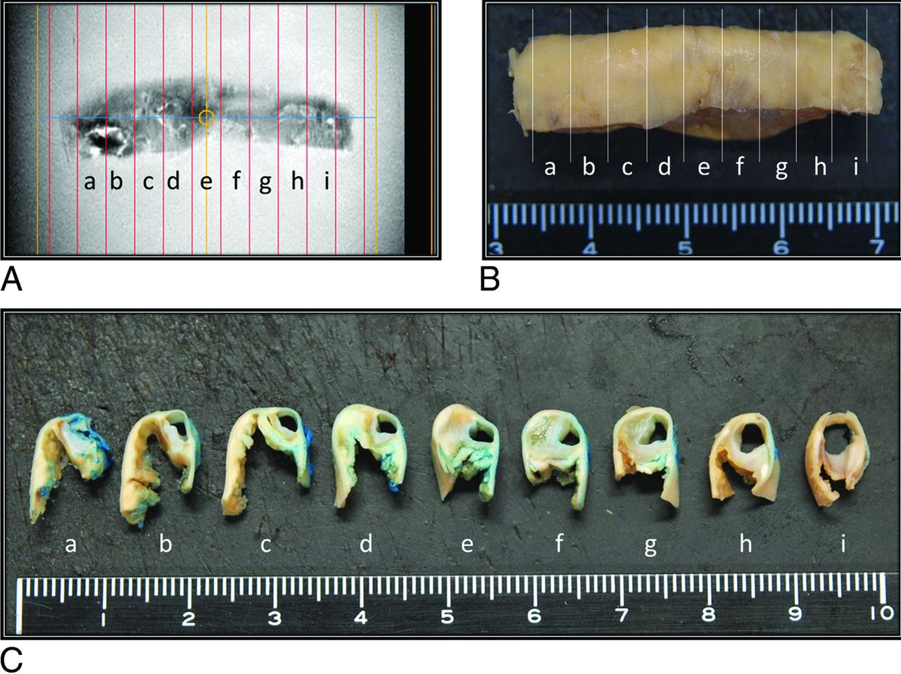
- FIG 1.
Resected carotid artery specimen from a 72-year-old man. Tissue preparation for MR imaging and histology studies. Formalin-fixed carotid endarterectomy tissue specimens (B and C) were cut into 3-mm-thick sections and matched at 3-mm intervals against the corresponding MR image (A). Blue dye was used for plaque orientation (C).
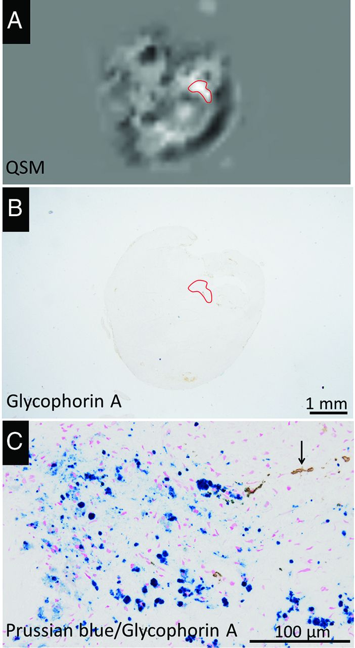
- FIG 2.
Representative QSM (A) and MPRAGE (B) images of carotid plaques and corresponding histologic images (C–F) in a 77-year-old man. Carotid plaque with both hyper- and hypointense areas on the QSM image (A) and corresponding MPRAGE image (B). The hyperintense ROI (A, yellow outline) corresponds with histologic evidence of intraplaque hemorrhage and erythrocytes in the atherosclerotic necrotic core (C, H&E stain). The glycophorin-A-positive erythrocytes and erythrocyte fragments are abundant in the plaque (D). In the hyperintense ROI (yellow outline) on the QSM image (A), Prussian blue staining shows no iron deposition (E). Two hypointense ROIs (blue outline) on the QSM image (A) correspond with basophilic calcification in the plaque (F, H&E stain). The corresponding blue and yellow outlines are placed on the MPRAGE image (B).
- FIG 3.
Representative QSM image (A) of carotid plaques of a 79-year-old man and corresponding histologic images (B and C). Carotid plaque with a hyperintense area on the QSM image (A, red outline), which contained few GYPA-positive erythrocyte components in the corresponding histologic ROI (B, red outline). There are many intracellular hemosiderin deposits (blue) and a few erythrocytes (brown, arrow) in the ROI (C).
- FIG 4.
Correlative analysis of erythrocytes and iron deposits on histologic sections from a 73-year-old man (A–D) and the SV on the QSM image of whole plaque (E). Areas positive for GYPA staining (A) and color extraction (B) were relatively well-correlated with the distribution of hyperintensity on the QSM image (E). There are a few small areas positive for Prussian blue staining (C) and color extraction (D). Positively stained areas were extracted under specific protocols using the hue of the color and its lightness and saturation. Data are expressed as the ratio (percentage) of extracted green areas relative to the whole plaque (dashed outline in B and D). The ROI on the QSM image (yellow outline, E) is matched with the histologic ROI for measuring the mean SV of the whole plaque.
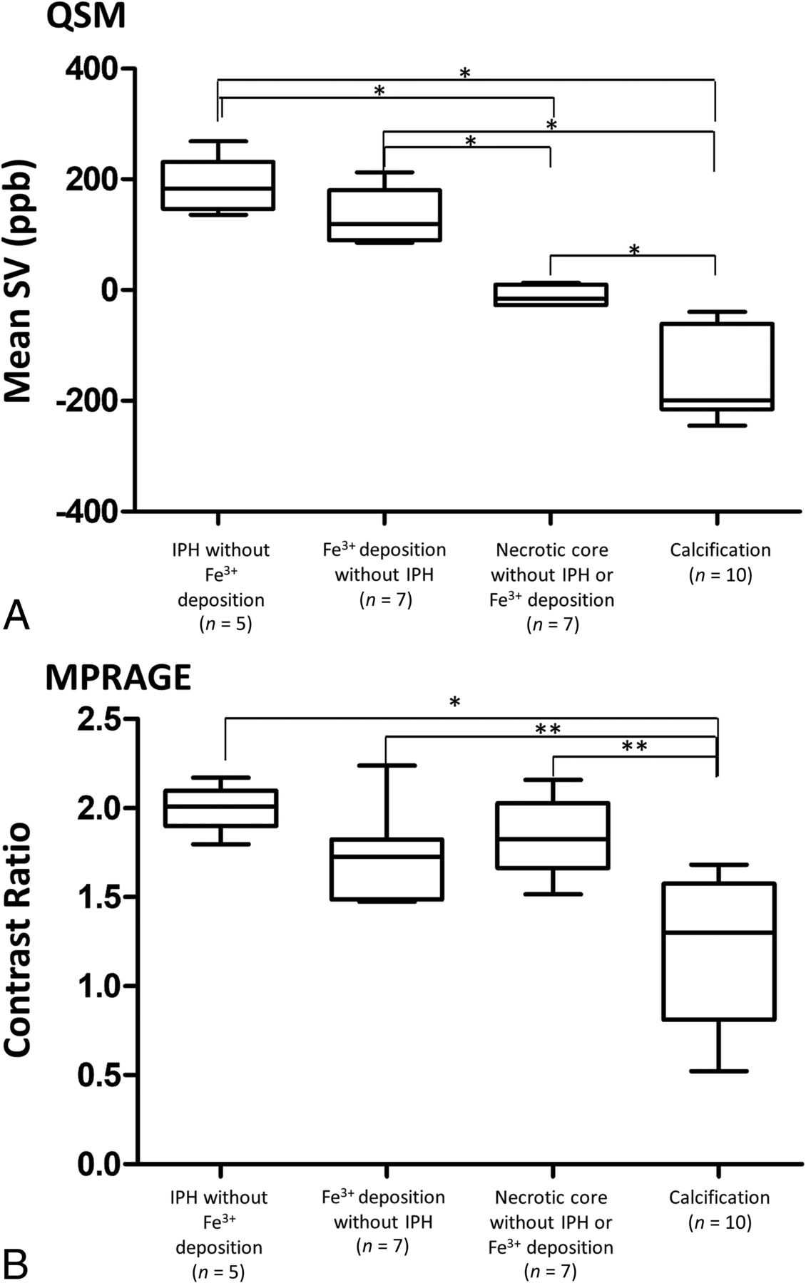
- FIG 5.
Comparison of the mean SV on QSM images (A) and the CR on MPRAGE images (B) among 4 typical atherosclerotic components. Analysis was with 1-way ANOVA with the Scheffe test (asterisk, P < .001; double asterisks, P < .01).
- FIG 6.
Relationship between the mean magnetic SV and areas positive for GYPA (A, erythrocytes) or Fe3+ (B). Relationship between Prussian blue–stained areas and GYPA-positive areas (C).


Tables
Comparison of histologically identified plaque components in hyper- and hypointense areas on QSM imagesa
Plaque Components Hyperintense Area (n = 26) Hypointense Area (n = 9) P Value Intraplaque hemorrhage 19 (73) 0 (0) .0002 Iron deposition 20 (76.9) 0 (0) <.0001 Calcification 7 (26.9) 8 (88.9) .002 Fibrous matrix deposits 9 (34.6) 9 (100) .001 Dense inflammatory infiltrates 10 (38.5) 4 (44.4) 1.0 Necrotic core without intraplaque hemorrhage 5 (19.2) 1 (11.1) 1.0 ↵a Data in parentheses are percentages.


Jump to section
Related Articles
Cited By...
- Diagnostic Accuracy of Preoperative Quantitative Susceptibility Mapping for Detecting Histologic Intraplaque Hemorrhage in Cervical ICA Stenosis in Patients Undergoing Carotid Endarterectomy
- Microstructural and mechanical insight into atherosclerotic plaques- an ex vivo DTI study to better assess plaque vulnerability
- Quantitative susceptibility mapping of carotid arterial tissue ex vivo: assessing sensitivity to vessel microstructural composition