Advertisement

Main menu

  • Home
  • Content
    • Current Issue
    • Accepted Manuscripts
    • Article Preview
    • Past Issue Archive
    • Video Articles
    • AJNR Case Collection
    • Case of the Week Archive
    • Case of the Month Archive
    • Classic Case Archive
  • Special Collections
    • AJNR Awards
    • Low-Field MRI
    • Alzheimer Disease
    • ASNR Foundation Special Collection
    • Photon-Counting CT
    • View All
  • Multimedia
    • AJNR Podcasts
    • AJNR SCANtastic
    • Trainee Corner
    • MRI Safety Corner
    • Imaging Protocols
  • For Authors
    • Submit a Manuscript
    • Submit a Video Article
    • Submit an eLetter to the Editor/Response
    • Manuscript Submission Guidelines
    • Statistical Tips
    • Fast Publishing of Accepted Manuscripts
    • Graphical Abstract Preparation
    • Imaging Protocol Submission
    • Author Policies
  • About Us
    • About AJNR
    • Editorial Board
    • Editorial Board Alumni
  • More
    • Become a Reviewer/Academy of Reviewers
    • Subscribers
    • Permissions
    • Alerts
    • Feedback
    • Advertisers
    • ASNR Home

User menu

  • Alerts
  • Log in

Search

  • Advanced search
American Journal of Neuroradiology

American Journal of Neuroradiology

ASHNR American Society of Functional Neuroradiology ASHNR American Society of Pediatric Neuroradiology ASSR
  • Alerts
  • Log in

Advanced Search

  • Home
  • Content
    • Current Issue
    • Accepted Manuscripts
    • Article Preview
    • Past Issue Archive
    • Video Articles
    • AJNR Case Collection
    • Case of the Week Archive
    • Case of the Month Archive
    • Classic Case Archive
  • Special Collections
    • AJNR Awards
    • Low-Field MRI
    • Alzheimer Disease
    • ASNR Foundation Special Collection
    • Photon-Counting CT
    • View All
  • Multimedia
    • AJNR Podcasts
    • AJNR SCANtastic
    • Trainee Corner
    • MRI Safety Corner
    • Imaging Protocols
  • For Authors
    • Submit a Manuscript
    • Submit a Video Article
    • Submit an eLetter to the Editor/Response
    • Manuscript Submission Guidelines
    • Statistical Tips
    • Fast Publishing of Accepted Manuscripts
    • Graphical Abstract Preparation
    • Imaging Protocol Submission
    • Author Policies
  • About Us
    • About AJNR
    • Editorial Board
    • Editorial Board Alumni
  • More
    • Become a Reviewer/Academy of Reviewers
    • Subscribers
    • Permissions
    • Alerts
    • Feedback
    • Advertisers
    • ASNR Home
  • Follow AJNR on Twitter
  • Visit AJNR on Facebook
  • Follow AJNR on Instagram
  • Join AJNR on LinkedIn
  • RSS Feeds

AJNR Awards, New Junior Editors, and more. Read the latest AJNR updates

Letter
Open Access

Ocular MR Imaging as a Substitute for Ultrasound during the COVID-19 Pandemic

Y.M. Purcell, O. Bergès, J. Savatovsky and A. Lecler
American Journal of Neuroradiology December 2020, 41 (12) E95-E96; DOI: https://doi.org/10.3174/ajnr.A6819
Y.M. Purcell
aNeuroradiology DepartmentFondation Ophtalmologique Adolphe de RothschildParis, France
  • Find this author on Google Scholar
  • Find this author on PubMed
  • Search for this author on this site
  • ORCID record for Y.M. Purcell
O. Bergès
aNeuroradiology DepartmentFondation Ophtalmologique Adolphe de RothschildParis, France
  • Find this author on Google Scholar
  • Find this author on PubMed
  • Search for this author on this site
  • ORCID record for O. Bergès
J. Savatovsky
aNeuroradiology DepartmentFondation Ophtalmologique Adolphe de RothschildParis, France
  • Find this author on Google Scholar
  • Find this author on PubMed
  • Search for this author on this site
  • ORCID record for J. Savatovsky
A. Lecler
aNeuroradiology DepartmentFondation Ophtalmologique Adolphe de RothschildParis, France
  • Find this author on Google Scholar
  • Find this author on PubMed
  • Search for this author on this site
  • ORCID record for A. Lecler
  • Article
  • Figures & Data
  • Info & Metrics
  • Responses
  • References
  • PDF
Loading

The novel coronavirus disease 2019 (COVID-19) pandemic is a public health emergency of international concern, bringing a multitude of new challenges in caring for patients. There is considerable variability in the prevalence of the virus as well as the availability of personal protective equipment (PPE) worldwide.

Ultrasonography examinations involve close, often prolonged (>10 minutes) physical contact with patients and confer a high risk of viral transmission by respiratory droplets. This is particularly the case for ocular ultrasound, with the practitioner positioned directly above the patient’s head in close proximity to the upper respiratory tract and nasopharnyx.1 Furthermore, Severe Acute Respiratory Syndrome coronavirus 2 (SARS-CoV-2) viral RNA has been detected in ocular swab samples in patients with conjunctivitis and in ocular fluids in the early incubation period before the onset of characteristic respiratory symptoms. Therefore, ocular fluids may not only be the point of virus entry but also a source of contagion to other patients and eye care professionals.2

Strict protective measures by each ultrasonography operator are necessary when performing an examination on patients positive for COVID-19. The American Institute of Ultrasound in Medicine advises that all patients be considered as possibly infected with COVID-19 for the purposes of protecting personnel.3 Radiologic and ophthalmologic societies recommend that all examinations be strictly limited to an absolute minimum. Given the limited supply of PPE, health care professionals are at high risk of contamination, including those in ophthalmology, previously considered a low-risk subspecialty.4

In response to the COVID-19 pandemic, we, as an institution, have devised a high-resolution MR imaging protocol as a substitute for ocular ultrasonography in the setting of ophthalmologic emergencies. This is an initiative by the radiology department in collaboration with our ophthalmology colleagues, as a risk-mitigation strategy to protect staff while maintaining optimum patient care.1 Local institutional review board authorization was granted.

All MR imaging examinations are performed at 3T. Our protocol is centered on the orbits with high-resolution 3D T1-weighted sequences before and after intravenous injection of gadolinium chelate and a 3D T2-weighted sequence. We obtain high in-plane spatial resolution of the order of 0.45–0.55 mm, which is obtainable on most commercially available MR imaging scanners. Practically, this allows reconstructions in any plane approximating the accuracy of B-mode ultrasound at 10 MHz, which can localize anomalies according to standard field or meridian nomenclature.

Urgent or emergent examinations are determined on an individual basis but include those entities requiring immediate clinical or surgical intervention, often with sight-threatening complications, including ocular trauma, retinal detachment, choroidal detachment, vitreous hemorrhage, and ocular tumors. We illustrate the value of ocular MR imaging in some of these settings (Figs 1–3).

FIG 1.
  • Download figure
  • Open in new tab
  • Download powerpoint
FIG 1.

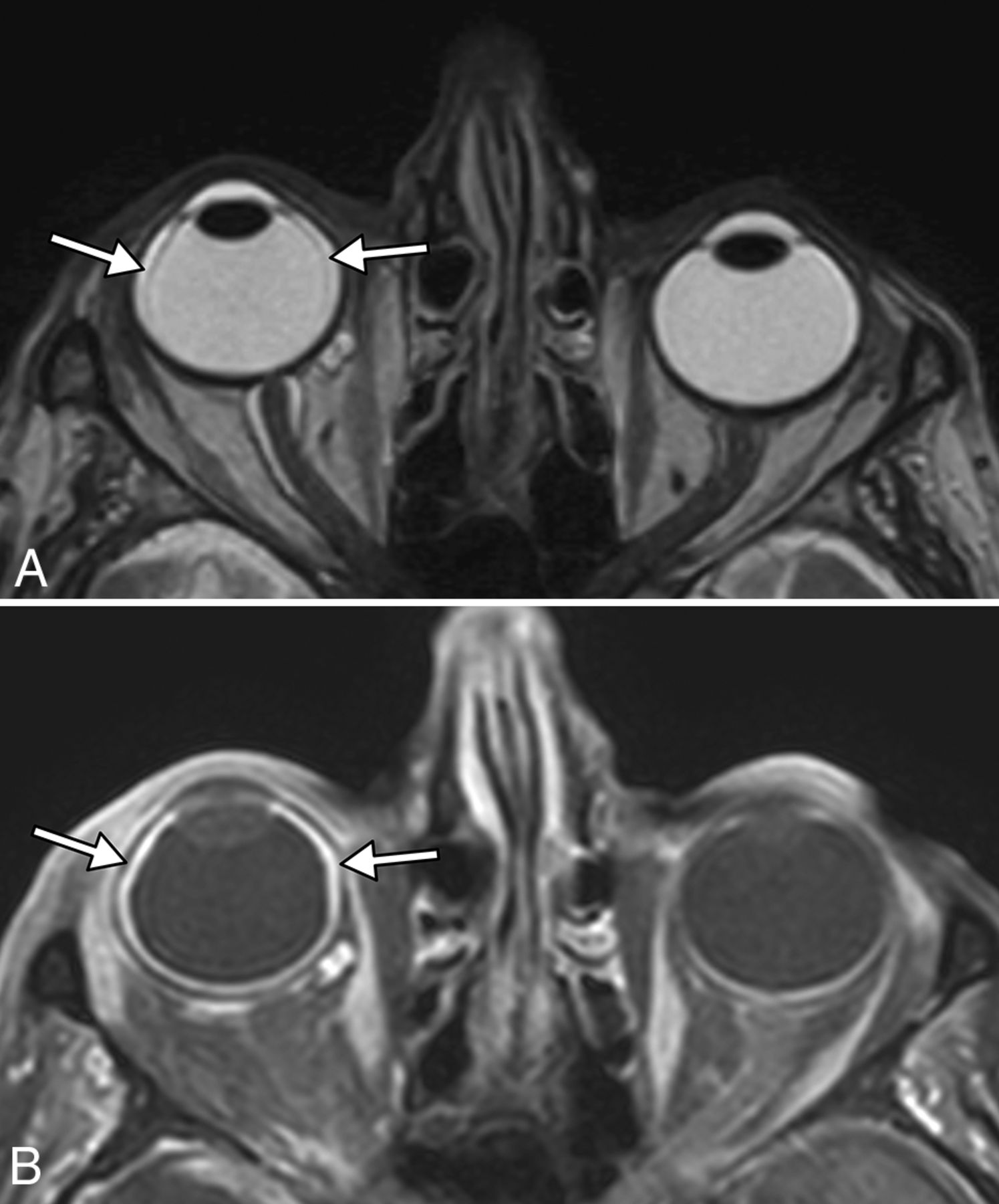
Axial MR imaging T2-weighted sequence (A) demonstrating a thin membrane (arrows) in the right globe extending anteriorly to the ciliary body, with corresponding enhancement on postcontrast T1-weighted sequence (B, arrows), consistent with a thin peripheral ciliochoroidal detachment. Postcontrast imaging is useful to confirm choroidal detachment because the choroid displays frank enhancement as opposed to the retina or sclera.

FIG 2.
  • Download figure
  • Open in new tab
  • Download powerpoint
FIG 2.

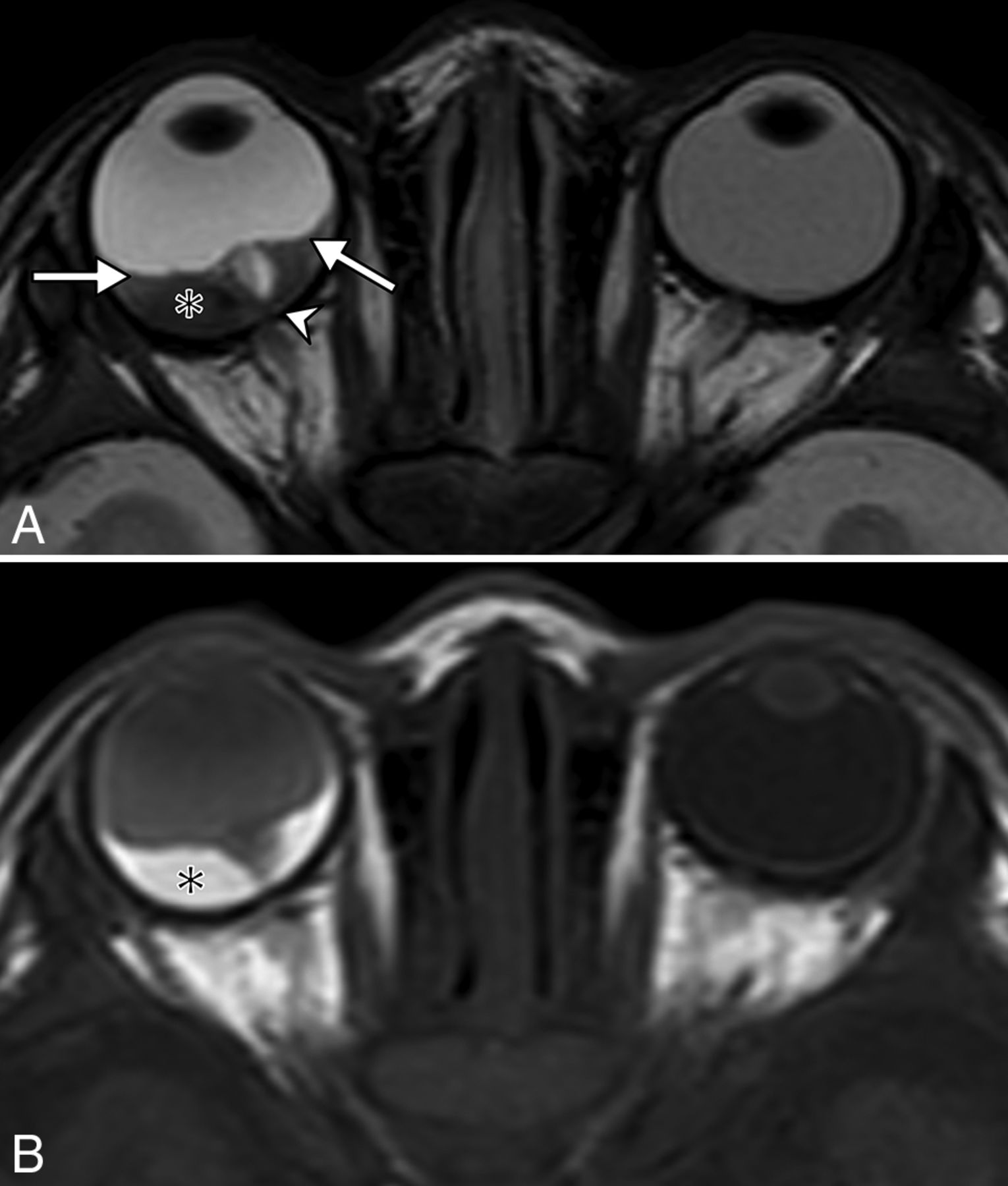
Axial MR imaging T2-weighted sequence (A) demonstrating a V-shaped membrane (arrows) converging on the right optic disc posteriorly (arrowhead), limited by the ora serrata anteriorly. This is the classic appearance of total retinal detachment, confirmed at surgery. There is an underlying subretinal hematoma (asterisk); these are intrinsically T1-hyperintense (B). Note the slightly T1-hyperintense vitreous compared to the contralateral side, consistent with vitreous hemorrhage, initially seen at slit-lamp examination.

FIG 3.
  • Download figure
  • Open in new tab
  • Download powerpoint
FIG 3.

Axial MR imaging T2-weighted (A) and T1-weighted post–IV gadolinium (B) sequences demonstrating the classic V-shaped membrane of a retinal detachment (arrow), converging on the optic disc posterior to the right globe, due to an underlying, exophytic, enhancing mass in the nasal quadrant (asterisk). There are also inflammatory changes in the anterior chamber (arrowhead).

In conclusion, we show that ocular MR imaging can be a useful diagnostic tool as a substitute for ultrasound and a temporary measure during the COVID-19 pandemic. The sensitivity and specificity of MR imaging compared with ultrasound for the investigation of ocular emergencies is not currently known; therefore, further studies are required.

Footnotes

  • Disclosures: Julien Savatovsky—UNRELATED: Consultancy: Bayer AG; Payment for Lectures Including Service on Speakers Bureaus: Philips, Medtronic; Travel/Accommodations/Meeting Expenses Unrelated to Activities Listed: Bayer AG.

Indicates open access to non-subscribers at www.ajnr.org

References

  1. 1.↵
    1. Ji-Peng OL,
    2. Shantha J ,
    3. Wong TY, et al
    . Preparedness among ophthalmologists: during and beyond the COVID-19 pandemic. Ophthalmology 2020;127:569–72 doi:10.1016/j.ophtha.2020.03.037 pmid:32327128
    CrossRefPubMed
  2. 2.↵
    1. Colavita F,
    2. Lapa D,
    3. Carletti F, et al
    . SARS-CoV-2 isolation from ocular secretions of a patient with COVID-19 in Italy with prolonged viral RNA detection. Ann Intern Med 2020;173:242–43 doi:10.7326/M20-1176 pmid:32302380
    CrossRefPubMed
  3. 3.↵
    American Institute of Ultrasound in Medicine. Quick Guide on COVID-19 Protections—Patient and Ultrasound Provider Protection. 2020. https://aium.s3.amazonaws.com/covid19/Covid19_Quick_Guide_PUPP.pdf. Accessed April 24, 2020
  4. 4.↵
    1. Qiao C,
    2. Zhang H,
    3. He M, et al
    . Symptomatic COVID-19 Infection in Eye Professionals in Wuhan China. Ophthalmology 2020;127(9):1268–1270 doi:10.1016/j.ophtha.2020.04.026 pmid:32359839
    CrossRefPubMed
  • © 2020 by American Journal of Neuroradiology
PreviousNext
Back to top

In this issue

American Journal of Neuroradiology: 41 (12)
American Journal of Neuroradiology
Vol. 41, Issue 12
1 Dec 2020
  • Table of Contents
  • Index by author
  • Complete Issue (PDF)
Advertisement
Print
Download PDF
Email Article

Thank you for your interest in spreading the word on American Journal of Neuroradiology.

NOTE: We only request your email address so that the person you are recommending the page to knows that you wanted them to see it, and that it is not junk mail. We do not capture any email address.

Enter multiple addresses on separate lines or separate them with commas.
Ocular MR Imaging as a Substitute for Ultrasound during the COVID-19 Pandemic
(Your Name) has sent you a message from American Journal of Neuroradiology
(Your Name) thought you would like to see the American Journal of Neuroradiology web site.
CAPTCHA
This question is for testing whether or not you are a human visitor and to prevent automated spam submissions.
Cite this article
Y.M. Purcell, O. Bergès, J. Savatovsky, A. Lecler
Ocular MR Imaging as a Substitute for Ultrasound during the COVID-19 Pandemic
American Journal of Neuroradiology Dec 2020, 41 (12) E95-E96; DOI: 10.3174/ajnr.A6819

Citation Manager Formats

  • BibTeX
  • Bookends
  • EasyBib
  • EndNote (tagged)
  • EndNote 8 (xml)
  • Medlars
  • Mendeley
  • Papers
  • RefWorks Tagged
  • Ref Manager
  • RIS
  • Zotero
0 Responses
Respond to this article
Share
Bookmark this article
Ocular MR Imaging as a Substitute for Ultrasound during the COVID-19 Pandemic
Y.M. Purcell, O. Bergès, J. Savatovsky, A. Lecler
American Journal of Neuroradiology Dec 2020, 41 (12) E95-E96; DOI: 10.3174/ajnr.A6819
del.icio.us logo Twitter logo Facebook logo Mendeley logo
  • Tweet Widget
  • Facebook Like
  • Google Plus One
Purchase

Jump to section

  • Article
    • Footnotes
    • References
  • Figures & Data
  • Info & Metrics
  • Responses
  • References
  • PDF

Related Articles

  • No related articles found.
  • PubMed
  • Google Scholar

Cited By...

  • No citing articles found.
  • Crossref
  • Google Scholar

This article has not yet been cited by articles in journals that are participating in Crossref Cited-by Linking.

Similar Articles

Advertisement

Indexed Content

  • Current Issue
  • Accepted Manuscripts
  • Article Preview
  • Past Issues
  • Editorials
  • Editor's Choice
  • Fellows' Journal Club
  • Letters to the Editor
  • Video Articles

Cases

  • Case Collection
  • Archive - Case of the Week
  • Archive - Case of the Month
  • Archive - Classic Case

More from AJNR

  • Trainee Corner
  • Imaging Protocols
  • MRI Safety Corner
  • Book Reviews

Multimedia

  • AJNR Podcasts
  • AJNR Scantastics

Resources

  • Turnaround Time
  • Submit a Manuscript
  • Submit a Video Article
  • Submit an eLetter to the Editor/Response
  • Manuscript Submission Guidelines
  • Statistical Tips
  • Fast Publishing of Accepted Manuscripts
  • Graphical Abstract Preparation
  • Imaging Protocol Submission
  • Evidence-Based Medicine Level Guide
  • Publishing Checklists
  • Author Policies
  • Become a Reviewer/Academy of Reviewers
  • News and Updates

About Us

  • About AJNR
  • Editorial Board
  • Editorial Board Alumni
  • Alerts
  • Permissions
  • Not an AJNR Subscriber? Join Now
  • Advertise with Us
  • Librarian Resources
  • Feedback
  • Terms and Conditions
  • AJNR Editorial Board Alumni

American Society of Neuroradiology

  • Not an ASNR Member? Join Now

© 2025 by the American Society of Neuroradiology All rights, including for text and data mining, AI training, and similar technologies, are reserved.
Print ISSN: 0195-6108 Online ISSN: 1936-959X

Powered by HighWire