
Article Figures & Data
Figures
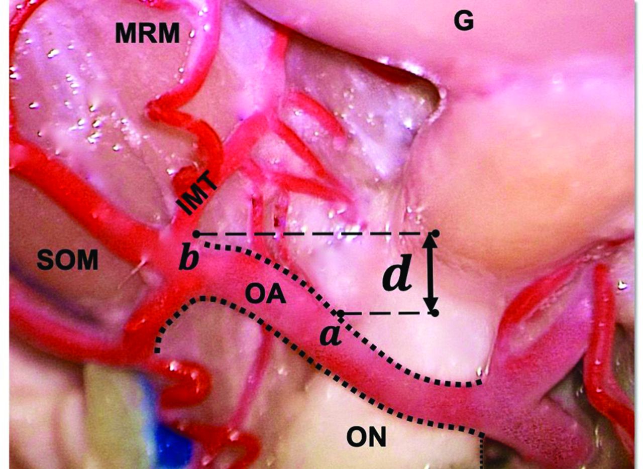
- FIG 1.
Dorsal view of the right orbit and its contents, with the medial rectus muscle (MRM) and superior oblique muscle (SOM) reflected laterally. G indicates globe; a, OA-ON cross-point; b, branch point of IMT; d, distance (in millimeters) from the OA-ON crosspoint and the IMT branch point.
- FIG 2.
Axial T1-weighted MR image in a 75-year-old woman with squamous cell carcinoma (white arrowhead) in the posterior intraconal right orbit. The OA (white arrow) is seen crossing the medial margin of the ON (asterisk).
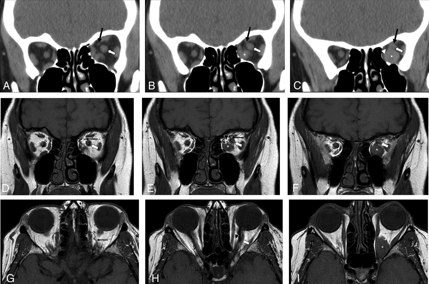
- FIG 3.
Orbital imaging in a 69-year-old woman with a venous malformation in the posterior intraconal left orbit (asterisk). Coronal NECT images (A–C, anterior to posterior) show the OA (black arrow) crossing the medial margin of the ON (white arrow, B). There is incomplete effacement of the fat at the level of the lesion within the posteromedial intraconal space on CT (white arrowhead). Coronal T1-weighted (D–F, anterior to posterior) and axial T1-weighted MR images (G–I, superior to inferior) of the orbits show the OA (black arrow) coursing above the ON (white arrow). The OA is seen crossing the medial margin of the ON (E). There is incomplete effacement of the posteromedial intraconal fat on MR imaging at the level of the lesion (black arrowhead).
- FIG 4.
CECT of the orbits in a 74-year-old man with a venous malformation in the posterior intraconal right orbit. Coronal images (A and B, anterior to posterior) demonstrate the OA (black arrow) crossing the medial margin of the ON (white arrowhead, A) anterior to the lesion (asterisk). Axial images (C and D, superior to inferior) also demonstrate the OA (white arrow) crossing the medial margin of the ON (white arrowhead, C) anterior to the lesion (asterisk, D). Effacement of the fat in the posteromedial intraconal space is demonstrated at the level of the lesion (D).
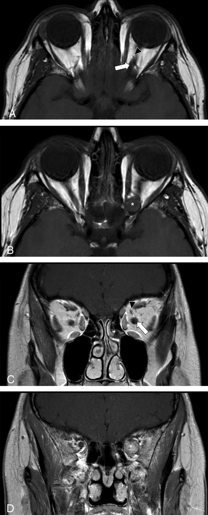
- FIG 5.
Axial T1-weighted precontrast (A and B, superior to inferior) and coronal T1-weighted postcontrast (C and D, anterior to posterior) images of the orbits in a 39-year-old woman with a venous malformation in the posterior intraconal left orbit. The OA (black arrowhead) can be seen crossing the medial margin of the ON (white arrow, A and C) anterior to the lesion (asterisk). There is full effacement of the posterior intraconal fat at the level of the lesion (B and D).


Jump to section
Related Articles
Cited By...
- No citing articles found.