Advertisement

Main menu

  • Home
  • Content
    • Current Issue
    • Accepted Manuscripts
    • Article Preview
    • Past Issue Archive
    • Video Articles
    • AJNR Case Collection
    • Case of the Week Archive
    • Case of the Month Archive
    • Classic Case Archive
  • Special Collections
    • Advancing NeuroMRI with High-Relaxivity Contrast Agents
    • AJNR Awards
    • Low-Field MRI
    • Alzheimer Disease
    • ASNR Foundation Special Collection
    • Photon-Counting CT
    • View All
  • Multimedia
    • AJNR Podcasts
    • AJNR SCANtastic
    • Trainee Corner
    • MRI Safety Corner
    • Imaging Protocols
  • For Authors
    • Submit a Manuscript
    • Submit a Video Article
    • Submit an eLetter to the Editor/Response
    • Manuscript Submission Guidelines
    • Statistical Tips
    • Fast Publishing of Accepted Manuscripts
    • Graphical Abstract Preparation
    • Imaging Protocol Submission
    • Author Policies
  • About Us
    • About AJNR
    • Editorial Board
    • Editorial Board Alumni
  • More
    • Become a Reviewer/Academy of Reviewers
    • Subscribers
    • Permissions
    • Alerts
    • Feedback
    • Advertisers
    • ASNR Home

User menu

  • Alerts
  • Log in

Search

  • Advanced search
American Journal of Neuroradiology

American Journal of Neuroradiology

ASHNR American Society of Functional Neuroradiology ASHNR American Society of Pediatric Neuroradiology ASSR
  • Alerts
  • Log in

Advanced Search

  • Home
  • Content
    • Current Issue
    • Accepted Manuscripts
    • Article Preview
    • Past Issue Archive
    • Video Articles
    • AJNR Case Collection
    • Case of the Week Archive
    • Case of the Month Archive
    • Classic Case Archive
  • Special Collections
    • Advancing NeuroMRI with High-Relaxivity Contrast Agents
    • AJNR Awards
    • Low-Field MRI
    • Alzheimer Disease
    • ASNR Foundation Special Collection
    • Photon-Counting CT
    • View All
  • Multimedia
    • AJNR Podcasts
    • AJNR SCANtastic
    • Trainee Corner
    • MRI Safety Corner
    • Imaging Protocols
  • For Authors
    • Submit a Manuscript
    • Submit a Video Article
    • Submit an eLetter to the Editor/Response
    • Manuscript Submission Guidelines
    • Statistical Tips
    • Fast Publishing of Accepted Manuscripts
    • Graphical Abstract Preparation
    • Imaging Protocol Submission
    • Author Policies
  • About Us
    • About AJNR
    • Editorial Board
    • Editorial Board Alumni
  • More
    • Become a Reviewer/Academy of Reviewers
    • Subscribers
    • Permissions
    • Alerts
    • Feedback
    • Advertisers
    • ASNR Home
  • Follow AJNR on Twitter
  • Visit AJNR on Facebook
  • Follow AJNR on Instagram
  • Join AJNR on LinkedIn
  • RSS Feeds

AJNR Awards, New Junior Editors, and more. Read the latest AJNR updates


Improved Turnaround Times | Median time to first decision: 12 days

Research ArticleAdult Brain
Open Access

Cortical Distribution of Fragile Periventricular Anastomotic Collateral Vessels in Moyamoya Disease: An Exploratory Cross-Sectional Study of Japanese Patients with Moyamoya Disease

A. Miyakoshi, T. Funaki, Y. Fushimi, T. Nakae, M. Okawa, T. Kikuchi, H. Kataoka, K. Yoshida, Y. Mineharu, M. Matsuhashi, E. Nakatani and S. Miyamoto
American Journal of Neuroradiology December 2020, 41 (12) 2243-2249; DOI: https://doi.org/10.3174/ajnr.A6861
A. Miyakoshi
aFrom the Department of Neurosurgery (A.M., T.F., M.O., T.K., H.K., K.Y., Y.M., S.M.)
dDepartment of Neurosurgery (A.M.)
  • Find this author on Google Scholar
  • Find this author on PubMed
  • Search for this author on this site
  • ORCID record for A. Miyakoshi
T. Funaki
aFrom the Department of Neurosurgery (A.M., T.F., M.O., T.K., H.K., K.Y., Y.M., S.M.)
  • Find this author on Google Scholar
  • Find this author on PubMed
  • Search for this author on this site
  • ORCID record for T. Funaki
Y. Fushimi
bDiagnostic Imaging and Nuclear Medicine (Y.F.)
  • Find this author on Google Scholar
  • Find this author on PubMed
  • Search for this author on this site
  • ORCID record for Y. Fushimi
T. Nakae
fDepartment of Neurosurgery (T.N.), Shiga General Hospital, Shiga, Japan
  • Find this author on Google Scholar
  • Find this author on PubMed
  • Search for this author on this site
  • ORCID record for T. Nakae
M. Okawa
aFrom the Department of Neurosurgery (A.M., T.F., M.O., T.K., H.K., K.Y., Y.M., S.M.)
  • Find this author on Google Scholar
  • Find this author on PubMed
  • Search for this author on this site
  • ORCID record for M. Okawa
T. Kikuchi
aFrom the Department of Neurosurgery (A.M., T.F., M.O., T.K., H.K., K.Y., Y.M., S.M.)
  • Find this author on Google Scholar
  • Find this author on PubMed
  • Search for this author on this site
  • ORCID record for T. Kikuchi
H. Kataoka
aFrom the Department of Neurosurgery (A.M., T.F., M.O., T.K., H.K., K.Y., Y.M., S.M.)
  • Find this author on Google Scholar
  • Find this author on PubMed
  • Search for this author on this site
  • ORCID record for H. Kataoka
K. Yoshida
aFrom the Department of Neurosurgery (A.M., T.F., M.O., T.K., H.K., K.Y., Y.M., S.M.)
  • Find this author on Google Scholar
  • Find this author on PubMed
  • Search for this author on this site
  • ORCID record for K. Yoshida
Y. Mineharu
aFrom the Department of Neurosurgery (A.M., T.F., M.O., T.K., H.K., K.Y., Y.M., S.M.)
  • Find this author on Google Scholar
  • Find this author on PubMed
  • Search for this author on this site
  • ORCID record for Y. Mineharu
M. Matsuhashi
cEpilepsy, Movement Disorders and Physiology (M.M.), Kyoto University Graduate School of Medicine, Kyoto, Japan
  • Find this author on Google Scholar
  • Find this author on PubMed
  • Search for this author on this site
  • ORCID record for M. Matsuhashi
E. Nakatani
eDivision of Statistical Analysis (E.N.), Shizuoka General Hospital, Shizuoka, Japan
  • Find this author on Google Scholar
  • Find this author on PubMed
  • Search for this author on this site
  • ORCID record for E. Nakatani
S. Miyamoto
aFrom the Department of Neurosurgery (A.M., T.F., M.O., T.K., H.K., K.Y., Y.M., S.M.)
  • Find this author on Google Scholar
  • Find this author on PubMed
  • Search for this author on this site
  • ORCID record for S. Miyamoto
  • Article
  • Figures & Data
  • Supplemental
  • Info & Metrics
  • Responses
  • References
  • PDF
Loading

Article Figures & Data

Figures

  • Tables
  • FIG 1.
    • Download figure
    • Open in new tab
    • Download powerpoint
    FIG 1.

    Schematic illustration showing a coronal plane of the left cerebral hemisphere and 3 subtypes of collateral anastomoses: lenticulostriate, thalamic, and choroidal anastomoses. A indicates artery; Med, medullary; Subepend, subependymal; TGA, thalamogeniculate artery; TPA, thalamoperforating artery; TTA, thalamotuberal artery. Reprinted with permission from Funaki et al.18

  • FIG 2.
    • Download figure
    • Open in new tab
    • Download powerpoint
    FIG 2.

    Flowchart for patient inclusion. Asterisk indicates that the patients have the anastomoses for which signals could be identified all the way from the origin to the cerebral cortex without interruption on MR angiography.

  • FIG 3.
    • Download figure
    • Open in new tab
    • Download powerpoint
    FIG 3.

    Cortical distributions for each type of collateral depicted on a standard brain surface model (A, All types; B, Lenticulostriate anastomosis; C, Thalamic anastomosis; D, Choroidal anastomosis). Each dot shows the point of anastomosis outflow to the cortex (blue dot, lenticulostriate anastomosis; black dot, thalamic anastomosis; red dot, choroidal anastomosis). Unfilled circles represent outflow on the insular cortex. Pale dots represent outflow located in a sulcus. CS indicates central sulcus.

  • FIG 4.
    • Download figure
    • Open in new tab
    • Download powerpoint
    FIG 4.

    Cortical distributions of each type of collateral depicted on a standard brain surface model. Each dot shows the point where each anastomosis outflowed to the cortex (blue dot, lenticulostriate anastomosis; black dot, thalamic anastomosis; red dot, choroidal anastomosis). White circles (circles without fill) show dots located on the insular cortex. A, All dots are depicted on a brain surface model. B, The view from the medial surface of a 3D brain surface model. Dots were extracted that exist within 15 mm from the midline.

  • FIG 5.
    • Download figure
    • Open in new tab
    • Download powerpoint
    FIG 5.

    Mediolateral (x-coordinate) distribution of outflow to the cortex for each type of anastomosis. The value for the midline of images is 181, and the value for the lateral edge of images is 361. Values for x-coordinates are shown as density estimations. The asterisk indicates P < .001.

  • FIG 6.
    • Download figure
    • Open in new tab
    • Download powerpoint
    FIG 6.

    Posteroanterior (y-coordinate) distribution of outflow to the cortex for each type of anastomosis. The value for the posterior edge of images is 1, and the value for the frontal edge of images is 395. Values for y-coordinates are shown as density estimations. The asterisk indicates P < .001.
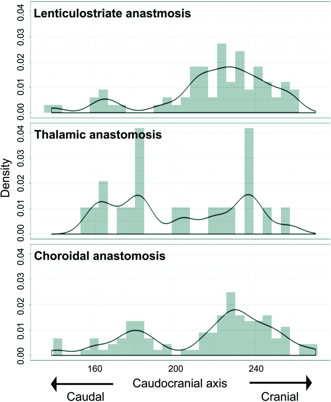
  • FIG 7.
    • Download figure
    • Open in new tab
    • Download powerpoint
    FIG 7.

    Caudocranial (z-coordinate) distribution of outflow to the cortex for each type of anastomosis. The value for the caudal edge of images is 1, and the value for the cranial edge of images is 345. Values for z-coordinates are shown as density estimations.

Tables

  • Figures
  • Patient characteristics

    Included Patients with Moyamoya Disease (n = 46)
    Median age (range) (yr)28.5 (3–56)
    Male sex (No.) (%)14 (30.4)
    Clinical presentation (No.) (%)
     Ischemic symptoms28 (60.9)
     Hemorrhagic symptoms11 (23.9)
     Other symptoms7 (15.2)
PreviousNext
Back to top

In this issue

American Journal of Neuroradiology: 41 (12)
American Journal of Neuroradiology
Vol. 41, Issue 12
1 Dec 2020
  • Table of Contents
  • Index by author
  • Complete Issue (PDF)
Advertisement
Print
Download PDF
Email Article

Thank you for your interest in spreading the word on American Journal of Neuroradiology.

NOTE: We only request your email address so that the person you are recommending the page to knows that you wanted them to see it, and that it is not junk mail. We do not capture any email address.

Enter multiple addresses on separate lines or separate them with commas.
Cortical Distribution of Fragile Periventricular Anastomotic Collateral Vessels in Moyamoya Disease: An Exploratory Cross-Sectional Study of Japanese Patients with Moyamoya Disease
(Your Name) has sent you a message from American Journal of Neuroradiology
(Your Name) thought you would like to see the American Journal of Neuroradiology web site.
CAPTCHA
This question is for testing whether or not you are a human visitor and to prevent automated spam submissions.
Cite this article
A. Miyakoshi, T. Funaki, Y. Fushimi, T. Nakae, M. Okawa, T. Kikuchi, H. Kataoka, K. Yoshida, Y. Mineharu, M. Matsuhashi, E. Nakatani, S. Miyamoto
Cortical Distribution of Fragile Periventricular Anastomotic Collateral Vessels in Moyamoya Disease: An Exploratory Cross-Sectional Study of Japanese Patients with Moyamoya Disease
American Journal of Neuroradiology Dec 2020, 41 (12) 2243-2249; DOI: 10.3174/ajnr.A6861

Citation Manager Formats

  • BibTeX
  • Bookends
  • EasyBib
  • EndNote (tagged)
  • EndNote 8 (xml)
  • Medlars
  • Mendeley
  • Papers
  • RefWorks Tagged
  • Ref Manager
  • RIS
  • Zotero
0 Responses
Respond to this article
Share
Bookmark this article
Cortical Distribution of Fragile Periventricular Anastomotic Collateral Vessels in Moyamoya Disease: An Exploratory Cross-Sectional Study of Japanese Patients with Moyamoya Disease
A. Miyakoshi, T. Funaki, Y. Fushimi, T. Nakae, M. Okawa, T. Kikuchi, H. Kataoka, K. Yoshida, Y. Mineharu, M. Matsuhashi, E. Nakatani, S. Miyamoto
American Journal of Neuroradiology Dec 2020, 41 (12) 2243-2249; DOI: 10.3174/ajnr.A6861
del.icio.us logo Twitter logo Facebook logo Mendeley logo
  • Tweet Widget
  • Facebook Like
  • Google Plus One
Purchase

Jump to section

  • Article
    • Abstract
    • ABBREVIATIONS:
    • MATERIALS AND METHODS
    • RESULTS
    • DISCUSSION
    • CONCLUSIONS
    • Footnotes
    • References
  • Figures & Data
  • Supplemental
  • Info & Metrics
  • Responses
  • References
  • PDF

Related Articles

  • PubMed
  • Google Scholar

Cited By...

  • Larger Posterior Revascularization Associated with Reduction of Choroidal Anastomosis in Moyamoya Disease: A Quantitative Angiographic Analysis
  • Crossref
  • Google Scholar

This article has not yet been cited by articles in journals that are participating in Crossref Cited-by Linking.

More in this TOC Section

  • Diagnostic Neuroradiology of Monoclonal Antibodies
  • Cerebral ADC Changes in Fabry Disease
  • ML for Glioma Molecular Subtype Prediction
Show more Adult Brain

Similar Articles

Advertisement

Indexed Content

  • Current Issue
  • Accepted Manuscripts
  • Article Preview
  • Past Issues
  • Editorials
  • Editor's Choice
  • Fellows' Journal Club
  • Letters to the Editor
  • Video Articles

Cases

  • Case Collection
  • Archive - Case of the Week
  • Archive - Case of the Month
  • Archive - Classic Case

More from AJNR

  • Trainee Corner
  • Imaging Protocols
  • MRI Safety Corner
  • Book Reviews

Multimedia

  • AJNR Podcasts
  • AJNR Scantastics

Resources

  • Turnaround Time
  • Submit a Manuscript
  • Submit a Video Article
  • Submit an eLetter to the Editor/Response
  • Manuscript Submission Guidelines
  • Statistical Tips
  • Fast Publishing of Accepted Manuscripts
  • Graphical Abstract Preparation
  • Imaging Protocol Submission
  • Evidence-Based Medicine Level Guide
  • Publishing Checklists
  • Author Policies
  • Become a Reviewer/Academy of Reviewers
  • News and Updates

About Us

  • About AJNR
  • Editorial Board
  • Editorial Board Alumni
  • Alerts
  • Permissions
  • Not an AJNR Subscriber? Join Now
  • Advertise with Us
  • Librarian Resources
  • Feedback
  • Terms and Conditions
  • AJNR Editorial Board Alumni

American Society of Neuroradiology

  • Not an ASNR Member? Join Now

© 2025 by the American Society of Neuroradiology All rights, including for text and data mining, AI training, and similar technologies, are reserved.
Print ISSN: 0195-6108 Online ISSN: 1936-959X

Powered by HighWire