Advertisement

Main menu

  • Home
  • Content
    • Current Issue
    • Accepted Manuscripts
    • Article Preview
    • Past Issue Archive
    • Video Articles
    • AJNR Case Collection
    • Case of the Week Archive
    • Case of the Month Archive
    • Classic Case Archive
  • Special Collections
    • AJNR Awards
    • Low-Field MRI
    • Alzheimer Disease
    • ASNR Foundation Special Collection
    • Photon-Counting CT
    • View All
  • Multimedia
    • AJNR Podcasts
    • AJNR SCANtastic
    • Trainee Corner
    • MRI Safety Corner
    • Imaging Protocols
  • For Authors
    • Submit a Manuscript
    • Submit a Video Article
    • Submit an eLetter to the Editor/Response
    • Manuscript Submission Guidelines
    • Statistical Tips
    • Fast Publishing of Accepted Manuscripts
    • Graphical Abstract Preparation
    • Imaging Protocol Submission
    • Author Policies
  • About Us
    • About AJNR
    • Editorial Board
    • Editorial Board Alumni
  • More
    • Become a Reviewer/Academy of Reviewers
    • Subscribers
    • Permissions
    • Alerts
    • Feedback
    • Advertisers
    • ASNR Home

User menu

  • Alerts
  • Log in

Search

  • Advanced search
American Journal of Neuroradiology

American Journal of Neuroradiology

ASHNR American Society of Functional Neuroradiology ASHNR American Society of Pediatric Neuroradiology ASSR
  • Alerts
  • Log in

Advanced Search

  • Home
  • Content
    • Current Issue
    • Accepted Manuscripts
    • Article Preview
    • Past Issue Archive
    • Video Articles
    • AJNR Case Collection
    • Case of the Week Archive
    • Case of the Month Archive
    • Classic Case Archive
  • Special Collections
    • AJNR Awards
    • Low-Field MRI
    • Alzheimer Disease
    • ASNR Foundation Special Collection
    • Photon-Counting CT
    • View All
  • Multimedia
    • AJNR Podcasts
    • AJNR SCANtastic
    • Trainee Corner
    • MRI Safety Corner
    • Imaging Protocols
  • For Authors
    • Submit a Manuscript
    • Submit a Video Article
    • Submit an eLetter to the Editor/Response
    • Manuscript Submission Guidelines
    • Statistical Tips
    • Fast Publishing of Accepted Manuscripts
    • Graphical Abstract Preparation
    • Imaging Protocol Submission
    • Author Policies
  • About Us
    • About AJNR
    • Editorial Board
    • Editorial Board Alumni
  • More
    • Become a Reviewer/Academy of Reviewers
    • Subscribers
    • Permissions
    • Alerts
    • Feedback
    • Advertisers
    • ASNR Home
  • Follow AJNR on Twitter
  • Visit AJNR on Facebook
  • Follow AJNR on Instagram
  • Join AJNR on LinkedIn
  • RSS Feeds

AJNR Awards, New Junior Editors, and more. Read the latest AJNR updates

Research ArticleSpine Imaging and Spine Image-Guided Interventions
Open Access

Automatic Spinal Cord Gray Matter Quantification: A Novel Approach

C. Tsagkas, A. Horvath, A. Altermatt, S. Pezold, M. Weigel, T. Haas, M. Amann, L. Kappos, T. Sprenger, O. Bieri, P. Cattin and K. Parmar
American Journal of Neuroradiology September 2019, 40 (9) 1592-1600; DOI: https://doi.org/10.3174/ajnr.A6157
C. Tsagkas
aFrom the Neurologic Clinic and Policlinic (C.T., M.A., L.K., T.S., K.P.), Department of Medicine and Biomedical Engineering
bTranslational Imaging in Neurology Basel (C.T., A.A., M.A., M.W., L.K., K.P.), Department of Medicine and Biomedical Engineering
eMedical Image Analysis Center (C.T., A.A., M.A.), Basel, Switzerland
  • Find this author on Google Scholar
  • Find this author on PubMed
  • Search for this author on this site
  • ORCID record for C. Tsagkas
A. Horvath
fDepartment of Biomedical Engineering (A.H., A.A., S.P., M.W., O.B., P.C.), University of Basel, Allschwil, Switzerland
  • Find this author on Google Scholar
  • Find this author on PubMed
  • Search for this author on this site
  • ORCID record for A. Horvath
A. Altermatt
bTranslational Imaging in Neurology Basel (C.T., A.A., M.A., M.W., L.K., K.P.), Department of Medicine and Biomedical Engineering
eMedical Image Analysis Center (C.T., A.A., M.A.), Basel, Switzerland
fDepartment of Biomedical Engineering (A.H., A.A., S.P., M.W., O.B., P.C.), University of Basel, Allschwil, Switzerland
  • Find this author on Google Scholar
  • Find this author on PubMed
  • Search for this author on this site
  • ORCID record for A. Altermatt
S. Pezold
fDepartment of Biomedical Engineering (A.H., A.A., S.P., M.W., O.B., P.C.), University of Basel, Allschwil, Switzerland
  • Find this author on Google Scholar
  • Find this author on PubMed
  • Search for this author on this site
  • ORCID record for S. Pezold
M. Weigel
bTranslational Imaging in Neurology Basel (C.T., A.A., M.A., M.W., L.K., K.P.), Department of Medicine and Biomedical Engineering
cDivision of Radiological Physics (M.W., T.H., O.B.), Department of Radiology
fDepartment of Biomedical Engineering (A.H., A.A., S.P., M.W., O.B., P.C.), University of Basel, Allschwil, Switzerland
  • Find this author on Google Scholar
  • Find this author on PubMed
  • Search for this author on this site
  • ORCID record for M. Weigel
T. Haas
cDivision of Radiological Physics (M.W., T.H., O.B.), Department of Radiology
  • Find this author on Google Scholar
  • Find this author on PubMed
  • Search for this author on this site
  • ORCID record for T. Haas
M. Amann
aFrom the Neurologic Clinic and Policlinic (C.T., M.A., L.K., T.S., K.P.), Department of Medicine and Biomedical Engineering
bTranslational Imaging in Neurology Basel (C.T., A.A., M.A., M.W., L.K., K.P.), Department of Medicine and Biomedical Engineering
dDivision of Diagnostic and Interventional Neuroradiology (M.A.), Department of Radiology, University Hospital Basel, University of Basel, Basel, Switzerland
eMedical Image Analysis Center (C.T., A.A., M.A.), Basel, Switzerland
  • Find this author on Google Scholar
  • Find this author on PubMed
  • Search for this author on this site
  • ORCID record for M. Amann
L. Kappos
aFrom the Neurologic Clinic and Policlinic (C.T., M.A., L.K., T.S., K.P.), Department of Medicine and Biomedical Engineering
bTranslational Imaging in Neurology Basel (C.T., A.A., M.A., M.W., L.K., K.P.), Department of Medicine and Biomedical Engineering
  • Find this author on Google Scholar
  • Find this author on PubMed
  • Search for this author on this site
  • ORCID record for L. Kappos
T. Sprenger
aFrom the Neurologic Clinic and Policlinic (C.T., M.A., L.K., T.S., K.P.), Department of Medicine and Biomedical Engineering
gDepartment of Neurology (T.S.), DKD HELIOS Klinik, Wiesbaden, Germany.
  • Find this author on Google Scholar
  • Find this author on PubMed
  • Search for this author on this site
  • ORCID record for T. Sprenger
O. Bieri
cDivision of Radiological Physics (M.W., T.H., O.B.), Department of Radiology
fDepartment of Biomedical Engineering (A.H., A.A., S.P., M.W., O.B., P.C.), University of Basel, Allschwil, Switzerland
  • Find this author on Google Scholar
  • Find this author on PubMed
  • Search for this author on this site
  • ORCID record for O. Bieri
P. Cattin
fDepartment of Biomedical Engineering (A.H., A.A., S.P., M.W., O.B., P.C.), University of Basel, Allschwil, Switzerland
  • Find this author on Google Scholar
  • Find this author on PubMed
  • Search for this author on this site
  • ORCID record for P. Cattin
K. Parmar
aFrom the Neurologic Clinic and Policlinic (C.T., M.A., L.K., T.S., K.P.), Department of Medicine and Biomedical Engineering
bTranslational Imaging in Neurology Basel (C.T., A.A., M.A., M.W., L.K., K.P.), Department of Medicine and Biomedical Engineering
  • Find this author on Google Scholar
  • Find this author on PubMed
  • Search for this author on this site
  • ORCID record for K. Parmar
  • Article
  • Figures & Data
  • Supplemental
  • Info & Metrics
  • Responses
  • References
  • PDF
Loading

Article Figures & Data

Figures

  • Tables
  • Fig 1.
    • Download figure
    • Open in new tab
    • Download powerpoint
    Fig 1.

    Exemplary axial AMIRA slice of 1 representative volunteer at the C4 level. A–H, Eight images of different tissue contrast acquired by the AMIRA sequence, shown in chronologic order from lowest-to-highest TI. I, Average image from A to E in full view, which delivers a high contrast-to-noise-ratio for GM/WM. J, Average image from F to H, which delivers a high contrast-to-noise ratio for SC/CSF. K, Same average image as in I but histogram-equalized and zoomed.

  • Fig 2.
    • Download figure
    • Open in new tab
    • Download powerpoint
    Fig 2.

    Flow chart of the automatic segmentation pipeline. As a first step of the algorithm to align the 12 slices, the images are center-cropped and slice-wise successively coregistered rostral to caudal using translations in pixel-size steps to prevent further interpolation. Then, the algorithm automatically locates and delineates the ring-shaped CSF from its surroundings and extracts the cross-sectional SC surface. Finally, it uses the previously segmented SC surface as a mask for GM/WM differentiation. The iterative steps of CSF segmentation are shown as a zoomed-in view. GM segmentation uses essentially the same steps and is thus not shown in detail.
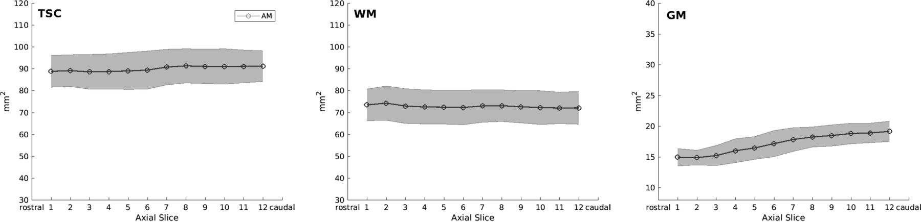
  • Fig 3.
    • Download figure
    • Open in new tab
    • Download powerpoint
    Fig 3.

    Cross-sectional areas of total spinal cord, white matter, and gray matter per axial slice as measured by automatic segmentations. Notice the slight increase of total spinal cord (TSC) and the marked GM cross-sectional area increase caudally, which corresponds to the cervical SC enlargement. The light gray area depicts the limits of ±1 SD.

  • Fig 4.
    • Download figure
    • Open in new tab
    • Download powerpoint
    Fig 4.

    Comparison between the reproducibility of manual and automatic measurements (AM) of spinal cord gray matter and white matter per axial slice. Intrasession and intersession reproducibility is assessed in terms of Dice coefficients (graphics on the left) and coefficients of variation (graphics on the right). Manual and automatic intersession reproducibility is shown in dark gray, whereas manual and automatic intrasession reproducibility is shown in light gray. Error bars display mean values ± 0.2 SDs.
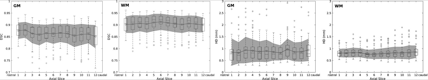
  • Fig 5.
    • Download figure
    • Open in new tab
    • Download powerpoint
    Fig 5.

    Accuracy measurements in terms of Dice coefficients (graphics on the left) and Hausdorff distances (graphics on the right) of white matter and gray matter per slice. Overlaid boxplots display median values as well as 25th and 75th percentile values. Gray areas depict the mean standard error values ± 1 SD.
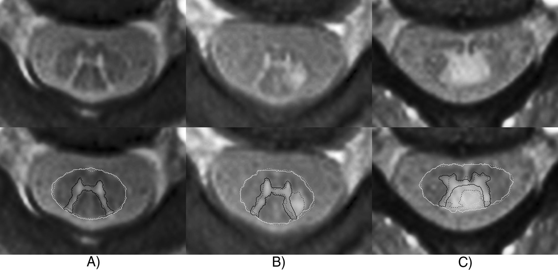
  • Fig 6.
    • Download figure
    • Open in new tab
    • Download powerpoint
    Fig 6.

    Examples of segmentations of representative patients with MS. The thick continuous line indicates automatic segmentation; the dashed line, manual reference standard. A, A 54-year-old female patient with MS. Rostral cervical SC slices of the C1/C2 level without focal lesions. Automatic segmentation highly corresponds to the manual reference standard. B, A 32-year-old male patient with MS. Rostral cervical SC slice of the C2 level with a focal posterolateral lesion fused with the left posterior gray matter horn. Automatic segmentation misclassifies the focal lesion as SC GM. C, A 33-year-old female patient with MS. A cervical SC slice of the C3/C4 level with a focal posterior lesion fusing with the posterior SC GM horns and the central SC GM commissure. Automatic segmentation misclassifies the focal lesion as SC GM and CSF.

Tables

  • Figures
  • SC GM, WM, and TSC accuracy of automatic and semiautomatic segmentations against the manual reference standard segmentationsa

    ParameterGMWMTSC
    AM (on 88% of acquired slices, nondiscarded from initial analysis)DSC0.86 ± 0.04 (0.87)0.90 ± 0.03 (0.91)0.95 ± 0.03 (0.95)
    HD (mm)0.90 ± 0.44 (0.72)0.82 ± 0.22 (0.75)0.64 ± 0.27 (0.67)
    SAM (on 12% of acquired slices, discarded from initial analysis)DSC0.83 ± 0.04 (0.84)0.96 ± 0.01 (0.96)
    HD (mm)1.11 ± 0.55 (0.93)0.64 ± 0.15 (0.67)
    AM (on nondiscarded samples) and SAM (on discarded slices), mixed (100% of acquired slices)DSC0.86 ± 0.04 (0.86)0.91 ± 0.04 (0.92)0.96 ± 0.03 (0.96)
    HD (mm)0.91 ± 0.46 (0.81)0.80 ± 0.22 (0.75)0.60 ± 0.29 (0.55)
    • Note:—SAM indicates semiautomatic segmentation; TSC, total spinal cord; AM, automatic segmentations; DSC, Dice coefficient; HD, Hausdorff distances.

    • ↵a All values are shown as mean ± SD (median).

PreviousNext
Back to top

In this issue

American Journal of Neuroradiology: 40 (9)
American Journal of Neuroradiology
Vol. 40, Issue 9
1 Sep 2019
  • Table of Contents
  • Index by author
  • Complete Issue (PDF)
Advertisement
Print
Download PDF
Email Article

Thank you for your interest in spreading the word on American Journal of Neuroradiology.

NOTE: We only request your email address so that the person you are recommending the page to knows that you wanted them to see it, and that it is not junk mail. We do not capture any email address.

Enter multiple addresses on separate lines or separate them with commas.
Automatic Spinal Cord Gray Matter Quantification: A Novel Approach
(Your Name) has sent you a message from American Journal of Neuroradiology
(Your Name) thought you would like to see the American Journal of Neuroradiology web site.
CAPTCHA
This question is for testing whether or not you are a human visitor and to prevent automated spam submissions.
Cite this article
C. Tsagkas, A. Horvath, A. Altermatt, S. Pezold, M. Weigel, T. Haas, M. Amann, L. Kappos, T. Sprenger, O. Bieri, P. Cattin, K. Parmar
Automatic Spinal Cord Gray Matter Quantification: A Novel Approach
American Journal of Neuroradiology Sep 2019, 40 (9) 1592-1600; DOI: 10.3174/ajnr.A6157

Citation Manager Formats

  • BibTeX
  • Bookends
  • EasyBib
  • EndNote (tagged)
  • EndNote 8 (xml)
  • Medlars
  • Mendeley
  • Papers
  • RefWorks Tagged
  • Ref Manager
  • RIS
  • Zotero
0 Responses
Respond to this article
Share
Bookmark this article
Automatic Spinal Cord Gray Matter Quantification: A Novel Approach
C. Tsagkas, A. Horvath, A. Altermatt, S. Pezold, M. Weigel, T. Haas, M. Amann, L. Kappos, T. Sprenger, O. Bieri, P. Cattin, K. Parmar
American Journal of Neuroradiology Sep 2019, 40 (9) 1592-1600; DOI: 10.3174/ajnr.A6157
del.icio.us logo Twitter logo Facebook logo Mendeley logo
  • Tweet Widget
  • Facebook Like
  • Google Plus One
Purchase

Jump to section

  • Article
    • Abstract
    • ABBREVIATIONS:
    • Materials and Methods
    • Results
    • Discussion
    • Conclusions
    • Footnotes
    • REFERENCES
  • Figures & Data
  • Supplemental
  • Info & Metrics
  • Responses
  • References
  • PDF

Related Articles

  • PubMed
  • Google Scholar

Cited By...

  • Fully Automatic Method for Reliable Spinal Cord Compartment Segmentation in Multiple Sclerosis
  • What are the gray and white matter volumes of the human spinal cord?
  • Crossref
  • Google Scholar

This article has not yet been cited by articles in journals that are participating in Crossref Cited-by Linking.

More in this TOC Section

  • Dynamic CT Myelography: Patient Positioning
  • Evaluation of SIH MR Scoring Systems in Normals
  • Management Outcomes For VO Spine Biopsy
Show more SPINE IMAGING AND SPINE IMAGE-GUIDED INTERVENTIONS

Similar Articles

Advertisement

Indexed Content

  • Current Issue
  • Accepted Manuscripts
  • Article Preview
  • Past Issues
  • Editorials
  • Editor's Choice
  • Fellows' Journal Club
  • Letters to the Editor
  • Video Articles

Cases

  • Case Collection
  • Archive - Case of the Week
  • Archive - Case of the Month
  • Archive - Classic Case

More from AJNR

  • Trainee Corner
  • Imaging Protocols
  • MRI Safety Corner
  • Book Reviews

Multimedia

  • AJNR Podcasts
  • AJNR Scantastics

Resources

  • Turnaround Time
  • Submit a Manuscript
  • Submit a Video Article
  • Submit an eLetter to the Editor/Response
  • Manuscript Submission Guidelines
  • Statistical Tips
  • Fast Publishing of Accepted Manuscripts
  • Graphical Abstract Preparation
  • Imaging Protocol Submission
  • Evidence-Based Medicine Level Guide
  • Publishing Checklists
  • Author Policies
  • Become a Reviewer/Academy of Reviewers
  • News and Updates

About Us

  • About AJNR
  • Editorial Board
  • Editorial Board Alumni
  • Alerts
  • Permissions
  • Not an AJNR Subscriber? Join Now
  • Advertise with Us
  • Librarian Resources
  • Feedback
  • Terms and Conditions
  • AJNR Editorial Board Alumni

American Society of Neuroradiology

  • Not an ASNR Member? Join Now

© 2025 by the American Society of Neuroradiology All rights, including for text and data mining, AI training, and similar technologies, are reserved.
Print ISSN: 0195-6108 Online ISSN: 1936-959X

Powered by HighWire