Advertisement

Main menu

  • Home
  • Content
    • Current Issue
    • Accepted Manuscripts
    • Article Preview
    • Past Issue Archive
    • Video Articles
    • AJNR Case Collection
    • Case of the Week Archive
    • Case of the Month Archive
    • Classic Case Archive
  • Special Collections
    • Advancing NeuroMRI with High-Relaxivity Contrast Agents
    • AJNR Awards
    • Low-Field MRI
    • Alzheimer Disease
    • ASNR Foundation Special Collection
    • Photon-Counting CT
    • View All
  • Multimedia
    • AJNR Podcasts
    • AJNR SCANtastic
    • Trainee Corner
    • MRI Safety Corner
    • Imaging Protocols
  • For Authors
    • Submit a Manuscript
    • Submit a Video Article
    • Submit an eLetter to the Editor/Response
    • Manuscript Submission Guidelines
    • Statistical Tips
    • Fast Publishing of Accepted Manuscripts
    • Graphical Abstract Preparation
    • Imaging Protocol Submission
    • Author Policies
  • About Us
    • About AJNR
    • Editorial Board
    • Editorial Board Alumni
  • More
    • Become a Reviewer/Academy of Reviewers
    • Subscribers
    • Permissions
    • Alerts
    • Feedback
    • Advertisers
    • ASNR Home

User menu

  • Alerts
  • Log in

Search

  • Advanced search
American Journal of Neuroradiology

American Journal of Neuroradiology

ASHNR American Society of Functional Neuroradiology ASHNR American Society of Pediatric Neuroradiology ASSR
  • Alerts
  • Log in

Advanced Search

  • Home
  • Content
    • Current Issue
    • Accepted Manuscripts
    • Article Preview
    • Past Issue Archive
    • Video Articles
    • AJNR Case Collection
    • Case of the Week Archive
    • Case of the Month Archive
    • Classic Case Archive
  • Special Collections
    • Advancing NeuroMRI with High-Relaxivity Contrast Agents
    • AJNR Awards
    • Low-Field MRI
    • Alzheimer Disease
    • ASNR Foundation Special Collection
    • Photon-Counting CT
    • View All
  • Multimedia
    • AJNR Podcasts
    • AJNR SCANtastic
    • Trainee Corner
    • MRI Safety Corner
    • Imaging Protocols
  • For Authors
    • Submit a Manuscript
    • Submit a Video Article
    • Submit an eLetter to the Editor/Response
    • Manuscript Submission Guidelines
    • Statistical Tips
    • Fast Publishing of Accepted Manuscripts
    • Graphical Abstract Preparation
    • Imaging Protocol Submission
    • Author Policies
  • About Us
    • About AJNR
    • Editorial Board
    • Editorial Board Alumni
  • More
    • Become a Reviewer/Academy of Reviewers
    • Subscribers
    • Permissions
    • Alerts
    • Feedback
    • Advertisers
    • ASNR Home
  • Follow AJNR on Twitter
  • Visit AJNR on Facebook
  • Follow AJNR on Instagram
  • Join AJNR on LinkedIn
  • RSS Feeds

AJNR Awards, New Junior Editors, and more. Read the latest AJNR updates


Improved Turnaround Times | Median time to first decision: 12 days

Research ArticlePediatric Neuroimaging
Open Access

Global and Regional Changes in Cortical Development Assessed by MRI in Fetuses with Isolated Nonsevere Ventriculomegaly Correlate with Neonatal Neurobehavior

N. Hahner, O.M. Benkarim, M. Aertsen, M. Perez-Cruz, G. Piella, G. Sanroma, N. Bargallo, J. Deprest, M.A. Gonzalez Ballester, E. Gratacos and E. Eixarch
American Journal of Neuroradiology September 2019, 40 (9) 1567-1574; DOI: https://doi.org/10.3174/ajnr.A6165
N. Hahner
aFrom the Fetal i+D Fetal Medicine Research Center (N.H., M.P.-C., E.G., E.E.), BCNatal–Barcelona Center for Maternal-Fetal and Neonatal Medicine (Hospital Clínic and Hospital Sant Joan de Déu), Institut Clínic de Ginecologia, Obstetricia i Neonatologia, Institut d'Investigacions Biomèdiques August Pi i Sunyer, Universitat de Barcelona, Barcelona, Spain
  • Find this author on Google Scholar
  • Find this author on PubMed
  • Search for this author on this site
  • ORCID record for N. Hahner
O.M. Benkarim
eBCN MedTech (O.M.B., G.P., G.S., M.A.G.B.), Universitat Pompeu Fabra, Barcelona, Spain
  • Find this author on Google Scholar
  • Find this author on PubMed
  • Search for this author on this site
  • ORCID record for O.M. Benkarim
M. Aertsen
gDepartments of Radiology (M.A.)
  • Find this author on Google Scholar
  • Find this author on PubMed
  • Search for this author on this site
  • ORCID record for M. Aertsen
M. Perez-Cruz
aFrom the Fetal i+D Fetal Medicine Research Center (N.H., M.P.-C., E.G., E.E.), BCNatal–Barcelona Center for Maternal-Fetal and Neonatal Medicine (Hospital Clínic and Hospital Sant Joan de Déu), Institut Clínic de Ginecologia, Obstetricia i Neonatologia, Institut d'Investigacions Biomèdiques August Pi i Sunyer, Universitat de Barcelona, Barcelona, Spain
  • Find this author on Google Scholar
  • Find this author on PubMed
  • Search for this author on this site
  • ORCID record for M. Perez-Cruz
G. Piella
eBCN MedTech (O.M.B., G.P., G.S., M.A.G.B.), Universitat Pompeu Fabra, Barcelona, Spain
  • Find this author on Google Scholar
  • Find this author on PubMed
  • Search for this author on this site
  • ORCID record for G. Piella
G. Sanroma
eBCN MedTech (O.M.B., G.P., G.S., M.A.G.B.), Universitat Pompeu Fabra, Barcelona, Spain
  • Find this author on Google Scholar
  • Find this author on PubMed
  • Search for this author on this site
  • ORCID record for G. Sanroma
N. Bargallo
cMagnetic Resonance Image Core Facility (N.B.), Institut d'Investigacions Biomediques August Pi i Sunyer, Barcelona, Spain
dDepartment of Radiology (N.B.), Centre de Diagnòstic per la Imatge, Hospital Clínic, Barcelona, Spain
  • Find this author on Google Scholar
  • Find this author on PubMed
  • Search for this author on this site
  • ORCID record for N. Bargallo
J. Deprest
hObstetrics (J.D.), UZ Leuven, Leuven, Belgium
iInstitute for Women's Health (J.D.), University College London, London, UK.
  • Find this author on Google Scholar
  • Find this author on PubMed
  • Search for this author on this site
  • ORCID record for J. Deprest
M.A. Gonzalez Ballester
eBCN MedTech (O.M.B., G.P., G.S., M.A.G.B.), Universitat Pompeu Fabra, Barcelona, Spain
fICREA (M.A.G.B.), Barcelona, Spain
  • Find this author on Google Scholar
  • Find this author on PubMed
  • Search for this author on this site
  • ORCID record for M.A. Gonzalez Ballester
E. Gratacos
aFrom the Fetal i+D Fetal Medicine Research Center (N.H., M.P.-C., E.G., E.E.), BCNatal–Barcelona Center for Maternal-Fetal and Neonatal Medicine (Hospital Clínic and Hospital Sant Joan de Déu), Institut Clínic de Ginecologia, Obstetricia i Neonatologia, Institut d'Investigacions Biomèdiques August Pi i Sunyer, Universitat de Barcelona, Barcelona, Spain
bCentre for Biomedical Research on Rare Diseases (E.G., E.E.), Barcelona, Spain
  • Find this author on Google Scholar
  • Find this author on PubMed
  • Search for this author on this site
  • ORCID record for E. Gratacos
E. Eixarch
aFrom the Fetal i+D Fetal Medicine Research Center (N.H., M.P.-C., E.G., E.E.), BCNatal–Barcelona Center for Maternal-Fetal and Neonatal Medicine (Hospital Clínic and Hospital Sant Joan de Déu), Institut Clínic de Ginecologia, Obstetricia i Neonatologia, Institut d'Investigacions Biomèdiques August Pi i Sunyer, Universitat de Barcelona, Barcelona, Spain
bCentre for Biomedical Research on Rare Diseases (E.G., E.E.), Barcelona, Spain
  • Find this author on Google Scholar
  • Find this author on PubMed
  • Search for this author on this site
  • ORCID record for E. Eixarch
  • Article
  • Figures & Data
  • Supplemental
  • Info & Metrics
  • Responses
  • References
  • PDF
Loading

Article Figures & Data

Figures

  • Tables
  • Fig 1.
    • Download figure
    • Open in new tab
    • Download powerpoint
    Fig 1.

    Assessment of sulcal depth and cortical grading. Sulcal depth: The bold white interhemispheric line is used as reference for all the measurements and the thin white line shows the measurement of the sulcus of interest, including the insula and Sylvian fissure (dotted line) in the axial transthalamic plane (A), parieto-occipital sulcus in the axial transventricular plane (B), cingulate sulcus in the coronal transthalamic view (C), and the calcarine sulcus in the coronal transcerebellar plane (D). Cortical grading: Curved lines indicate the areas, and arrows point out the sulci of interest. In the transthalamic plane frontal and temporal area (E), the Sylvian fissure and superior temporal sulcus were evaluated. In the transventricular plane (F), the parieto-occipital sulcus and frontal area were assessed, and in the plane superior to it (G), the central sulcus and frontal and parietal areas were assessed. The mesial area and cingulate sulcus were evaluated in the coronal transthalamic plane (H), and the calcarine sulcus was assessed in the coronal transcerebellar plane (I). Scheme: grading scale for Sylvian fissure (J), cortical areas (K) and sulci (L) by Pistorius et al.20

  • Fig 2.
    • Download figure
    • Open in new tab
    • Download powerpoint
    Fig 2.

    Steps of fetal brain MR imaging reconstruction and tissue segmentation. Columns A and B, The raw stacks, including the image obtained as a reference (Column A, axial, and Column B, coronal) and the orthogonal views (note the low quality of these images). Column C, The 3 orthogonal planes of the final reconstruction. Column D, The segmentation into cortical gray matter (green), white matter (blue), lateral ventricles (yellow), CSF outside the ventricles (red), cerebellum (turquoise), and brain stem (purple).
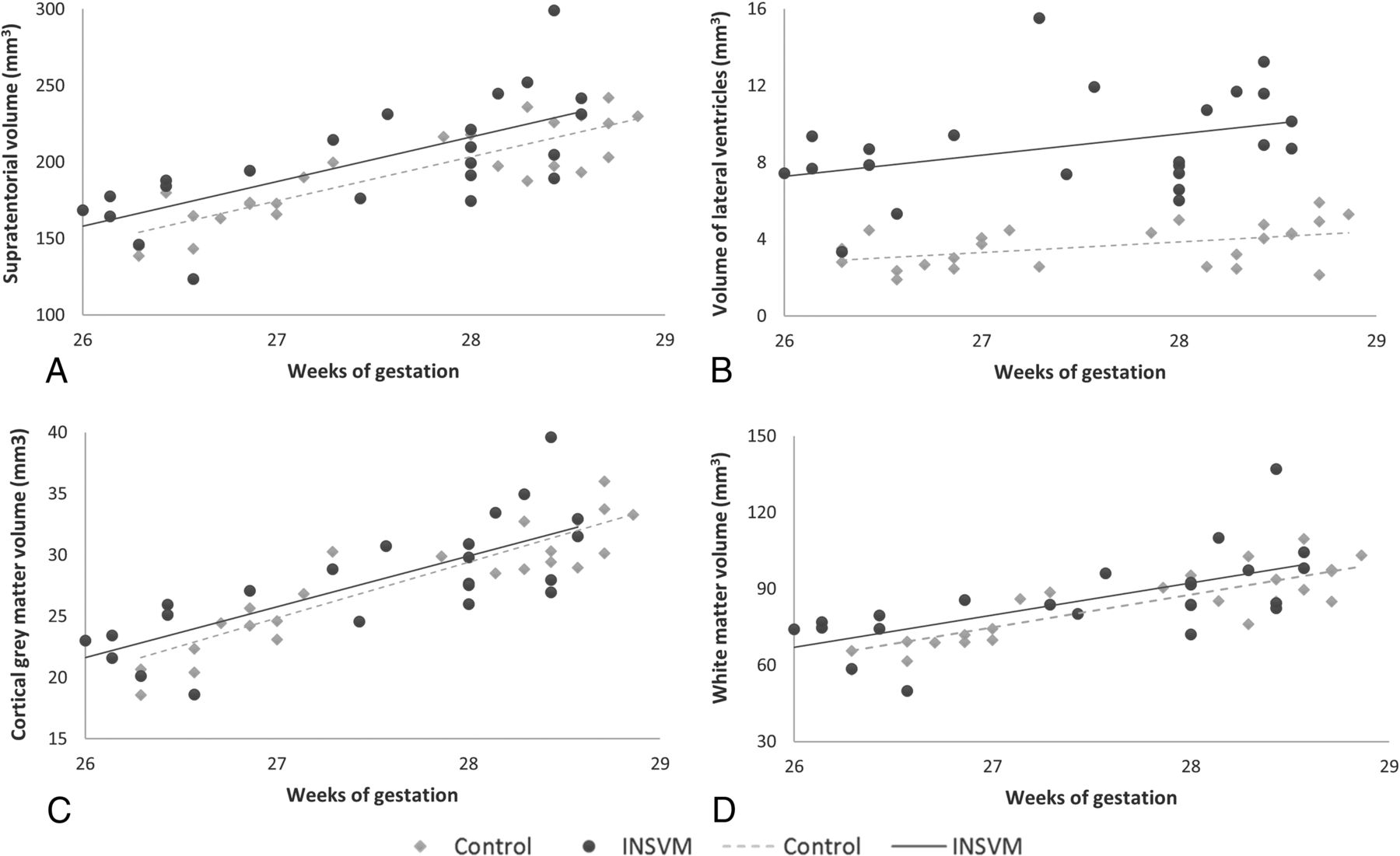
  • Fig 3.
    • Download figure
    • Open in new tab
    • Download powerpoint
    Fig 3.

    Brain tissue volumes in study groups. Cerebral tissue volumes including both hemispheres in INSVM (circles) and control (rhombuses) cohorts. A, Supratentorial volumes. B, Volumes of the lateral ventricles. C, Cortical gray matter volumes. D, White matter volumes. Volume measures are in cubic millimeters.

Tables

  • Figures
    • View popup
    Table 1:

    Clinical characteristics of the study groupsa

    Controls (n = 29)INSVM (n = 32)P
    Maternal age (yr)33.7 ± 4.232.4 ± 5.5.32
    Birth weight (g)3414 ± 5173406 ± 553.96
    Gestational age at birth (wk)39.8 ± 0.939.8 ± 1.2.81
    Gestational age at MRI (wk)27.7 ± 0.927.7 ± 0.9.91
    Lateral ventricular widtha4.6 ± 1.410.6 ± 1.1<.01b
    Fetus sex.02d
        Male58.6% (17)87.5% (28)
        Female41.4% (12)12.5% (4)
    Laterality of ventriculomegaly
        Bilateral–34.4% (11)
        Unilateral left–34.4% (11)
        Unilateral right–31.2% (10)
    Evolution of lateral ventricular widthc
        Regressive–25% (8)
        Stable–75% (24)
        Progressive–0% (0)
    Classification according to atrial width
        Mild (10.0–11.9 mm)–84.3% (27)
        Moderate (12.0–13.9 mm)–16.7% (5)
    • Note:— – indicates that these characteristics do not apply for the control group.

    • ↵a Comparison of clinical characteristics between the control and case cohort. Results are expressed as means or percentage and number of subjects as appropriate.

    • ↵b Measurement of the more dilated lateral ventricle by ultrasound at diagnosis.

    • ↵c Evolution of lateral ventricular width until term of pregnancy.

    • ↵d Significant (≤.05).

    • View popup
    Table 2:

    Sulcal depth and grading scores of the study groupsa

    VariableControls (n = 29)INSVM (n = 32)P
    Left hemisphere depths
        Insula29.4 ± 1.529.7 ± 1.8.03b
        Sylvian fissure16.7 ± 1.617.2 ± 2.4.15
        Parieto-occipital sulcus10.2 ± 2.38.1 ± 2.6<.01b
        Cingulate sulcus3.8 ± 1.34.3 ± 2.5.29
        Calcarine sulcus12.5 ± 2.710.9 ± 2.1<.01b
    Right hemisphere depths
        Insula29.6 ± 1.429.7 ± 1.7.04b
        Sylvian fissure16.8 ± 1.916.0 ± 1.6.01b
        Parieto-occipital sulcus11.1 ± 2.49.0 ± 2.8<.01b
        Cingulate sulcus3.6 ± 1.14.0 ± 1.5.33
        Calcarine sulcus13.5 ± 2.211.8 ± 4.8<.02b
    Sum of grading scores
        Left hemisphere21.1 ± 5.318.2 ± 5.1.01b
        Right hemisphere21.8 ± 5.119.6 ± 5.3.03b
        Total cortex42.9 ± 10.237.8 ± 9.9.01b
    • ↵a Comparison of sulcal depth measurements between the control and case cohort. Depth measurements (in millimeters) were normalized by BPD and multiplied by 100. Results are expressed as means. P values were adjusted by gestational age at examination and sex.

    • ↵b Values ≤ .05.

PreviousNext
Back to top

In this issue

American Journal of Neuroradiology: 40 (9)
American Journal of Neuroradiology
Vol. 40, Issue 9
1 Sep 2019
  • Table of Contents
  • Index by author
  • Complete Issue (PDF)
Advertisement
Print
Download PDF
Email Article

Thank you for your interest in spreading the word on American Journal of Neuroradiology.

NOTE: We only request your email address so that the person you are recommending the page to knows that you wanted them to see it, and that it is not junk mail. We do not capture any email address.

Enter multiple addresses on separate lines or separate them with commas.
Global and Regional Changes in Cortical Development Assessed by MRI in Fetuses with Isolated Nonsevere Ventriculomegaly Correlate with Neonatal Neurobehavior
(Your Name) has sent you a message from American Journal of Neuroradiology
(Your Name) thought you would like to see the American Journal of Neuroradiology web site.
CAPTCHA
This question is for testing whether or not you are a human visitor and to prevent automated spam submissions.
Cite this article
N. Hahner, O.M. Benkarim, M. Aertsen, M. Perez-Cruz, G. Piella, G. Sanroma, N. Bargallo, J. Deprest, M.A. Gonzalez Ballester, E. Gratacos, E. Eixarch
Global and Regional Changes in Cortical Development Assessed by MRI in Fetuses with Isolated Nonsevere Ventriculomegaly Correlate with Neonatal Neurobehavior
American Journal of Neuroradiology Sep 2019, 40 (9) 1567-1574; DOI: 10.3174/ajnr.A6165

Citation Manager Formats

  • BibTeX
  • Bookends
  • EasyBib
  • EndNote (tagged)
  • EndNote 8 (xml)
  • Medlars
  • Mendeley
  • Papers
  • RefWorks Tagged
  • Ref Manager
  • RIS
  • Zotero
0 Responses
Respond to this article
Share
Bookmark this article
Global and Regional Changes in Cortical Development Assessed by MRI in Fetuses with Isolated Nonsevere Ventriculomegaly Correlate with Neonatal Neurobehavior
N. Hahner, O.M. Benkarim, M. Aertsen, M. Perez-Cruz, G. Piella, G. Sanroma, N. Bargallo, J. Deprest, M.A. Gonzalez Ballester, E. Gratacos, E. Eixarch
American Journal of Neuroradiology Sep 2019, 40 (9) 1567-1574; DOI: 10.3174/ajnr.A6165
del.icio.us logo Twitter logo Facebook logo Mendeley logo
  • Tweet Widget
  • Facebook Like
  • Google Plus One
Purchase

Jump to section

  • Article
    • Abstract
    • ABBREVIATIONS:
    • Materials and Methods
    • Results
    • Discussion
    • Conclusions
    • Acknowledgments
    • Footnotes
    • REFERENCES
  • Figures & Data
  • Supplemental
  • Info & Metrics
  • Responses
  • References
  • PDF

Related Articles

  • PubMed
  • Google Scholar

Cited By...

  • Longitudinal MRI Evaluation of Brain Development in Fetuses with Congenital Diaphragmatic Hernia around the Time of Fetal Endotracheal Occlusion
  • Effects of gestational age at birth on perinatal structural brain development in healthy term-born babies
  • Crossref
  • Google Scholar

This article has not yet been cited by articles in journals that are participating in Crossref Cited-by Linking.

More in this TOC Section

  • Pediatric Glioma:Imaging Clusters,Molecular Traits
  • Clinical SVR of Fetal Brain MRI
  • FRACTURE MR in Congenital Vertebral Anomalies
Show more PEDIATRIC NEUROIMAGING

Similar Articles

Advertisement

Indexed Content

  • Current Issue
  • Accepted Manuscripts
  • Article Preview
  • Past Issues
  • Editorials
  • Editor's Choice
  • Fellows' Journal Club
  • Letters to the Editor
  • Video Articles

Cases

  • Case Collection
  • Archive - Case of the Week
  • Archive - Case of the Month
  • Archive - Classic Case

More from AJNR

  • Trainee Corner
  • Imaging Protocols
  • MRI Safety Corner
  • Book Reviews

Multimedia

  • AJNR Podcasts
  • AJNR Scantastics

Resources

  • Turnaround Time
  • Submit a Manuscript
  • Submit a Video Article
  • Submit an eLetter to the Editor/Response
  • Manuscript Submission Guidelines
  • Statistical Tips
  • Fast Publishing of Accepted Manuscripts
  • Graphical Abstract Preparation
  • Imaging Protocol Submission
  • Evidence-Based Medicine Level Guide
  • Publishing Checklists
  • Author Policies
  • Become a Reviewer/Academy of Reviewers
  • News and Updates

About Us

  • About AJNR
  • Editorial Board
  • Editorial Board Alumni
  • Alerts
  • Permissions
  • Not an AJNR Subscriber? Join Now
  • Advertise with Us
  • Librarian Resources
  • Feedback
  • Terms and Conditions
  • AJNR Editorial Board Alumni

American Society of Neuroradiology

  • Not an ASNR Member? Join Now

© 2025 by the American Society of Neuroradiology All rights, including for text and data mining, AI training, and similar technologies, are reserved.
Print ISSN: 0195-6108 Online ISSN: 1936-959X

Powered by HighWire