Advertisement

Main menu

  • Home
  • Content
    • Current Issue
    • Accepted Manuscripts
    • Article Preview
    • Past Issue Archive
    • Video Articles
    • AJNR Case Collection
    • Case of the Week Archive
    • Case of the Month Archive
    • Classic Case Archive
  • Special Collections
    • AJNR Awards
    • Low-Field MRI
    • Alzheimer Disease
    • ASNR Foundation Special Collection
    • Photon-Counting CT
    • View All
  • Multimedia
    • AJNR Podcasts
    • AJNR SCANtastic
    • Trainee Corner
    • MRI Safety Corner
    • Imaging Protocols
  • For Authors
    • Submit a Manuscript
    • Submit a Video Article
    • Submit an eLetter to the Editor/Response
    • Manuscript Submission Guidelines
    • Statistical Tips
    • Fast Publishing of Accepted Manuscripts
    • Graphical Abstract Preparation
    • Imaging Protocol Submission
    • Author Policies
  • About Us
    • About AJNR
    • Editorial Board
    • Editorial Board Alumni
  • More
    • Become a Reviewer/Academy of Reviewers
    • Subscribers
    • Permissions
    • Alerts
    • Feedback
    • Advertisers
    • ASNR Home

User menu

  • Alerts
  • Log in

Search

  • Advanced search
American Journal of Neuroradiology

American Journal of Neuroradiology

ASHNR American Society of Functional Neuroradiology ASHNR American Society of Pediatric Neuroradiology ASSR
  • Alerts
  • Log in

Advanced Search

  • Home
  • Content
    • Current Issue
    • Accepted Manuscripts
    • Article Preview
    • Past Issue Archive
    • Video Articles
    • AJNR Case Collection
    • Case of the Week Archive
    • Case of the Month Archive
    • Classic Case Archive
  • Special Collections
    • AJNR Awards
    • Low-Field MRI
    • Alzheimer Disease
    • ASNR Foundation Special Collection
    • Photon-Counting CT
    • View All
  • Multimedia
    • AJNR Podcasts
    • AJNR SCANtastic
    • Trainee Corner
    • MRI Safety Corner
    • Imaging Protocols
  • For Authors
    • Submit a Manuscript
    • Submit a Video Article
    • Submit an eLetter to the Editor/Response
    • Manuscript Submission Guidelines
    • Statistical Tips
    • Fast Publishing of Accepted Manuscripts
    • Graphical Abstract Preparation
    • Imaging Protocol Submission
    • Author Policies
  • About Us
    • About AJNR
    • Editorial Board
    • Editorial Board Alumni
  • More
    • Become a Reviewer/Academy of Reviewers
    • Subscribers
    • Permissions
    • Alerts
    • Feedback
    • Advertisers
    • ASNR Home
  • Follow AJNR on Twitter
  • Visit AJNR on Facebook
  • Follow AJNR on Instagram
  • Join AJNR on LinkedIn
  • RSS Feeds

AJNR Awards, New Junior Editors, and more. Read the latest AJNR updates

Research ArticleExtracranial Vascular

Carotid Plaque CTA Analysis in Symptomatic Subjects with Bilateral Intraplaque Hemorrhage: A Preliminary Analysis

L. Saba, G. Lanzino, P. Lucatelli, F. Lavra, R. Sanfilippo, R. Montisci, J.S. Suri and C. Yuan
American Journal of Neuroradiology September 2019, 40 (9) 1538-1545; DOI: https://doi.org/10.3174/ajnr.A6160
L. Saba
aFrom the Department of Radiology (L.S., F.L., R.S., R.M.), Azienda Ospedaliero Universitaria di Cagliari, Monserrato (Cagliari), Italy
  • Find this author on Google Scholar
  • Find this author on PubMed
  • Search for this author on this site
  • ORCID record for L. Saba
G. Lanzino
bDepartment of Neurologic Surgery (G.L.), Mayo Clinic, Rochester, Minnesota
  • Find this author on Google Scholar
  • Find this author on PubMed
  • Search for this author on this site
  • ORCID record for G. Lanzino
P. Lucatelli
cDepartment of Radiological, Oncological and Anatomopathological Sciences-Radiology (P.L.), Sapienza University of Rome, Rome, Italy
  • Find this author on Google Scholar
  • Find this author on PubMed
  • Search for this author on this site
  • ORCID record for P. Lucatelli
F. Lavra
aFrom the Department of Radiology (L.S., F.L., R.S., R.M.), Azienda Ospedaliero Universitaria di Cagliari, Monserrato (Cagliari), Italy
  • Find this author on Google Scholar
  • Find this author on PubMed
  • Search for this author on this site
  • ORCID record for F. Lavra
R. Sanfilippo
aFrom the Department of Radiology (L.S., F.L., R.S., R.M.), Azienda Ospedaliero Universitaria di Cagliari, Monserrato (Cagliari), Italy
  • Find this author on Google Scholar
  • Find this author on PubMed
  • Search for this author on this site
  • ORCID record for R. Sanfilippo
R. Montisci
aFrom the Department of Radiology (L.S., F.L., R.S., R.M.), Azienda Ospedaliero Universitaria di Cagliari, Monserrato (Cagliari), Italy
  • Find this author on Google Scholar
  • Find this author on PubMed
  • Search for this author on this site
  • ORCID record for R. Montisci
J.S. Suri
dDiagnostic and Monitoring Division (J.S.S.), Atheropoint, Roseville, California
eDepartment of Electrical Engineering (J.S.S.), University of Idaho, Moscow, Idaho
  • Find this author on Google Scholar
  • Find this author on PubMed
  • Search for this author on this site
  • ORCID record for J.S. Suri
C. Yuan
fCenter for Biomedical Imaging Research (C.Y.), Department of Biomedical Engineering, Tsinghua University School of Medicine, Beijing, China
gDepartment of Radiology (C.Y.), University of Washington, Seattle, Washington.
  • Find this author on Google Scholar
  • Find this author on PubMed
  • Search for this author on this site
  • ORCID record for C. Yuan
  • Article
  • Figures & Data
  • Info & Metrics
  • Responses
  • References
  • PDF
Loading

Article Figures & Data

Figures

  • Tables
  • Fig 1.
    • Download figure
    • Open in new tab
    • Download powerpoint
    Fig 1.

    Flow chart.

  • Fig 2.
    • Download figure
    • Open in new tab
    • Download powerpoint
    Fig 2.

    Example of semiautomatic segmentation of the carotid artery plaque of a 63-year-old subject. Both right (A–C) and left (D–F) carotid arteries are shown, and the red lines in the axial (B and D) and coronal planes (C and F) show the IPH components.

  • Fig 3.
    • Download figure
    • Open in new tab
    • Download powerpoint
    Fig 3.

    Boxplot analysis of the volume of the plaque components (A) and percentages (B) for symptomatic (blue plots) and asymptomatic (red plots) sides.

  • Fig 4.
    • Download figure
    • Open in new tab
    • Download powerpoint
    Fig 4.

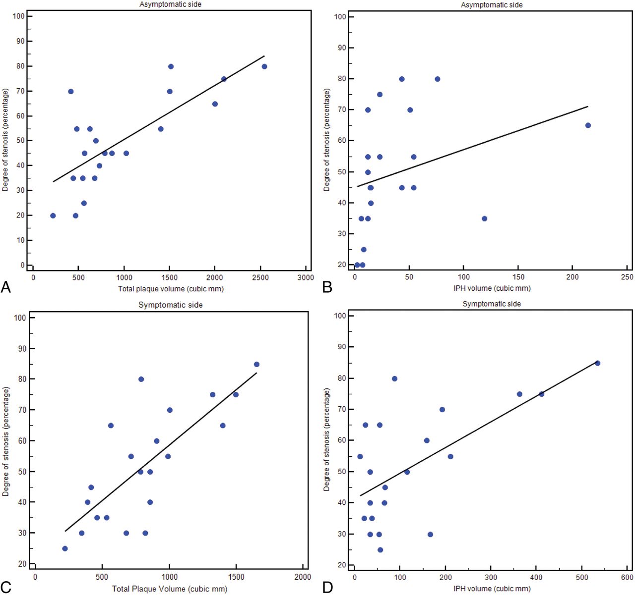
    Regression analysis diagrams between total plaque and IPH volume on asymptomatic sides (A and B, respectively) and between total plaque and IPH volume on symptomatic sides (C and D, respectively).

  • Fig 5.
    • Download figure
    • Open in new tab
    • Download powerpoint
    Fig 5.

    ROC curve analysis of the volume of the plaque components (A) and percentages (B) versus the presence of symptoms.

  • Fig 6.
    • Download figure
    • Open in new tab
    • Download powerpoint
    Fig 6.

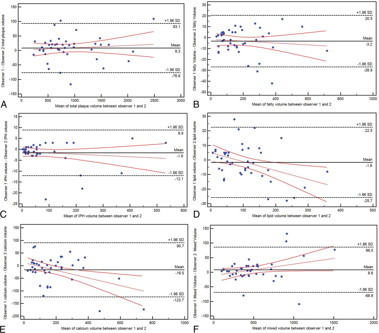
    Bland-Altman plot analysis for the interobserver agreement in total volume (A) and fatty (B), IPH (C), lipid (D), calcified (E), and mixed (F) tissue.

Tables

  • Figures
    • View popup
    Table 1:

    Clinical characteristics of the 21 subjects with bilateral IPH

    Mean or No. (%)
    Age (yr)70 ± 11
    Male sex15 (71.4%)
    Symptomatic side (right)9 (42.8%)
    Smoking11 (52.4%)
    Hypertension14 (67%)
    Hyperlipidemia13 (61.9%)
    Diabetes mellitus8 (38%)
    History of CHD9 (42.8%)
    Family history of CVD9 (42.8%)
    • Note:—CHD indicates coronary heart disease; CVD, cardiovascular disease.

    • View popup
    Table 2:

    Wilcoxon analysisa

    IpsilateralContralateralP Value
    Total plaque788 (548–941)689 (551–1189).543
    Volume (mm3)b
        IPH65 (36–161)15 (12–46).001
        Fatty154 (80–279)141 (91–205).375
        Lipid86 (44–115)103 (62–157).095
        Mixed460 (352–555)424 (313–713).339
        Calcium118 (66–206)119 (99–261).182
    Percentage
        IPH15% (6%–19%)3% (2%–5%).001
        Fatty23% (18%–30%)18% (14%–21%).011
        Lipid11% (7%–13%)15% (10%–16%).016
        Mixed57% (52%–63%)61% (56%–65%).312
        Calcium17% (12%–22%)18% (15%–26%).498
    IPH/fatty ratio0.596 (0.344–0.754)0.171 (0.126–0.1991).001
    • ↵a Wilcoxon Analysis between carotid plaque components and subcomponents between symptomatic and asymptomatic side. Data are expressed in cubic mm.

    • ↵b Voxel size is 0.5 mm isotropic.

    • View popup
    Table 3:

    ROC curve analysis

    AUCSE95% CIP Value
    Percentage
        IPH (<25 HU)0.8890.05060.754–0.965.001
        Fatty0.7070.08320.547–0.837.013
        Lipid0.710.08190.549–0.839.01
        Mixed0.5950.0910.433–0.744.295
        Calcium0.5510.09150.390–0.705.577
    Volume
        IPH (<25 HU)0.7960.06950.643–0.904.001
        Fatty0.5530.0910.392–0.707.558
        Lipid0.6510.08570.488–0.791.077
        Mixed0.5290.09440.370–0.685.755
        Calcium0.5820.09070.420–0.732.368
    IPH/fatty ratio0.5820.09070.420–0.732.368
    • Note:—AUC indicates area under the curve; SE, standard error.

PreviousNext
Back to top

In this issue

American Journal of Neuroradiology: 40 (9)
American Journal of Neuroradiology
Vol. 40, Issue 9
1 Sep 2019
  • Table of Contents
  • Index by author
  • Complete Issue (PDF)
Advertisement
Print
Download PDF
Email Article

Thank you for your interest in spreading the word on American Journal of Neuroradiology.

NOTE: We only request your email address so that the person you are recommending the page to knows that you wanted them to see it, and that it is not junk mail. We do not capture any email address.

Enter multiple addresses on separate lines or separate them with commas.
Carotid Plaque CTA Analysis in Symptomatic Subjects with Bilateral Intraplaque Hemorrhage: A Preliminary Analysis
(Your Name) has sent you a message from American Journal of Neuroradiology
(Your Name) thought you would like to see the American Journal of Neuroradiology web site.
CAPTCHA
This question is for testing whether or not you are a human visitor and to prevent automated spam submissions.
Cite this article
L. Saba, G. Lanzino, P. Lucatelli, F. Lavra, R. Sanfilippo, R. Montisci, J.S. Suri, C. Yuan
Carotid Plaque CTA Analysis in Symptomatic Subjects with Bilateral Intraplaque Hemorrhage: A Preliminary Analysis
American Journal of Neuroradiology Sep 2019, 40 (9) 1538-1545; DOI: 10.3174/ajnr.A6160

Citation Manager Formats

  • BibTeX
  • Bookends
  • EasyBib
  • EndNote (tagged)
  • EndNote 8 (xml)
  • Medlars
  • Mendeley
  • Papers
  • RefWorks Tagged
  • Ref Manager
  • RIS
  • Zotero
0 Responses
Respond to this article
Share
Bookmark this article
Carotid Plaque CTA Analysis in Symptomatic Subjects with Bilateral Intraplaque Hemorrhage: A Preliminary Analysis
L. Saba, G. Lanzino, P. Lucatelli, F. Lavra, R. Sanfilippo, R. Montisci, J.S. Suri, C. Yuan
American Journal of Neuroradiology Sep 2019, 40 (9) 1538-1545; DOI: 10.3174/ajnr.A6160
del.icio.us logo Twitter logo Facebook logo Mendeley logo
  • Tweet Widget
  • Facebook Like
  • Google Plus One
Purchase

Jump to section

  • Article
    • Abstract
    • ABBREVIATIONS:
    • Materials and Methods
    • Results
    • Discussion
    • Conclusions
    • Footnotes
    • REFERENCES
  • Figures & Data
  • Info & Metrics
  • Responses
  • References
  • PDF

Related Articles

  • No related articles found.
  • PubMed
  • Google Scholar

Cited By...

  • Impact Analysis of Different CT Configurations of Carotid Artery Plaque Calcifications on Cerebrovascular Events
  • Perivascular Fat Density and Contrast Plaque Enhancement: Does a Correlation Exist?
  • Crossref (11)
  • Google Scholar

This article has been cited by the following articles in journals that are participating in Crossref Cited-by Linking.

  • Carotid Artery Plaque Calcifications: Lessons From Histopathology to Diagnostic Imaging
    Luca Saba, Valentina Nardi, Riccardo Cau, Ajay Gupta, Hooman Kamel, Jasjit S. Suri, Antonella Balestrieri, Terenzio Congiu, Anthony P.H. Butler, Steven Gieseg, Daniela Fanni, Giulia Cerrone, Roberto Sanfilippo, Josep Puig, Qi Yang, Lorenzo Mannelli, Gavino Faa, Giuseppe Lanzino
    Stroke 2022 53 1
  • State-of-the-art CT and MR imaging and assessment of atherosclerotic carotid artery disease: standardization of scanning protocols and measurements—a consensus document by the European Society of Cardiovascular Radiology (ESCR)
    Luca Saba, Christian Loewe, Thomas Weikert, Michelle C. Williams, Nicola Galea, Ricardo P. J. Budde, Rozemarijn Vliegenthart, Birgitta K. Velthuis, Marco Francone, Jens Bremerich, Luigi Natale, Konstantin Nikolaou, Jean-Nicolas Dacher, Charles Peebles, Federico Caobelli, Alban Redheuil, Marc Dewey, Karl-Friedrich Kreitner, Rodrigo Salgado
    European Radiology 2022 33 2
  • Perivascular Fat Density and Contrast Plaque Enhancement: Does a Correlation Exist?
    L. Saba, S. Zucca, A. Gupta, G. Micheletti, J.S. Suri, A. Balestrieri, M. Porcu, P. Crivelli, G. Lanzino, Y. Qi, V. Nardi, G. Faa, R. Montisci
    American Journal of Neuroradiology 2020 41 8
  • Impact Analysis of Different CT Configurations of Carotid Artery Plaque Calcifications on Cerebrovascular Events
    L. Saba, H. Chen, R. Cau, G.D. Rubeis, G. Zhu, F. Pisu, B. Jang, G. Lanzino, J.S. Suri, Y. Qi, M. Wintermark
    American Journal of Neuroradiology 2022 43 2
  • State-of-the-art CT and MR imaging and assessment of atherosclerotic carotid artery disease: the reporting—a consensus document by the European Society of Cardiovascular Radiology (ESCR)
    Luca Saba, Christian Loewe, Thomas Weikert, Michelle C. Williams, Nicola Galea, Ricardo P. J. Budde, Rozemarijn Vliegenthart, Birgitta K. Velthuis, Marco Francone, Jens Bremerich, Luigi Natale, Konstantin Nikolaou, Jean-Nicolas Dacher, Charles Peebles, Federico Caobelli, Alban Redheuil, Marc Dewey, Karl-Friedrich Kreitner, Rodrigo Salgado
    European Radiology 2022 33 2
  • Extracranial Carotid Plaque Calcification and Cerebrovascular Ischemia: A Systematic Review and Meta-Analysis
    Moayad Homssi, Atin Saha, Diana Delgado, Arindam RoyChoudhury, Charlene Thomas, Matthew Lin, Hediyeh Baradaran, Hooman Kamel, Ajay Gupta
    Stroke 2023 54 10
  • Association Between Spotty Calcification in Nonstenosing Extracranial Carotid Artery Plaque and Ipsilateral Ischemic Stroke
    Moayad Homssi, Amar Vora, Cenai Zhang, Hediyeh Baradaran, Hooman Kamel, Ajay Gupta
    Journal of the American Heart Association 2023 12 10
  • Intraplaque CTA characteristics as predictors of symptomatology: a semiautomated volumetric analysis
    John C. Benson, Luis Savastano, Valentina Nardi, Giuseppe Lanzino, Amir Lerman, Waleed Brinjikji
    Emergency Radiology 2022 29 1
  • CT and MR Imaging of Carotid Wall and Plaque
    Luca Saba, Lorenzo Mannelli, Antonella Balestrieri, Alessandra Serra, PierPaolo Bassareo, Alessandro Murgia, Carola Politi, Michele Porcu, Paola Crivelli, Giulio Micheletti, Maurizio Conti, Jasjit S Suri
    Journal of Neurosonology and Neuroimaging 2019 11 2
  • Application of VascuCAP Software in the Analysis of Carotid Atherosclerotic Plaques in Stroke Patients
    慧 赵
    Advances in Clinical Medicine 2025 15 02

More in this TOC Section

  • Proximal Vertebral Artery Variants and Embryology
  • High-Risk Plaque Features in Carotid MRI
  • Nonstenotic Carotid Plaques and Stroke Review
Show more EXTRACRANIAL VASCULAR

Similar Articles

Advertisement

Indexed Content

  • Current Issue
  • Accepted Manuscripts
  • Article Preview
  • Past Issues
  • Editorials
  • Editor's Choice
  • Fellows' Journal Club
  • Letters to the Editor
  • Video Articles

Cases

  • Case Collection
  • Archive - Case of the Week
  • Archive - Case of the Month
  • Archive - Classic Case

More from AJNR

  • Trainee Corner
  • Imaging Protocols
  • MRI Safety Corner
  • Book Reviews

Multimedia

  • AJNR Podcasts
  • AJNR Scantastics

Resources

  • Turnaround Time
  • Submit a Manuscript
  • Submit a Video Article
  • Submit an eLetter to the Editor/Response
  • Manuscript Submission Guidelines
  • Statistical Tips
  • Fast Publishing of Accepted Manuscripts
  • Graphical Abstract Preparation
  • Imaging Protocol Submission
  • Evidence-Based Medicine Level Guide
  • Publishing Checklists
  • Author Policies
  • Become a Reviewer/Academy of Reviewers
  • News and Updates

About Us

  • About AJNR
  • Editorial Board
  • Editorial Board Alumni
  • Alerts
  • Permissions
  • Not an AJNR Subscriber? Join Now
  • Advertise with Us
  • Librarian Resources
  • Feedback
  • Terms and Conditions
  • AJNR Editorial Board Alumni

American Society of Neuroradiology

  • Not an ASNR Member? Join Now

© 2025 by the American Society of Neuroradiology All rights, including for text and data mining, AI training, and similar technologies, are reserved.
Print ISSN: 0195-6108 Online ISSN: 1936-959X

Powered by HighWire