Advertisement

Main menu

  • Home
  • Content
    • Current Issue
    • Accepted Manuscripts
    • Article Preview
    • Past Issue Archive
    • Video Articles
    • AJNR Case Collection
    • Case of the Week Archive
    • Case of the Month Archive
    • Classic Case Archive
  • Special Collections
    • AJNR Awards
    • Low-Field MRI
    • Alzheimer Disease
    • ASNR Foundation Special Collection
    • Photon-Counting CT
    • View All
  • Multimedia
    • AJNR Podcasts
    • AJNR SCANtastic
    • Trainee Corner
    • MRI Safety Corner
    • Imaging Protocols
  • For Authors
    • Submit a Manuscript
    • Submit a Video Article
    • Submit an eLetter to the Editor/Response
    • Manuscript Submission Guidelines
    • Statistical Tips
    • Fast Publishing of Accepted Manuscripts
    • Graphical Abstract Preparation
    • Imaging Protocol Submission
    • Author Policies
  • About Us
    • About AJNR
    • Editorial Board
    • Editorial Board Alumni
  • More
    • Become a Reviewer/Academy of Reviewers
    • Subscribers
    • Permissions
    • Alerts
    • Feedback
    • Advertisers
    • ASNR Home

User menu

  • Alerts
  • Log in

Search

  • Advanced search
American Journal of Neuroradiology

American Journal of Neuroradiology

ASHNR American Society of Functional Neuroradiology ASHNR American Society of Pediatric Neuroradiology ASSR
  • Alerts
  • Log in

Advanced Search

  • Home
  • Content
    • Current Issue
    • Accepted Manuscripts
    • Article Preview
    • Past Issue Archive
    • Video Articles
    • AJNR Case Collection
    • Case of the Week Archive
    • Case of the Month Archive
    • Classic Case Archive
  • Special Collections
    • AJNR Awards
    • Low-Field MRI
    • Alzheimer Disease
    • ASNR Foundation Special Collection
    • Photon-Counting CT
    • View All
  • Multimedia
    • AJNR Podcasts
    • AJNR SCANtastic
    • Trainee Corner
    • MRI Safety Corner
    • Imaging Protocols
  • For Authors
    • Submit a Manuscript
    • Submit a Video Article
    • Submit an eLetter to the Editor/Response
    • Manuscript Submission Guidelines
    • Statistical Tips
    • Fast Publishing of Accepted Manuscripts
    • Graphical Abstract Preparation
    • Imaging Protocol Submission
    • Author Policies
  • About Us
    • About AJNR
    • Editorial Board
    • Editorial Board Alumni
  • More
    • Become a Reviewer/Academy of Reviewers
    • Subscribers
    • Permissions
    • Alerts
    • Feedback
    • Advertisers
    • ASNR Home
  • Follow AJNR on Twitter
  • Visit AJNR on Facebook
  • Follow AJNR on Instagram
  • Join AJNR on LinkedIn
  • RSS Feeds

AJNR Awards, New Junior Editors, and more. Read the latest AJNR updates

Research ArticleAdult Brain
Open Access

A Novel Collateral Imaging Method Derived from Time-Resolved Dynamic Contrast-Enhanced MR Angiography in Acute Ischemic Stroke: A Pilot Study

H.G. Roh, E.Y. Kim, I.S. Kim, H.J. Lee, J.J. Park, S.B. Lee, J.W. Choi, Y.S. Jeon, M. Park, S.U. Kim and H.J. Kim
American Journal of Neuroradiology June 2019, 40 (6) 946-953; DOI: https://doi.org/10.3174/ajnr.A6068
H.G. Roh
aFrom the Departments of Radiology (H.G.R., J.W.C.)
  • Find this author on Google Scholar
  • Find this author on PubMed
  • Search for this author on this site
  • ORCID record for H.G. Roh
E.Y. Kim
dDepartment of Radiology (E.Y.K.), Gachon University Gil Medical Center, Incheon, Korea
  • Find this author on Google Scholar
  • Find this author on PubMed
  • Search for this author on this site
  • ORCID record for E.Y. Kim
I.S. Kim
eSiemens Healthineers Ltd (I.S.K.), Seoul, Korea
  • Find this author on Google Scholar
  • Find this author on PubMed
  • Search for this author on this site
  • ORCID record for I.S. Kim
H.J. Lee
fDepartments of Neurosurgery (H.J.L., S.U.K.)
  • Find this author on Google Scholar
  • Find this author on PubMed
  • Search for this author on this site
  • ORCID record for H.J. Lee
J.J. Park
bNeurology (J.J.P.)
  • Find this author on Google Scholar
  • Find this author on PubMed
  • Search for this author on this site
  • ORCID record for J.J. Park
S.B. Lee
gNeurology (S.B.L.)
  • Find this author on Google Scholar
  • Find this author on PubMed
  • Search for this author on this site
  • ORCID record for S.B. Lee
J.W. Choi
aFrom the Departments of Radiology (H.G.R., J.W.C.)
  • Find this author on Google Scholar
  • Find this author on PubMed
  • Search for this author on this site
  • ORCID record for J.W. Choi
Y.S. Jeon
cNeurosurgery (Y.S.J.), Konkuk University Medical Center, Konkuk University School of Medicine, Seoul, Korea
  • Find this author on Google Scholar
  • Find this author on PubMed
  • Search for this author on this site
  • ORCID record for Y.S. Jeon
M. Park
iDepartment of Radiology (M.P.), Gangnam Severance Hospital, Yonsei University College of Medicine, Seoul, Korea.
  • Find this author on Google Scholar
  • Find this author on PubMed
  • Search for this author on this site
  • ORCID record for M. Park
S.U. Kim
fDepartments of Neurosurgery (H.J.L., S.U.K.)
  • Find this author on Google Scholar
  • Find this author on PubMed
  • Search for this author on this site
  • ORCID record for S.U. Kim
H.J. Kim
hRadiology (H.J.K.), Daejeon St. Mary's Hospital, The Catholic University of Korea, Daejeon, Korea
  • Find this author on Google Scholar
  • Find this author on PubMed
  • Search for this author on this site
  • ORCID record for H.J. Kim
  • Article
  • Figures & Data
  • Info & Metrics
  • Responses
  • References
  • PDF
Loading

Article Figures & Data

Figures

  • Tables
  • Fig 1.
    • Download figure
    • Open in new tab
    • Download powerpoint
    Fig 1.

    MR imaging protocol for AIS diagnosis.

  • Fig 2.
    • Download figure
    • Open in new tab
    • Download powerpoint
    Fig 2.

    Postprocessing sequences for the mMRA collateral map (left) and the MRP collateral map (right). After being loaded and stored in a 4D matrix of DICOM source images of DCE-MRA and DSC-MRP for approximately 1 minute, subtracted MIP images with both the time and spatial information are displayed. We apply ROIs on the MCA in nonischemic hemisphere and superior sagittal sinus. Arterial and venous signal intensity–time curves are generated by plotting signal changes in the ROIs, and 4 phases (arterial phase = from the beginning of arrival of the contrast in the MCA to the arterial bolus peak; capillary phase = from just past the arterial bolus peak to just before the venous bolus peak in the superior sagittal sinus; early venous phase = first half of the venous phase from the venous bolus peak to the starting point of the venous plateau; late venous phase = second half of the venous phase) of collateral maps are determined according to the signal intensity–time curves automatically. In mMRA collateral maps, an axial reformatting process for the original coronal images is added before generating the collateral map.
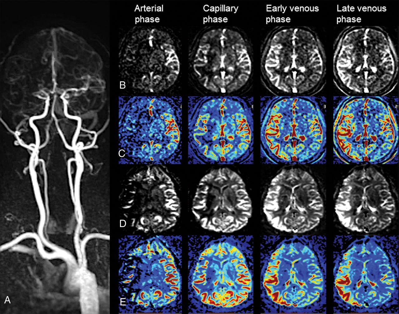
  • Fig 3.
    • Download figure
    • Open in new tab
    • Download powerpoint
    Fig 3.

    mMRA and MRP collateral maps at 2 hours 18 minutes and 2 hours 21 minutes after symptom onset in a 61-year-old woman with occlusion of the right MCA. The 3-month mRS score of the patient was 2. A, DCE-MRA shows occlusion of the right M1 MCA. Gray-scale (B) and color-coded (C) mMRA collateral maps. On the mMRA collateral maps, the collateral-perfusion delay involves more than one-half of the MCA territory, including the subcortical structures in the capillary phase and no collateral-perfusion delay in the early venous phase. The collateral score is 3 (intermediate to good). Gray-scale (D) and color-coded (E) MRP collateral maps. The MRP collateral map shows the same collateral score as the mMRA collateral map.

  • Fig 4.
    • Download figure
    • Open in new tab
    • Download powerpoint
    Fig 4.

    mMRA and MRP collateral maps at 5 hours 44 minutes and 5 hours 47 minutes after symptom onset in a 72-year-old man with occlusion of the left ICA and MCA. This patient died 27 days after admission. A, DCE-MRA shows occlusion of the left proximal ICA and MCA. Gray-scale (B) and color-coded (C) mMRA collateral maps. On the mMRA collateral maps, the collateral-perfusion delay involves more than one-half of the MCA territory, including the subcortical structures in the capillary phase, and persists until the late venous phase. The collateral score is 0 (very poor). Gray-scale (D) and color-coded (E) MRP collateral maps. The MRP collateral map shows the same collateral score as the mMRA collateral map.

  • Fig 5.
    • Download figure
    • Open in new tab
    • Download powerpoint
    Fig 5.

    Eight topographic regions of the MCA territory for MR collateral imaging (collateral map). I = insular ribbon; S = subcortical structures (basal ganglia and internal capsule); M1, M2, and M3 are the anterior, lateral, and posterior MCA territories, respectively, at the basal ganglia level; M4, M5, and M6 are the anterior, lateral, and posterior MCA territories, respectively, at the supraganglionic level.
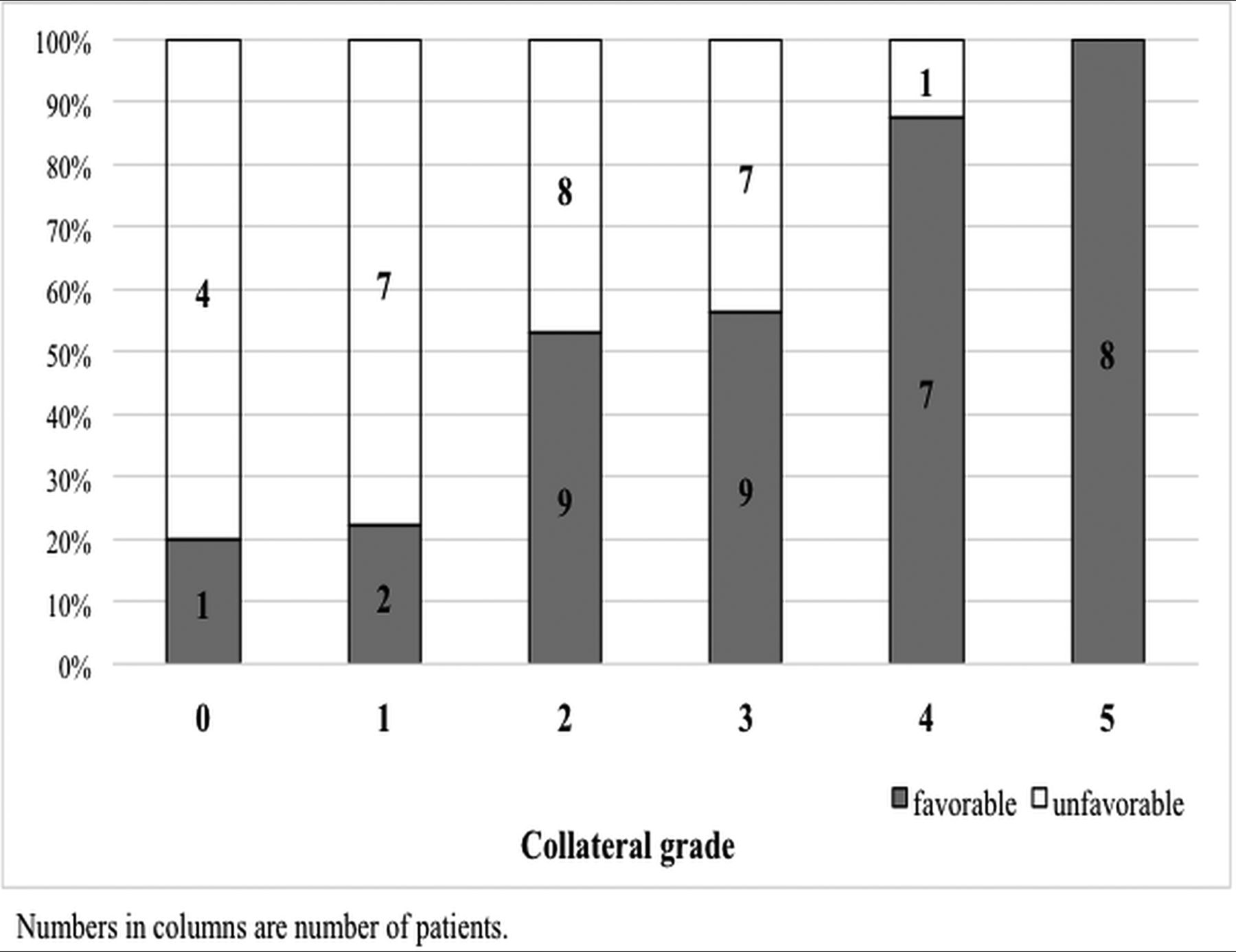
  • Fig 6.
    • Download figure
    • Open in new tab
    • Download powerpoint
    Fig 6.

    A 100% stacked column chart of the functional outcomes and collateral grades of patients.

Tables

  • Figures
    • View popup
    Table 1:

    Acute stroke MRI acquisition parameters

    ParameterAcute Stroke MRI
    SS EPI-DWISWIDCE-MRA (TWIST)DSC-MRPT2-FLAIR
    TR/TE (ms)4800/7128/202.62/0.951600/309000/95
    TI (ms)2500
    Turbo factor21
    Flip angle15°21°90°150°
    Bandwidth (Hz/pixel)16721207801446206
    Slice thickness/gap (mm)5/22/01.2/05/25/2
    FOV (mm)240 × 240240 × 195400 × 300240 × 240240 × 217
    Matrix size (mm)130 × 130384 × 156320 × 182128 × 128256 × 174
    B-value (s/mm2)0, 1000
    Measurements1130601
    GRAPPA223 × 22
    Temporal resolution (sec)1.61.6
    Dynamic reconstruction modeForward share
    K-space sampling (center/periphery) (%)15/20
    • Note:—SS indicates single-shot; TWIST, time-resolved imaging with stochastic trajectories; GRAPPA, generalized autocalibrating partially parallel acquisition.

    • View popup
    Table 2:

    Collateral grading system for analysis of the collateral map

    ScoreDescription of Collateral Status
    5 (Excellent)No or smalla collateral-perfusion delay in the MCA territory in the capillary phase regardless of the collateral status in the arterial phase
    4 (Good)Collateral-perfusion delay equal to or less than one-half of MCA territory in the capillary phase and no or small delay in the early venous phase
    3 (Intermediate to good)1) Collateral-perfusion delay equal to or less than one-half of the MCA territory in the capillary phase and equal or less than one-half in the early venous phase
    2) Collateral-perfusion delay more than one-half of the MCA territory in the capillary phase and no or small delay in the early venous phase
    2 (Intermediate to poor)Collateral-perfusion delay more than one-half of the MCA territory in the capillary phase and equal to or less than one-half in the early venous phase
    1 (Poor)Collateral-perfusion delay more than one-half of the MCA territory in the early venous phase and equal to or less than one-half in the late venous phase
    0 (Very poor)Collateral-perfusion delay/defect more than one-half of the MCA territory in the late venous phase regardless of perfusion status at previous phases
    • ↵a “Small” indicates an area <1 of 8 MCA regions (Fig 5).

PreviousNext
Back to top

In this issue

American Journal of Neuroradiology: 40 (6)
American Journal of Neuroradiology
Vol. 40, Issue 6
1 Jun 2019
  • Table of Contents
  • Index by author
  • Complete Issue (PDF)
Advertisement
Print
Download PDF
Email Article

Thank you for your interest in spreading the word on American Journal of Neuroradiology.

NOTE: We only request your email address so that the person you are recommending the page to knows that you wanted them to see it, and that it is not junk mail. We do not capture any email address.

Enter multiple addresses on separate lines or separate them with commas.
A Novel Collateral Imaging Method Derived from Time-Resolved Dynamic Contrast-Enhanced MR Angiography in Acute Ischemic Stroke: A Pilot Study
(Your Name) has sent you a message from American Journal of Neuroradiology
(Your Name) thought you would like to see the American Journal of Neuroradiology web site.
CAPTCHA
This question is for testing whether or not you are a human visitor and to prevent automated spam submissions.
Cite this article
H.G. Roh, E.Y. Kim, I.S. Kim, H.J. Lee, J.J. Park, S.B. Lee, J.W. Choi, Y.S. Jeon, M. Park, S.U. Kim, H.J. Kim
A Novel Collateral Imaging Method Derived from Time-Resolved Dynamic Contrast-Enhanced MR Angiography in Acute Ischemic Stroke: A Pilot Study
American Journal of Neuroradiology Jun 2019, 40 (6) 946-953; DOI: 10.3174/ajnr.A6068

Citation Manager Formats

  • BibTeX
  • Bookends
  • EasyBib
  • EndNote (tagged)
  • EndNote 8 (xml)
  • Medlars
  • Mendeley
  • Papers
  • RefWorks Tagged
  • Ref Manager
  • RIS
  • Zotero
0 Responses
Respond to this article
Share
Bookmark this article
A Novel Collateral Imaging Method Derived from Time-Resolved Dynamic Contrast-Enhanced MR Angiography in Acute Ischemic Stroke: A Pilot Study
H.G. Roh, E.Y. Kim, I.S. Kim, H.J. Lee, J.J. Park, S.B. Lee, J.W. Choi, Y.S. Jeon, M. Park, S.U. Kim, H.J. Kim
American Journal of Neuroradiology Jun 2019, 40 (6) 946-953; DOI: 10.3174/ajnr.A6068
del.icio.us logo Twitter logo Facebook logo Mendeley logo
  • Tweet Widget
  • Facebook Like
  • Google Plus One
Purchase

Jump to section

  • Article
    • Abstract
    • ABBREVIATIONS:
    • Materials and Methods
    • Results
    • Discussion
    • Conclusions
    • Footnotes
    • REFERENCES
  • Figures & Data
  • Info & Metrics
  • Responses
  • References
  • PDF

Related Articles

  • No related articles found.
  • PubMed
  • Google Scholar

Cited By...

  • Deep Learning-Based Synthetic TOF-MRA Generation Using Time-Resolved MRA in Fast Stroke Imaging
  • Crossref (20)
  • Google Scholar

This article has been cited by the following articles in journals that are participating in Crossref Cited-by Linking.

  • Cerebral Collateral Circulation in the Era of Reperfusion Therapies for Acute Ischemic Stroke
    Simone M. Uniken Venema, Jan Willem Dankbaar, Aad van der Lugt, Diederik W.J. Dippel, H. Bart van der Worp
    Stroke 2022 53 10
  • Multiphase MR Angiography Collateral Map: Functional Outcome after Acute Anterior Circulation Ischemic Stroke
    Hyun Jeong Kim, Sang Bong Lee, Jin Woo Choi, Yoo Sung Jeon, Hyung Jin Lee, Jeong Jin Park, Eung Yeop Kim, In Seong Kim, Taek Jun Lee, Yu Jin Jung, Seon Young Ryu, Young Il Chun, Ji Sung Lee, Hong Gee Roh
    Radiology 2020 295 1
  • Dynamic contrast-enhanced (DCE) imaging: state of the art and applications in whole-body imaging
    Domenico Albano, Federico Bruno, Andrea Agostini, Salvatore Alessio Angileri, Massimo Benenati, Giulia Bicchierai, Michaela Cellina, Vito Chianca, Diletta Cozzi, Ginevra Danti, Federica De Muzio, Letizia Di Meglio, Francesco Gentili, Giuliana Giacobbe, Giulia Grazzini, Irene Grazzini, Pasquale Guerriero, Carmelo Messina, Giuseppe Micci, Pierpaolo Palumbo, Maria Paola Rocco, Roberto Grassi, Vittorio Miele, Antonio Barile
    Japanese Journal of Radiology 2022 40 4
  • Collateral estimation by susceptibility-weighted imaging and prediction of functional outcomes after acute anterior circulation ischemic stroke
    Hyung Jin Lee, Hong Gee Roh, Sang Bong Lee, Yoo Sung Jeon, Jeong Jin Park, Taek-Jun Lee, Yu Jin Jung, Jin Woo Choi, Young Il Chun, Hee Jong Ki, Junsoo Cho, Ji Sung Lee, Hyun Jeong Kim
    Scientific Reports 2021 11 1
  • Imaging in Acute Anterior Circulation Ischemic Stroke: Current and Future
    Hyun Jeong Kim, Hong Gee Roh
    Neurointervention 2022 17 1
  • The collateral map: prediction of lesion growth and penumbra after acute anterior circulation ischemic stroke
    Jin Seok Yi, Hee Jong Ki, Yoo Sung Jeon, Jeong Jin Park, Taek-Jun Lee, Jin Tae Kwak, Sang Bong Lee, Hyung Jin Lee, In Seong Kim, Joo Hyun Kim, Ji Sung Lee, Hong Gee Roh, Hyun Jeong Kim
    European Radiology 2023 34 3
  • Collateral and permeability imaging derived from dynamic contrast material-enhanced MR angiography in prediction of PH 2 hemorrhagic transformation after acute ischemic stroke: a pilot study
    Taek-Jun Lee, Hong Gee Roh, Joo Hyun Kim, Sang Bong Lee, Jeong Jin Park, Hyung Jin Lee, Yoo Sung Jeon, Jin Woo Choi, Young Il Chun, Yu Jin Jung, Seon Young Ryu, Ji Sung Lee, Hyun Jeong Kim
    Neuroradiology 2021 63 9
  • Deep regression neural networks for collateral imaging from dynamic susceptibility contrast-enhanced magnetic resonance perfusion in acute ischemic stroke
    Minh Nguyen Nhat To, Hyun Jeong Kim, Hong Gee Roh, Yoon-Sik Cho, Jin Tae Kwak
    International Journal of Computer Assisted Radiology and Surgery 2020 15 1
  • Perfusion Maps Acquired From Dynamic Angiography MRI Using Deep Learning Approaches
    Muhammad Asaduddin, Hong Gee Roh, Hyun Jeong Kim, Eung Yeop Kim, Sung‐Hong Park
    Journal of Magnetic Resonance Imaging 2023 57 2
  • A 3D Multi-task Regression and Ordinal Regression Deep Neural Network for Collateral Imaging from Dynamic Susceptibility Contrast-Enhanced MR perfusion in Acute Ischemic Stroke
    Hoang Long Le, Hong Gee Roh, Hyun Jeong Kim, Jin Tae Kwak
    Computer Methods and Programs in Biomedicine 2022 225

More in this TOC Section

  • Diagnostic Neuroradiology of Monoclonal Antibodies
  • Cerebral ADC Changes in Fabry Disease
  • ML for Glioma Molecular Subtype Prediction
Show more ADULT BRAIN

Similar Articles

Advertisement

Indexed Content

  • Current Issue
  • Accepted Manuscripts
  • Article Preview
  • Past Issues
  • Editorials
  • Editor's Choice
  • Fellows' Journal Club
  • Letters to the Editor
  • Video Articles

Cases

  • Case Collection
  • Archive - Case of the Week
  • Archive - Case of the Month
  • Archive - Classic Case

More from AJNR

  • Trainee Corner
  • Imaging Protocols
  • MRI Safety Corner
  • Book Reviews

Multimedia

  • AJNR Podcasts
  • AJNR Scantastics

Resources

  • Turnaround Time
  • Submit a Manuscript
  • Submit a Video Article
  • Submit an eLetter to the Editor/Response
  • Manuscript Submission Guidelines
  • Statistical Tips
  • Fast Publishing of Accepted Manuscripts
  • Graphical Abstract Preparation
  • Imaging Protocol Submission
  • Evidence-Based Medicine Level Guide
  • Publishing Checklists
  • Author Policies
  • Become a Reviewer/Academy of Reviewers
  • News and Updates

About Us

  • About AJNR
  • Editorial Board
  • Editorial Board Alumni
  • Alerts
  • Permissions
  • Not an AJNR Subscriber? Join Now
  • Advertise with Us
  • Librarian Resources
  • Feedback
  • Terms and Conditions
  • AJNR Editorial Board Alumni

American Society of Neuroradiology

  • Not an ASNR Member? Join Now

© 2025 by the American Society of Neuroradiology All rights, including for text and data mining, AI training, and similar technologies, are reserved.
Print ISSN: 0195-6108 Online ISSN: 1936-959X

Powered by HighWire