Advertisement

Main menu

  • Home
  • Content
    • Current Issue
    • Accepted Manuscripts
    • Article Preview
    • Past Issue Archive
    • Video Articles
    • AJNR Case Collection
    • Case of the Week Archive
    • Case of the Month Archive
    • Classic Case Archive
  • Special Collections
    • AJNR Awards
    • Low-Field MRI
    • Alzheimer Disease
    • ASNR Foundation Special Collection
    • Photon-Counting CT
    • View All
  • Multimedia
    • AJNR Podcasts
    • AJNR SCANtastic
    • Trainee Corner
    • MRI Safety Corner
    • Imaging Protocols
  • For Authors
    • Submit a Manuscript
    • Submit a Video Article
    • Submit an eLetter to the Editor/Response
    • Manuscript Submission Guidelines
    • Statistical Tips
    • Fast Publishing of Accepted Manuscripts
    • Graphical Abstract Preparation
    • Imaging Protocol Submission
    • Author Policies
  • About Us
    • About AJNR
    • Editorial Board
    • Editorial Board Alumni
  • More
    • Become a Reviewer/Academy of Reviewers
    • Subscribers
    • Permissions
    • Alerts
    • Feedback
    • Advertisers
    • ASNR Home

User menu

  • Alerts
  • Log in

Search

  • Advanced search
American Journal of Neuroradiology

American Journal of Neuroradiology

ASHNR American Society of Functional Neuroradiology ASHNR American Society of Pediatric Neuroradiology ASSR
  • Alerts
  • Log in

Advanced Search

  • Home
  • Content
    • Current Issue
    • Accepted Manuscripts
    • Article Preview
    • Past Issue Archive
    • Video Articles
    • AJNR Case Collection
    • Case of the Week Archive
    • Case of the Month Archive
    • Classic Case Archive
  • Special Collections
    • AJNR Awards
    • Low-Field MRI
    • Alzheimer Disease
    • ASNR Foundation Special Collection
    • Photon-Counting CT
    • View All
  • Multimedia
    • AJNR Podcasts
    • AJNR SCANtastic
    • Trainee Corner
    • MRI Safety Corner
    • Imaging Protocols
  • For Authors
    • Submit a Manuscript
    • Submit a Video Article
    • Submit an eLetter to the Editor/Response
    • Manuscript Submission Guidelines
    • Statistical Tips
    • Fast Publishing of Accepted Manuscripts
    • Graphical Abstract Preparation
    • Imaging Protocol Submission
    • Author Policies
  • About Us
    • About AJNR
    • Editorial Board
    • Editorial Board Alumni
  • More
    • Become a Reviewer/Academy of Reviewers
    • Subscribers
    • Permissions
    • Alerts
    • Feedback
    • Advertisers
    • ASNR Home
  • Follow AJNR on Twitter
  • Visit AJNR on Facebook
  • Follow AJNR on Instagram
  • Join AJNR on LinkedIn
  • RSS Feeds

AJNR Awards, New Junior Editors, and more. Read the latest AJNR updates

Research ArticleSpine
Open Access

Deep Learning–Based Automatic Segmentation of Lumbosacral Nerves on CT for Spinal Intervention: A Translational Study

G. Fan, H. Liu, Z. Wu, Y. Li, C. Feng, D. Wang, J. Luo, W.M. Wells and S. He
American Journal of Neuroradiology June 2019, 40 (6) 1074-1081; DOI: https://doi.org/10.3174/ajnr.A6070
G. Fan
aFrom the Orthopedic Department, Shanghai Tenth People's Hospital (G.F., C.F., D.W., S.H.), Tongji University School of Medicine, Shanghai, China
bDepartment of Spine Surgery (G.F.), Third Affiliated Hospital of Sun Yatsen University, Guangzhou, China
cSurgical Planning Lab (G.F., J.L., W.M.W.), Radiology Department, Brigham and Women's Hospital, Harvard Medical School, Boston, Massachusetts
  • Find this author on Google Scholar
  • Find this author on PubMed
  • Search for this author on this site
  • ORCID record for G. Fan
H. Liu
dSpinal Pain Research Institute of Tongji University (H.L., C.F., D.W., S.H.), Shanghai, China
  • Find this author on Google Scholar
  • Find this author on PubMed
  • Search for this author on this site
  • ORCID record for H. Liu
Z. Wu
eSchool of Data and Computer Science (Z.W.), Sun Yat-sen University, Guangzhou, China
  • Find this author on Google Scholar
  • Find this author on PubMed
  • Search for this author on this site
  • ORCID record for Z. Wu
Y. Li
fShanghai Jiao Tong University School of Medicine (Y.L.), Shanghai, China
  • Find this author on Google Scholar
  • Find this author on PubMed
  • Search for this author on this site
  • ORCID record for Y. Li
C. Feng
aFrom the Orthopedic Department, Shanghai Tenth People's Hospital (G.F., C.F., D.W., S.H.), Tongji University School of Medicine, Shanghai, China
dSpinal Pain Research Institute of Tongji University (H.L., C.F., D.W., S.H.), Shanghai, China
  • Find this author on Google Scholar
  • Find this author on PubMed
  • Search for this author on this site
  • ORCID record for C. Feng
D. Wang
aFrom the Orthopedic Department, Shanghai Tenth People's Hospital (G.F., C.F., D.W., S.H.), Tongji University School of Medicine, Shanghai, China
dSpinal Pain Research Institute of Tongji University (H.L., C.F., D.W., S.H.), Shanghai, China
  • Find this author on Google Scholar
  • Find this author on PubMed
  • Search for this author on this site
  • ORCID record for D. Wang
J. Luo
cSurgical Planning Lab (G.F., J.L., W.M.W.), Radiology Department, Brigham and Women's Hospital, Harvard Medical School, Boston, Massachusetts
gGraduate School of Frontier Sciences (J.L.), University of Tokyo, Tokyo, Japan.
  • Find this author on Google Scholar
  • Find this author on PubMed
  • Search for this author on this site
  • ORCID record for J. Luo
W.M. Wells III
cSurgical Planning Lab (G.F., J.L., W.M.W.), Radiology Department, Brigham and Women's Hospital, Harvard Medical School, Boston, Massachusetts
  • Find this author on Google Scholar
  • Find this author on PubMed
  • Search for this author on this site
  • ORCID record for W.M. Wells III
S. He
aFrom the Orthopedic Department, Shanghai Tenth People's Hospital (G.F., C.F., D.W., S.H.), Tongji University School of Medicine, Shanghai, China
dSpinal Pain Research Institute of Tongji University (H.L., C.F., D.W., S.H.), Shanghai, China
  • Find this author on Google Scholar
  • Find this author on PubMed
  • Search for this author on this site
  • ORCID record for S. He
  • Article
  • Figures & Data
  • Supplemental
  • Info & Metrics
  • Responses
  • References
  • PDF
Loading

Article Figures & Data

Figures

  • Tables
  • Fig 1.
    • Download figure
    • Open in new tab
    • Download powerpoint
    Fig 1.

    Manual segmentation and 3D reconstruction on Slicer. A, manual labels. B, 3D reconstruction with a coronal image. C, Illustrations of the Dice score, Intersection-over-Union, and pixel accuracy.

  • Fig 2.
    • Download figure
    • Open in new tab
    • Download powerpoint
    Fig 2.

    Schematic of the network architecture.

  • Fig 3.
    • Download figure
    • Open in new tab
    • Download powerpoint
    Fig 3.

    Measurement of safe and Kambin triangles. A, Schematics of the Kambin triangle. B, Schematics of the safe triangle. C, Measurement of the Kambin triangle on a manually segmented image. D, Measurement of the safe triangle on automatically segmented images.

  • Fig 4.
    • Download figure
    • Open in new tab
    • Download powerpoint
    Fig 4.

    Automatic and manually labeled masks.
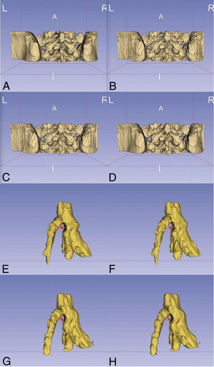
  • Fig 5.
    • Download figure
    • Open in new tab
    • Download powerpoint
    Fig 5.

    3D rendering of automatic masks and manually labeled masks of bones and nerves. A, 3D rendering of manual masks before preprocessing. B, 3D rendering of post-preprocessed masks. C, 3D rendering of automatically generated masks. D, Smoothed 3D rendering of automatically generated masks. E, 3D rendering of manual masks before preprocessing (arrow indicates the compressed dura). F, 3D rendering of post-preprocessed masks (arrow indicates the compressed dura). G, 3D rendering of automatically generated masks (arrow indicates the compressed dura). H, Smoothed 3D rendering of automatically generated masks (arrow indicates the compressed dura).

  • Fig 6.
    • Download figure
    • Open in new tab
    • Download powerpoint
    Fig 6.

    3D model–based viability assessment of a transforaminal epidural steroid injection. A, Inaccessible trajectory to the safe triangle on an axial CT slice. B, Minimal space of the safe triangle on the posterior 3D model. C, Accessible oblique trajectory to the safe triangle on the 3D model. D, Inaccessible trajectory to the Kambin triangle on axial CT slice. E, Accessible trajectory on the 3D model. F, Oblique trajectory-guided nonaxial CT plane.

Tables

  • Figures
    • View popup
    Table 1:

    Overview of the combined algorithm

    Algorithm 1: Combined Algorithm
    Require: X: CT volume, shape = D × H × W
    Require: xi=X(Li), (i=1, .…, k): CT voxel patch
    Require: yi = M(xi): yi is the output of the last layer (softmax activation function) of the model M, yi has 1 more dimension than xi, and this dimension has 3 channels. Each channel refers to the probability of the corresponding voxel belonging to background or bone or nerve, respectively.
    1) Initialize: Y ← 0
    2) For xi ∈ X,(i = 1, …, k) do
    3) Y(Li,:) + = yi
    4) End for
    5) S ← arg max(Y, axis = −1) (find the channel with the largest value in the last dimension)
    6) Return S (the automatic mask)
    • Note:—M indicates the model (network); L, location of the CT voxel patch x at the CT volumn X; Y, summed probability; max, maximum.

    • View popup
    Table 2:

    Segmentation accuracy in 10 testing casesa

    StructuresPixel Accuracy (%)IoU (%)Dice Score (%)
    Bones94.05 ± 6.6889.73 ± 4.3294.54 ± 2.43
    (82.0–99.9)(82.0–95.2)(90.1–97.5)
    Nerves91.43 ± 3.4882.71 ± 3.2590.51 ± 1.94
    (85.3–94.4)(76.3–87.4)(86.6–93.2)
    • ↵a Data are means and percentages unless otherwise noted.

    • View popup
    Table 3:

    Segmentation accuracy in 10 testing cases from the open dataseta

    StructuresPixel Accuracy (%)IoU (%)Dice Score (%)
    Bones99.62 ± 0.3581.40 ± 11.3389.34 ± 7.28
    (99.3–99.9)(60.5–93.3)(75.42–96.5)
    Nerves87.74 ± 4.8280.64 ± 3.3189.25 ± 2.00
    (79.4–93.1)(75.5–82.9)(88.1–93.4)
    • ↵a Data are means and percentages unless otherwise noted.

    • View popup
    Table 4:

    Measured area of the safe and Kambin trianglesa

    Area (mm2)Manual ImagesAutomatic ImagesP Value
    Kambin triangle37.80 ± 20.90 (15.11–87.51)36.41 ± 19.27 (11.46–78.63).302
    Safe triangle8.69 ± 2.24 (6.04–13.27)8.56 ± 3.25 (3.18–17.91).792
    • ↵a Data are means and percentages unless otherwise noted.

    • View popup
    Table 5:

    Test-retest reliability and interobserver reliability of multiple measurements

    Intraclass Correlation CoefficientTest-Retest ReliabilityInterobserver Reliability
    3D rendering of manual segmentation
        Kambin triangle0.9830.984
        Safe triangle0.8810.922
    3D rendering of automatic segmentation
        Kambin triangle0.9880.982
        Safe triangle0.9770.959
PreviousNext
Back to top

In this issue

American Journal of Neuroradiology: 40 (6)
American Journal of Neuroradiology
Vol. 40, Issue 6
1 Jun 2019
  • Table of Contents
  • Index by author
  • Complete Issue (PDF)
Advertisement
Print
Download PDF
Email Article

Thank you for your interest in spreading the word on American Journal of Neuroradiology.

NOTE: We only request your email address so that the person you are recommending the page to knows that you wanted them to see it, and that it is not junk mail. We do not capture any email address.

Enter multiple addresses on separate lines or separate them with commas.
Deep Learning–Based Automatic Segmentation of Lumbosacral Nerves on CT for Spinal Intervention: A Translational Study
(Your Name) has sent you a message from American Journal of Neuroradiology
(Your Name) thought you would like to see the American Journal of Neuroradiology web site.
CAPTCHA
This question is for testing whether or not you are a human visitor and to prevent automated spam submissions.
Cite this article
G. Fan, H. Liu, Z. Wu, Y. Li, C. Feng, D. Wang, J. Luo, W.M. Wells, S. He
Deep Learning–Based Automatic Segmentation of Lumbosacral Nerves on CT for Spinal Intervention: A Translational Study
American Journal of Neuroradiology Jun 2019, 40 (6) 1074-1081; DOI: 10.3174/ajnr.A6070

Citation Manager Formats

  • BibTeX
  • Bookends
  • EasyBib
  • EndNote (tagged)
  • EndNote 8 (xml)
  • Medlars
  • Mendeley
  • Papers
  • RefWorks Tagged
  • Ref Manager
  • RIS
  • Zotero
0 Responses
Respond to this article
Share
Bookmark this article
Deep Learning–Based Automatic Segmentation of Lumbosacral Nerves on CT for Spinal Intervention: A Translational Study
G. Fan, H. Liu, Z. Wu, Y. Li, C. Feng, D. Wang, J. Luo, W.M. Wells, S. He
American Journal of Neuroradiology Jun 2019, 40 (6) 1074-1081; DOI: 10.3174/ajnr.A6070
del.icio.us logo Twitter logo Facebook logo Mendeley logo
  • Tweet Widget
  • Facebook Like
  • Google Plus One
Purchase

Jump to section

  • Article
    • Abstract
    • ABBREVIATIONS:
    • Materials and Methods
    • Results
    • Discussion
    • Conclusions
    • Acknowledgments
    • Footnotes
    • REFERENCES
  • Figures & Data
  • Supplemental
  • Info & Metrics
  • Responses
  • References
  • PDF

Related Articles

  • PubMed
  • Google Scholar

Cited By...

  • Calcification of abdominal aorta is a high risk underappreciated cardiovascular disease factor in a general population
  • Crossref
  • Google Scholar

This article has not yet been cited by articles in journals that are participating in Crossref Cited-by Linking.

More in this TOC Section

  • CT-Guided C2 Dorsal Root Ganglion Radiofrequency Ablation for the Treatment of Cervicogenic Headache: Case Series and Clinical Outcomes
  • The Evaluation and Prediction of Laminoplasty Surgery Outcome in Patients with Degenerative Cervical Myelopathy Using Diffusion Tensor MRI
  • Subject-Specific Studies of CSF Bulk Flow Patterns in the Spinal Canal: Implications for the Dispersion of Solute Particles in Intrathecal Drug Delivery
Show more Spine

Similar Articles

Advertisement

Indexed Content

  • Current Issue
  • Accepted Manuscripts
  • Article Preview
  • Past Issues
  • Editorials
  • Editor's Choice
  • Fellows' Journal Club
  • Letters to the Editor
  • Video Articles

Cases

  • Case Collection
  • Archive - Case of the Week
  • Archive - Case of the Month
  • Archive - Classic Case

More from AJNR

  • Trainee Corner
  • Imaging Protocols
  • MRI Safety Corner
  • Book Reviews

Multimedia

  • AJNR Podcasts
  • AJNR Scantastics

Resources

  • Turnaround Time
  • Submit a Manuscript
  • Submit a Video Article
  • Submit an eLetter to the Editor/Response
  • Manuscript Submission Guidelines
  • Statistical Tips
  • Fast Publishing of Accepted Manuscripts
  • Graphical Abstract Preparation
  • Imaging Protocol Submission
  • Evidence-Based Medicine Level Guide
  • Publishing Checklists
  • Author Policies
  • Become a Reviewer/Academy of Reviewers
  • News and Updates

About Us

  • About AJNR
  • Editorial Board
  • Editorial Board Alumni
  • Alerts
  • Permissions
  • Not an AJNR Subscriber? Join Now
  • Advertise with Us
  • Librarian Resources
  • Feedback
  • Terms and Conditions
  • AJNR Editorial Board Alumni

American Society of Neuroradiology

  • Not an ASNR Member? Join Now

© 2025 by the American Society of Neuroradiology All rights, including for text and data mining, AI training, and similar technologies, are reserved.
Print ISSN: 0195-6108 Online ISSN: 1936-959X

Powered by HighWire