Advertisement

Main menu

  • Home
  • Content
    • Current Issue
    • Accepted Manuscripts
    • Article Preview
    • Past Issue Archive
    • Video Articles
    • AJNR Case Collection
    • Case of the Week Archive
    • Case of the Month Archive
    • Classic Case Archive
  • Special Collections
    • AJNR Awards
    • Low-Field MRI
    • Alzheimer Disease
    • ASNR Foundation Special Collection
    • Photon-Counting CT
    • View All
  • Multimedia
    • AJNR Podcasts
    • AJNR SCANtastic
    • Trainee Corner
    • MRI Safety Corner
    • Imaging Protocols
  • For Authors
    • Submit a Manuscript
    • Submit a Video Article
    • Submit an eLetter to the Editor/Response
    • Manuscript Submission Guidelines
    • Statistical Tips
    • Fast Publishing of Accepted Manuscripts
    • Graphical Abstract Preparation
    • Imaging Protocol Submission
    • Author Policies
  • About Us
    • About AJNR
    • Editorial Board
    • Editorial Board Alumni
  • More
    • Become a Reviewer/Academy of Reviewers
    • Subscribers
    • Permissions
    • Alerts
    • Feedback
    • Advertisers
    • ASNR Home

User menu

  • Alerts
  • Log in

Search

  • Advanced search
American Journal of Neuroradiology

American Journal of Neuroradiology

ASHNR American Society of Functional Neuroradiology ASHNR American Society of Pediatric Neuroradiology ASSR
  • Alerts
  • Log in

Advanced Search

  • Home
  • Content
    • Current Issue
    • Accepted Manuscripts
    • Article Preview
    • Past Issue Archive
    • Video Articles
    • AJNR Case Collection
    • Case of the Week Archive
    • Case of the Month Archive
    • Classic Case Archive
  • Special Collections
    • AJNR Awards
    • Low-Field MRI
    • Alzheimer Disease
    • ASNR Foundation Special Collection
    • Photon-Counting CT
    • View All
  • Multimedia
    • AJNR Podcasts
    • AJNR SCANtastic
    • Trainee Corner
    • MRI Safety Corner
    • Imaging Protocols
  • For Authors
    • Submit a Manuscript
    • Submit a Video Article
    • Submit an eLetter to the Editor/Response
    • Manuscript Submission Guidelines
    • Statistical Tips
    • Fast Publishing of Accepted Manuscripts
    • Graphical Abstract Preparation
    • Imaging Protocol Submission
    • Author Policies
  • About Us
    • About AJNR
    • Editorial Board
    • Editorial Board Alumni
  • More
    • Become a Reviewer/Academy of Reviewers
    • Subscribers
    • Permissions
    • Alerts
    • Feedback
    • Advertisers
    • ASNR Home
  • Follow AJNR on Twitter
  • Visit AJNR on Facebook
  • Follow AJNR on Instagram
  • Join AJNR on LinkedIn
  • RSS Feeds

AJNR Awards, New Junior Editors, and more. Read the latest AJNR updates

Research ArticlePediatrics

Focal Cortical Dysplasia and Refractory Epilepsy: Role of Multimodality Imaging and Outcome of Surgery

S. Jayalakshmi, S.K. Nanda, S. Vooturi, R. Vadapalli, P. Sudhakar, S. Madigubba and M. Panigrahi
American Journal of Neuroradiology May 2019, 40 (5) 892-898; DOI: https://doi.org/10.3174/ajnr.A6041
S. Jayalakshmi
aFrom the Departments of Neurology (S.J., S.K.N., S.V.)
  • Find this author on Google Scholar
  • Find this author on PubMed
  • Search for this author on this site
  • ORCID record for S. Jayalakshmi
S.K. Nanda
aFrom the Departments of Neurology (S.J., S.K.N., S.V.)
  • Find this author on Google Scholar
  • Find this author on PubMed
  • Search for this author on this site
  • ORCID record for S.K. Nanda
S. Vooturi
aFrom the Departments of Neurology (S.J., S.K.N., S.V.)
  • Find this author on Google Scholar
  • Find this author on PubMed
  • Search for this author on this site
  • ORCID record for S. Vooturi
R. Vadapalli
bRadiology (R.V.)
  • Find this author on Google Scholar
  • Find this author on PubMed
  • Search for this author on this site
  • ORCID record for R. Vadapalli
P. Sudhakar
cNuclear Medicine (P.S.)
  • Find this author on Google Scholar
  • Find this author on PubMed
  • Search for this author on this site
  • ORCID record for P. Sudhakar
S. Madigubba
dPathology (S.M.)
  • Find this author on Google Scholar
  • Find this author on PubMed
  • Search for this author on this site
  • ORCID record for S. Madigubba
M. Panigrahi
eNeurosurgery (M.P.), Krishna Institute of Medical Sciences, Secunderabad, India.
  • Find this author on Google Scholar
  • Find this author on PubMed
  • Search for this author on this site
  • ORCID record for M. Panigrahi
  • Article
  • Figures & Data
  • Info & Metrics
  • Responses
  • References
  • PDF
Loading

Article Figures & Data

Figures

  • Tables
  • Fig 1.
    • Download figure
    • Open in new tab
    • Download powerpoint
    Fig 1.

    A case of refractory epilepsy with daily seizures of 21 years' duration with moderate mental retardation. Brain MR imaging 3D-T1 volume fast-field echo sequences of axial (A), coronal (D), and coronal (E) 3D-FLAIR volume show a right middle frontal gyrus cortical laminar architectural abnormality with cortical thickening (arrow). The MR imaging abnormality was detected retrospectively after the FDG-PET (B and C) and MR imaging/FDG-PET coregistration (F) showed the focal hypermetabolic focus. The ictal SPECT (G and H) findings were inconclusive.
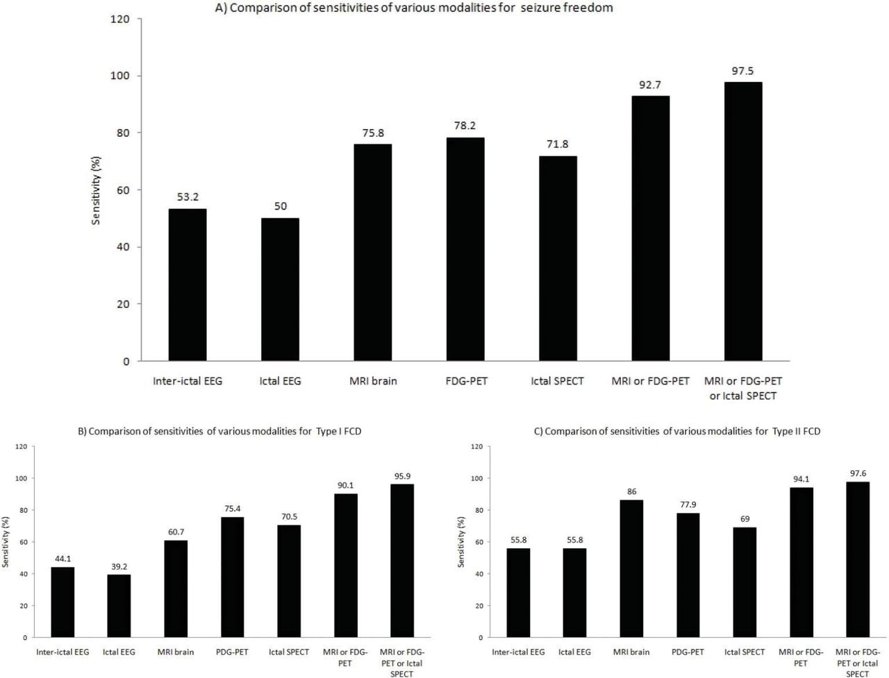
  • Fig 2.
    • Download figure
    • Open in new tab
    • Download powerpoint
    Fig 2.

    Comparison of sensitivities of various modalities for seizure freedom (A), type I FCD (B), and type II FCD (C).

Tables

  • Figures
    • View popup
    Table 1:

    Comparison of study variables for seizure freedom (n = 188)

    VariableSeizure-Free (n = 124)Persistent (n = 64)P Value
    Age at surgery (mean) (yr)18.68 ± 11.4314.66 ± 10.50.020
    Age of onset (mean) (yr)8.45 ± 8.186.91 ± 6.09.198
    Duration of epilepsy (mean) (%)10.72 ± 7.317.90 ± 6.56.012
    Women (%)56 (45.2%)36 (56.3%).168
    Febrile convulsions (%)15 (12.1%)8 (12.7%)1.000
    Multiple types of seizures (%)21 (16.9%)31 (48.4%)<.001
    Delayed development (%)23 (18.5%)24 (37.5%).007
    Daily seizures (%)63 (50.8%)43 (67.2%).043
    Presence of aura (%)35 (28.2%)8 (12.5%).017
    Regional spikes on interictal EEG (%)66 (54.1%)27 (42.2%).168
    Regional ictal EEG onset pattern (%)62 (50.0%)26 (46.2%).280
    Clear-cut FCD on MRI (%)94 (75.8%)42 (65.6%).169
    Focal FDG-PET pattern (%)97 (78.2%)47 (73.4%).472
    Focal ictal SPECT pattern (%)51 (71.8%)26 (66.7%).664
    Eloquent cortex location (%)16 (12.9%)20 (31.3%).003
    FCD type I (%)57 (47.5%)45 (73.8%).001
    FCD type II (%)63 (52.5%)16 (26.2%).001
    FCD type I and II (%)4 (3.2%)3 (4.6%).691
    Acute postoperative seizures (%)32 (25.8%)32 (50.0%).001
    Complete resection (%)105 (96.3%)28 (54.9%)<.001
    • View popup
    Table 2:

    Comparison of study variables for FCD types I and II (n = 181)

    VariableFCD Type I (n = 102)FCD Type II (n = 79)P Value
    Age at surgery (mean) (yr)16.54 ± 10.7618.11 ± 11.05.338
    Age of onset (mean) (yr)8.29 ± 7.887.76 ± 7.31.655
    Duration of epilepsy (mean) (%)9.08 ± 6.9110.82 ± 7.60.117
    Women (%)50 (49.0%)40 (50.6%).881
    Multilobar distribution of FCD (%)21 (20.6%)3 (3.8%)<.001
    Febrile convulsions (%)12 (11.9%)9 (11.4%)1.000
    Multiple types of seizures (%)31 (30.4%)19 (24.1%).403
    Delayed development (%)32 (31.4%)13 (16.5%).025
    Daily seizures (%)57 (55.9%)48 (60.8%).546
    Aura (%)18 (17.6%)23 (29.1%).076
    Regional ictal EEG onset pattern (%)40 (39.2%)45 (58.4%).024
    Clear-cut FCD on MRI (%)62 (60.8%)67 (84.8%)<.001
    Focal FDG-PET pattern (%)77 ( (75.5%)63 (79.7%).592
    Focal ictal SPECT pattern (%)48 (70.6%)29 (72.5%)1.000
    Eloquent cortex location (%)21 (20.6%)15 (19.0%).852
    Seizure-free (%)57 (55.8%)63 (79.7%).008
    Acute postoperative seizures (%)38 (37.3%)24 (30.4%).348
    Complete resection (%)63 (77.8%)65 (90.3%).048
PreviousNext
Back to top

In this issue

American Journal of Neuroradiology: 40 (5)
American Journal of Neuroradiology
Vol. 40, Issue 5
1 May 2019
  • Table of Contents
  • Index by author
  • Complete Issue (PDF)
Advertisement
Print
Download PDF
Email Article

Thank you for your interest in spreading the word on American Journal of Neuroradiology.

NOTE: We only request your email address so that the person you are recommending the page to knows that you wanted them to see it, and that it is not junk mail. We do not capture any email address.

Enter multiple addresses on separate lines or separate them with commas.
Focal Cortical Dysplasia and Refractory Epilepsy: Role of Multimodality Imaging and Outcome of Surgery
(Your Name) has sent you a message from American Journal of Neuroradiology
(Your Name) thought you would like to see the American Journal of Neuroradiology web site.
CAPTCHA
This question is for testing whether or not you are a human visitor and to prevent automated spam submissions.
Cite this article
S. Jayalakshmi, S.K. Nanda, S. Vooturi, R. Vadapalli, P. Sudhakar, S. Madigubba, M. Panigrahi
Focal Cortical Dysplasia and Refractory Epilepsy: Role of Multimodality Imaging and Outcome of Surgery
American Journal of Neuroradiology May 2019, 40 (5) 892-898; DOI: 10.3174/ajnr.A6041

Citation Manager Formats

  • BibTeX
  • Bookends
  • EasyBib
  • EndNote (tagged)
  • EndNote 8 (xml)
  • Medlars
  • Mendeley
  • Papers
  • RefWorks Tagged
  • Ref Manager
  • RIS
  • Zotero
0 Responses
Respond to this article
Share
Bookmark this article
Focal Cortical Dysplasia and Refractory Epilepsy: Role of Multimodality Imaging and Outcome of Surgery
S. Jayalakshmi, S.K. Nanda, S. Vooturi, R. Vadapalli, P. Sudhakar, S. Madigubba, M. Panigrahi
American Journal of Neuroradiology May 2019, 40 (5) 892-898; DOI: 10.3174/ajnr.A6041
del.icio.us logo Twitter logo Facebook logo Mendeley logo
  • Tweet Widget
  • Facebook Like
  • Google Plus One
Purchase

Jump to section

  • Article
    • Abstract
    • ABBREVIATIONS:
    • Materials and Methods
    • Results
    • Discussion
    • Conclusions
    • REFERENCES
  • Figures & Data
  • Info & Metrics
  • Responses
  • References
  • PDF

Related Articles

  • No related articles found.
  • PubMed
  • Google Scholar

Cited By...

  • Independent Evaluation of Deep Learning Models for Detecting Focal Cortical Dysplasia
  • Crossref
  • Google Scholar

This article has not yet been cited by articles in journals that are participating in Crossref Cited-by Linking.

More in this TOC Section

  • Early Ultrasonic Monitoring of Brain Growth and Later Neurodevelopmental Outcome in Very Preterm Infants
  • Diagnostic Value of Sylvian Fissure Hyperechogenicity in Fetal SAH
  • Feasibility and Added Value of Fetal DTI Tractography in the Evaluation of an Isolated Short Corpus Callosum: Preliminary Results
Show more Pediatrics

Similar Articles

Advertisement

Indexed Content

  • Current Issue
  • Accepted Manuscripts
  • Article Preview
  • Past Issues
  • Editorials
  • Editor's Choice
  • Fellows' Journal Club
  • Letters to the Editor
  • Video Articles

Cases

  • Case Collection
  • Archive - Case of the Week
  • Archive - Case of the Month
  • Archive - Classic Case

More from AJNR

  • Trainee Corner
  • Imaging Protocols
  • MRI Safety Corner
  • Book Reviews

Multimedia

  • AJNR Podcasts
  • AJNR Scantastics

Resources

  • Turnaround Time
  • Submit a Manuscript
  • Submit a Video Article
  • Submit an eLetter to the Editor/Response
  • Manuscript Submission Guidelines
  • Statistical Tips
  • Fast Publishing of Accepted Manuscripts
  • Graphical Abstract Preparation
  • Imaging Protocol Submission
  • Evidence-Based Medicine Level Guide
  • Publishing Checklists
  • Author Policies
  • Become a Reviewer/Academy of Reviewers
  • News and Updates

About Us

  • About AJNR
  • Editorial Board
  • Editorial Board Alumni
  • Alerts
  • Permissions
  • Not an AJNR Subscriber? Join Now
  • Advertise with Us
  • Librarian Resources
  • Feedback
  • Terms and Conditions
  • AJNR Editorial Board Alumni

American Society of Neuroradiology

  • Not an ASNR Member? Join Now

© 2025 by the American Society of Neuroradiology All rights, including for text and data mining, AI training, and similar technologies, are reserved.
Print ISSN: 0195-6108 Online ISSN: 1936-959X

Powered by HighWire