Advertisement

Main menu

  • Home
  • Content
    • Current Issue
    • Accepted Manuscripts
    • Article Preview
    • Past Issue Archive
    • Video Articles
    • AJNR Case Collection
    • Case of the Week Archive
    • Case of the Month Archive
    • Classic Case Archive
  • Special Collections
    • AJNR Awards
    • Low-Field MRI
    • Alzheimer Disease
    • ASNR Foundation Special Collection
    • Photon-Counting CT
    • View All
  • Multimedia
    • AJNR Podcasts
    • AJNR SCANtastic
    • Trainee Corner
    • MRI Safety Corner
    • Imaging Protocols
  • For Authors
    • Submit a Manuscript
    • Submit a Video Article
    • Submit an eLetter to the Editor/Response
    • Manuscript Submission Guidelines
    • Statistical Tips
    • Fast Publishing of Accepted Manuscripts
    • Graphical Abstract Preparation
    • Imaging Protocol Submission
    • Author Policies
  • About Us
    • About AJNR
    • Editorial Board
    • Editorial Board Alumni
  • More
    • Become a Reviewer/Academy of Reviewers
    • Subscribers
    • Permissions
    • Alerts
    • Feedback
    • Advertisers
    • ASNR Home

User menu

  • Alerts
  • Log in

Search

  • Advanced search
American Journal of Neuroradiology

American Journal of Neuroradiology

ASHNR American Society of Functional Neuroradiology ASHNR American Society of Pediatric Neuroradiology ASSR
  • Alerts
  • Log in

Advanced Search

  • Home
  • Content
    • Current Issue
    • Accepted Manuscripts
    • Article Preview
    • Past Issue Archive
    • Video Articles
    • AJNR Case Collection
    • Case of the Week Archive
    • Case of the Month Archive
    • Classic Case Archive
  • Special Collections
    • AJNR Awards
    • Low-Field MRI
    • Alzheimer Disease
    • ASNR Foundation Special Collection
    • Photon-Counting CT
    • View All
  • Multimedia
    • AJNR Podcasts
    • AJNR SCANtastic
    • Trainee Corner
    • MRI Safety Corner
    • Imaging Protocols
  • For Authors
    • Submit a Manuscript
    • Submit a Video Article
    • Submit an eLetter to the Editor/Response
    • Manuscript Submission Guidelines
    • Statistical Tips
    • Fast Publishing of Accepted Manuscripts
    • Graphical Abstract Preparation
    • Imaging Protocol Submission
    • Author Policies
  • About Us
    • About AJNR
    • Editorial Board
    • Editorial Board Alumni
  • More
    • Become a Reviewer/Academy of Reviewers
    • Subscribers
    • Permissions
    • Alerts
    • Feedback
    • Advertisers
    • ASNR Home
  • Follow AJNR on Twitter
  • Visit AJNR on Facebook
  • Follow AJNR on Instagram
  • Join AJNR on LinkedIn
  • RSS Feeds

AJNR Awards, New Junior Editors, and more. Read the latest AJNR updates

Research ArticlePediatrics
Open Access

Intravoxel Incoherent Motion MR Imaging of Pediatric Intracranial Tumors: Correlation with Histology and Diagnostic Utility

K. Kikuchi, A. Hiwatashi, O. Togao, K. Yamashita, R. Kamei, D. Momosaka, N. Hata, K. Iihara, S.O. Suzuki, T. Iwaki and H. Honda
American Journal of Neuroradiology May 2019, 40 (5) 878-884; DOI: https://doi.org/10.3174/ajnr.A6052
K. Kikuchi
aFrom the Departments of Clinical Radiology (K.K., A.H., O.T., K.Y., R.K., D.M., H.H.)
  • Find this author on Google Scholar
  • Find this author on PubMed
  • Search for this author on this site
  • ORCID record for K. Kikuchi
A. Hiwatashi
aFrom the Departments of Clinical Radiology (K.K., A.H., O.T., K.Y., R.K., D.M., H.H.)
  • Find this author on Google Scholar
  • Find this author on PubMed
  • Search for this author on this site
  • ORCID record for A. Hiwatashi
O. Togao
aFrom the Departments of Clinical Radiology (K.K., A.H., O.T., K.Y., R.K., D.M., H.H.)
  • Find this author on Google Scholar
  • Find this author on PubMed
  • Search for this author on this site
  • ORCID record for O. Togao
K. Yamashita
aFrom the Departments of Clinical Radiology (K.K., A.H., O.T., K.Y., R.K., D.M., H.H.)
  • Find this author on Google Scholar
  • Find this author on PubMed
  • Search for this author on this site
  • ORCID record for K. Yamashita
R. Kamei
aFrom the Departments of Clinical Radiology (K.K., A.H., O.T., K.Y., R.K., D.M., H.H.)
  • Find this author on Google Scholar
  • Find this author on PubMed
  • Search for this author on this site
  • ORCID record for R. Kamei
D. Momosaka
aFrom the Departments of Clinical Radiology (K.K., A.H., O.T., K.Y., R.K., D.M., H.H.)
  • Find this author on Google Scholar
  • Find this author on PubMed
  • Search for this author on this site
  • ORCID record for D. Momosaka
N. Hata
bNeurosurgery (N.H., K.I.)
  • Find this author on Google Scholar
  • Find this author on PubMed
  • Search for this author on this site
  • ORCID record for N. Hata
K. Iihara
bNeurosurgery (N.H., K.I.)
  • Find this author on Google Scholar
  • Find this author on PubMed
  • Search for this author on this site
  • ORCID record for K. Iihara
S.O. Suzuki
cNeuropathology (S.O.S., T.I.), Graduate School of Medical Sciences, Kyushu University, Fukuoka, Japan.
  • Find this author on Google Scholar
  • Find this author on PubMed
  • Search for this author on this site
  • ORCID record for S.O. Suzuki
T. Iwaki
cNeuropathology (S.O.S., T.I.), Graduate School of Medical Sciences, Kyushu University, Fukuoka, Japan.
  • Find this author on Google Scholar
  • Find this author on PubMed
  • Search for this author on this site
  • ORCID record for T. Iwaki
H. Honda
aFrom the Departments of Clinical Radiology (K.K., A.H., O.T., K.Y., R.K., D.M., H.H.)
  • Find this author on Google Scholar
  • Find this author on PubMed
  • Search for this author on this site
  • ORCID record for H. Honda
  • Article
  • Figures & Data
  • Supplemental
  • Info & Metrics
  • Responses
  • References
  • PDF
Loading

Article Figures & Data

Figures

  • Tables
  • Fig 1.
    • Download figure
    • Open in new tab
    • Download powerpoint
    Fig 1.

    Comparison of the IVIM parameters of high- (WHO grades III and IV) and low-grade (WHO grades I and II) tumors. D and ADC were lower in high-grade than in low-grade tumors (D, 0.85 ± 0.40 versus 1.53 ± 0.21 × 10⁻³ mm2/s, P = .0003; ADC, 1.04 ± 0.33 versus 1.60 ± 0.21 × 10⁻³ mm2/s, P = .0007; A). D was significantly lower than the ADC in high- (P = .0010) and low-grade (P = .0004; A) tumors. D* showed wide variability and no significant differences between the high- and low-grade groups (P = .8337; B). The f was higher in high- than in low-grade tumors (21.7 ± 8.2% versus 7.6 ± 4.3%, P = .0003; C).
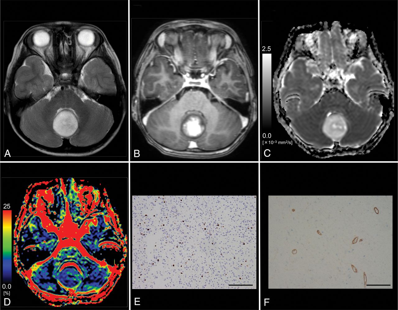
  • Fig 2.
    • Download figure
    • Open in new tab
    • Download powerpoint
    Fig 2.

    Images from an 11-year-old boy with histologically defined classic medulloblastoma (WHO grade IV). A, T2WI shows a heterogeneous, hyperintense mass in the vermis. B, Contrast-enhanced T1WI shows heterogeneous enhancement of the tumor. C, The D map shows the low D value (0.53 × 10⁻³ mm2/s) of the lesion, which is lower than the cutoff value (≤1.15 × 10⁻³ mm2/s; Table 3). D, The f map shows the f value (27.1%) of the lesion, which is higher than the cutoff value (>14.1%; Table 3). E, MIB-1 staining reveals a high MIB-1 index (72.6%). F, Immunohistochemical staining for anti-CD31 shows a high MVD (10.2%). Bar = 100 μm.

  • Fig 3.
    • Download figure
    • Open in new tab
    • Download powerpoint
    Fig 3.

    Images from a 6-year-old boy with pilocytic astrocytoma (WHO grade I). A, T2WI shows a homogeneous, hyperintense mass in the vermis. B, Contrast-enhanced T1WI shows mild, heterogeneous enhancement of the tumor. C, The D map shows the high D value (1.72 × 10⁻³ mm2/s) of the lesion, which is higher than the cutoff value (≤1.15 × 10⁻³ mm2/s; Table 3). D, The f map shows the low f value (4.2%) of the lesion, which is lower than the cutoff value (>14.1%; Table 3). E, MIB-1 staining reveals a low MIB-1 index (6.0%). F, Immunohistochemical staining for anti-CD31 reveals a low MVD (3.2%). Bar = 100 μm.

Tables

  • Figures
    • View popup
    Table 1:

    Patient demographics and tumor characteristics

    No.Age, SexHistologyIVIM ParametersADC (× 10−3 mm2/s)
    Pathologic DiagnosisWHO GradeMIB-1 (%)MVDD (× 10−3 mm2/s)f (%)D* (× 10−3 mm2/s)
    16 yr, MDiffuse midline glioma, H3 K27M-mutantIV59.23.11.5016.45.61.61
    24 yr, MMedulloblastoma, classic, histologically definedIV15.634.70.3037.713.70.56
    311 yr, MMedulloblastoma, classic, histologically definedIV72.610.20.5327.130.20.87
    43 yr, MAtypical teratoid/rhabdoid tumorIV50.011.00.8614.19.00.98
    52 yr, FAnaplastic ependymomaIII23.212.50.8719.318.41.02
    63 yr, MAnaplastic ependymomaIII19.64.31.1516.85.51.30
    76 yr, FAnaplastic ependymomaIII58.410.10.7120.741.70.96
    812 yr, MDiffuse astrocytoma, IDH wild-typeII2.12.11.228.433.31.29
    91 yr, MPilocytic astrocytomaI4.45.81.453.55.51.50
    103 yr, FPilocytic astrocytomaI6.74.01.827.210.21.88
    113 yr, MPilocytic astrocytomaI8.04.71.5210.048.31.64
    126 yr, MPilocytic astrocytomaI6.03.21.724.258.41.78
    1311 yr, FPilocytic astrocytomaI4.05.61.555.512.81.61
    142 yr, MSubependymomaI0.11.91.585.111.31.60
    152 mo, FChoroid plexus papillomaI0.113.41.4717.411.61.62
    1610 yr, MDysembryoplastic neuroepithelial tumorI2.71.31.7610.94.31.82
    1715 yr, FGangliogliomaI1.01.11.213.622.41.24
    • Note:—IDH indicates isocitrate dehydrogenase.

    • View popup
    Table 2:

    Comparison of parameters between high- and low-grade tumorsa

    ParametersHigh-Grade TumorLow-Grade TumorP Value
    D (× 10−3 mm2/s)0.30–1.50, 0.86, 0.85 ± 0.401.21–1.82, 1.54, 1.53 ± 0.21.0003b
    ADC (× 10−3 mm2/s)0.56–1.61, 0.98, 1.04 ± 0.331.24–1.88, 1.62, 1.60 ± 0.21.0007b
    D* (× 10−3 mm2/s)5.5–41.7, 13.7, 17.7 ± 13.74.3–58.4, 12.8, 21.8 ± 18.8.8337c
    f (%)14.1–37.7, 19.3, 21.7 ± 8.23.5–17.4, 6.4, 7.6 ± 4.3.0003b
    MVD (%)3.1–34.7, 10.2, 12.3 ± 10.51.1–13.4, 3.6, 4.3 ± 3.6.0431c
    MIB-1 (%)15.6–72.6, 50.0, 42.7 ± 22.80.1–8.0, 3.4, 3.5 ± 2.8<.0001b
    • ↵a Data are expressed as range, median, mean.

    • ↵b Unpaired test.

    • ↵c Mann-Whitney U test.

    • View popup
    Table 3:

    Diagnostic performance of parameters in differentiating high- and low-grade tumors

    ParametersCutoff ValueSensitivity (%)Specificity (%)AUC
    D (× 10−3 mm2/s)≤1.1585.785.70.943
    D* (× 10−3 mm2/s)>9.042.922.90.536
    f (%)>14.110090.00.957
    ADC (× 10−3 mm2/s)≤1.0271.471.40.907
    D + f≤1.50, >16.410090.00.986
    • Note:—AUC indicates area under the curve.

PreviousNext
Back to top

In this issue

American Journal of Neuroradiology: 40 (5)
American Journal of Neuroradiology
Vol. 40, Issue 5
1 May 2019
  • Table of Contents
  • Index by author
  • Complete Issue (PDF)
Advertisement
Print
Download PDF
Email Article

Thank you for your interest in spreading the word on American Journal of Neuroradiology.

NOTE: We only request your email address so that the person you are recommending the page to knows that you wanted them to see it, and that it is not junk mail. We do not capture any email address.

Enter multiple addresses on separate lines or separate them with commas.
Intravoxel Incoherent Motion MR Imaging of Pediatric Intracranial Tumors: Correlation with Histology and Diagnostic Utility
(Your Name) has sent you a message from American Journal of Neuroradiology
(Your Name) thought you would like to see the American Journal of Neuroradiology web site.
CAPTCHA
This question is for testing whether or not you are a human visitor and to prevent automated spam submissions.
Cite this article
K. Kikuchi, A. Hiwatashi, O. Togao, K. Yamashita, R. Kamei, D. Momosaka, N. Hata, K. Iihara, S.O. Suzuki, T. Iwaki, H. Honda
Intravoxel Incoherent Motion MR Imaging of Pediatric Intracranial Tumors: Correlation with Histology and Diagnostic Utility
American Journal of Neuroradiology May 2019, 40 (5) 878-884; DOI: 10.3174/ajnr.A6052

Citation Manager Formats

  • BibTeX
  • Bookends
  • EasyBib
  • EndNote (tagged)
  • EndNote 8 (xml)
  • Medlars
  • Mendeley
  • Papers
  • RefWorks Tagged
  • Ref Manager
  • RIS
  • Zotero
0 Responses
Respond to this article
Share
Bookmark this article
Intravoxel Incoherent Motion MR Imaging of Pediatric Intracranial Tumors: Correlation with Histology and Diagnostic Utility
K. Kikuchi, A. Hiwatashi, O. Togao, K. Yamashita, R. Kamei, D. Momosaka, N. Hata, K. Iihara, S.O. Suzuki, T. Iwaki, H. Honda
American Journal of Neuroradiology May 2019, 40 (5) 878-884; DOI: 10.3174/ajnr.A6052
del.icio.us logo Twitter logo Facebook logo Mendeley logo
  • Tweet Widget
  • Facebook Like
  • Google Plus One
Purchase

Jump to section

  • Article
    • Abstract
    • ABBREVIATIONS:
    • Materials and Methods
    • Results
    • Discussion
    • Conclusions
    • Footnotes
    • REFERENCES
  • Figures & Data
  • Supplemental
  • Info & Metrics
  • Responses
  • References
  • PDF

Related Articles

  • PubMed
  • Google Scholar

Cited By...

  • Perfusion Showdown: Comparison of Multiple MRI Perfusion Techniques in the Grading of Pediatric Brain Tumors
  • Interobserver Reliability on Intravoxel Incoherent Motion Imaging in Patients with Acute Ischemic Stroke
  • Grading of Pediatric Intracranial Tumors: Are Intravoxel Incoherent Motion and Diffusional Kurtosis Imaging Superior to Conventional DWI?
  • Radiomics of Pediatric Low-Grade Gliomas: Toward a Pretherapeutic Differentiation of BRAF-Mutated and BRAF-Fused Tumors
  • Crossref
  • Google Scholar

This article has not yet been cited by articles in journals that are participating in Crossref Cited-by Linking.

More in this TOC Section

  • Early Ultrasonic Monitoring of Brain Growth and Later Neurodevelopmental Outcome in Very Preterm Infants
  • Diagnostic Value of Sylvian Fissure Hyperechogenicity in Fetal SAH
  • Feasibility and Added Value of Fetal DTI Tractography in the Evaluation of an Isolated Short Corpus Callosum: Preliminary Results
Show more Pediatrics

Similar Articles

Advertisement

Indexed Content

  • Current Issue
  • Accepted Manuscripts
  • Article Preview
  • Past Issues
  • Editorials
  • Editor's Choice
  • Fellows' Journal Club
  • Letters to the Editor
  • Video Articles

Cases

  • Case Collection
  • Archive - Case of the Week
  • Archive - Case of the Month
  • Archive - Classic Case

More from AJNR

  • Trainee Corner
  • Imaging Protocols
  • MRI Safety Corner
  • Book Reviews

Multimedia

  • AJNR Podcasts
  • AJNR Scantastics

Resources

  • Turnaround Time
  • Submit a Manuscript
  • Submit a Video Article
  • Submit an eLetter to the Editor/Response
  • Manuscript Submission Guidelines
  • Statistical Tips
  • Fast Publishing of Accepted Manuscripts
  • Graphical Abstract Preparation
  • Imaging Protocol Submission
  • Evidence-Based Medicine Level Guide
  • Publishing Checklists
  • Author Policies
  • Become a Reviewer/Academy of Reviewers
  • News and Updates

About Us

  • About AJNR
  • Editorial Board
  • Editorial Board Alumni
  • Alerts
  • Permissions
  • Not an AJNR Subscriber? Join Now
  • Advertise with Us
  • Librarian Resources
  • Feedback
  • Terms and Conditions
  • AJNR Editorial Board Alumni

American Society of Neuroradiology

  • Not an ASNR Member? Join Now

© 2025 by the American Society of Neuroradiology All rights, including for text and data mining, AI training, and similar technologies, are reserved.
Print ISSN: 0195-6108 Online ISSN: 1936-959X

Powered by HighWire