
Article Figures & Data
Figures
- Fig 1.
Patient flow chart.
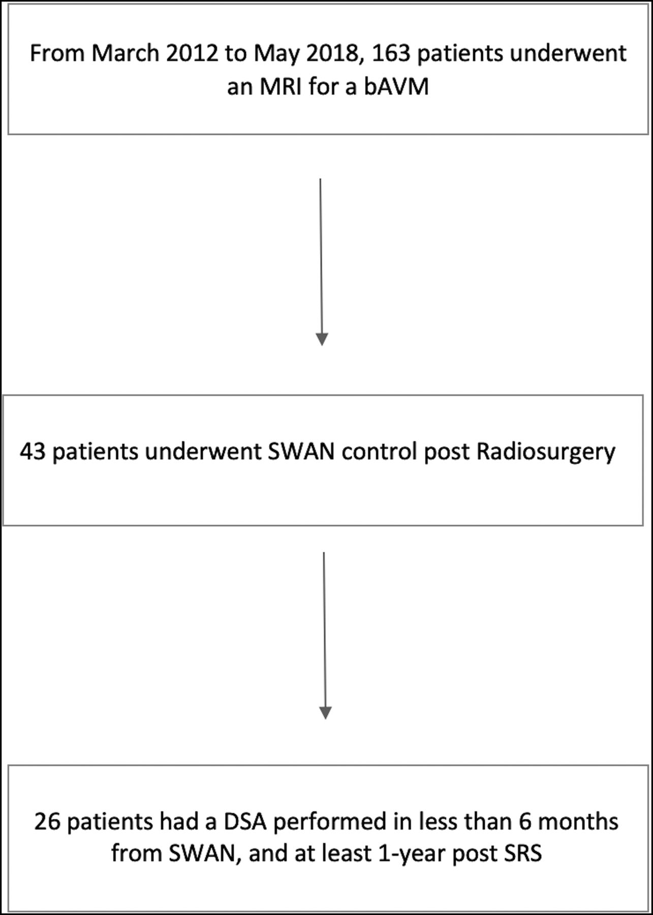
- Fig 2.
A 42-year-old man with a left cerebellar AVM, partially embolized with glue. A, SWAN imaging 3 years after SRS shows hyperintense vessels (white arrow) in the posterior part of the nidus that correspond to a nidal remnant confirmed by DSA (black arrow, B).
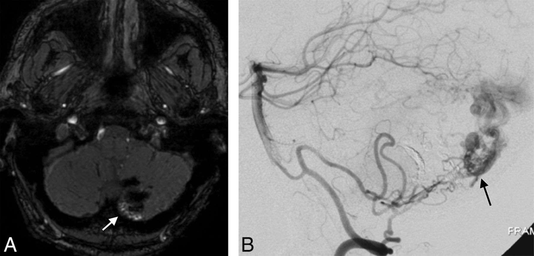
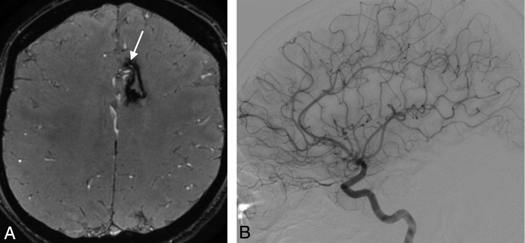
- Fig 3.
A 19-year-old woman with a right posterior frontal AVM with hemorrhagic presentation, partially embolized with glue. SWAN imaging 2.5 years after SRS shows the complete occlusion of the nidus (white arrow) confirmed by DSA (not shown).
- Fig 4.
A 32-year-old man with a right parietal AVM with hemorrhagic presentation, partially embolized with glue. SWAN imaging 3.5 years after SRS (white arrow, A) fails to show a very small residual nidus that was depicted by DSA (black arrow, B).
- Fig 5.
A 60-year-old man with a left parietal AVM with hemorrhagic presentation that was partially embolized with glue. A, SWAN imaging 4 years after SRS shows an amorphous area of hyperintensity within the nidus (white arrow) that was diagnosed as a nidus remnant, but the DSA findings were negative. B, CT shows extensive calcification (white arrow) inside the AVM scar.
- Fig 6.
A 19-year-old man with a left frontal AVM with hemorrhagic presentation that was partially embolized with glue. A, SWAN imaging 5 years after SRS shows hyperintense vessels (white arrow) near the AVM that were mistakenly diagnosed as a nidus remnant. B, Digital subtraction angiography findings were negative.


Tables
Characteristics Age (yr) 33 (22–42) Sex, male 17 (65.4%) bAVM location Supratentorial 24 (92.3%) Infratentorial 2 (7.7%) Spetzler-Martin grade I (I–II) Presentation Hemorrhage 20 (76.9%) Seizure 4 (15.4%) Headache 5 (19.2%) Neurologic symptoms 1 (3.8%) Type of treatment Embolization then radiosurgery 22 (84.6%) Radiosurgery 2 (7.7%) Surgery then embolization then radiosurgery 2 (7.7%) Time intervals Delay between last treatment and SWAN (mo)b 34 (27.7–46.8) Delay between SWAN and DSA (days)b 3 (3–65) DSA SWAN Reader 1 SWAN Reader 2 Consensus Reading Patent Obliterated Patent Obliterated Patent Obliterated Patent (n = 14) 11 3 11 2 12 2 Obliterated (n = 14) 5 9 3 12 2 12 Sensitivity (%) 68.7 78.6 85.7 Specificity (%) 75 85.7 85.7 PPV (%) 78.6 84.6 85.7 NPV (%) 64.3 80 85.7 Note:—PPV indicates positive predictive value; NPV, negative predictive value.
↵a Prevalence of nidus remnant after SRS for bAVM.


