Advertisement

Main menu

  • Home
  • Content
    • Current Issue
    • Accepted Manuscripts
    • Article Preview
    • Past Issue Archive
    • Video Articles
    • AJNR Case Collection
    • Case of the Week Archive
    • Case of the Month Archive
    • Classic Case Archive
  • Special Collections
    • Advancing NeuroMRI with High-Relaxivity Contrast Agents
    • AJNR Awards
    • Low-Field MRI
    • Alzheimer Disease
    • ASNR Foundation Special Collection
    • Photon-Counting CT
    • View All
  • Multimedia
    • AJNR Podcasts
    • AJNR SCANtastic
    • Trainee Corner
    • MRI Safety Corner
    • Imaging Protocols
  • For Authors
    • Submit a Manuscript
    • Submit a Video Article
    • Submit an eLetter to the Editor/Response
    • Manuscript Submission Guidelines
    • Statistical Tips
    • Fast Publishing of Accepted Manuscripts
    • Graphical Abstract Preparation
    • Imaging Protocol Submission
    • Author Policies
  • About Us
    • About AJNR
    • Editorial Board
    • Editorial Board Alumni
  • More
    • Become a Reviewer/Academy of Reviewers
    • Subscribers
    • Permissions
    • Alerts
    • Feedback
    • Advertisers
    • ASNR Home

User menu

  • Alerts
  • Log in

Search

  • Advanced search
American Journal of Neuroradiology

American Journal of Neuroradiology

ASHNR American Society of Functional Neuroradiology ASHNR American Society of Pediatric Neuroradiology ASSR
  • Alerts
  • Log in

Advanced Search

  • Home
  • Content
    • Current Issue
    • Accepted Manuscripts
    • Article Preview
    • Past Issue Archive
    • Video Articles
    • AJNR Case Collection
    • Case of the Week Archive
    • Case of the Month Archive
    • Classic Case Archive
  • Special Collections
    • Advancing NeuroMRI with High-Relaxivity Contrast Agents
    • AJNR Awards
    • Low-Field MRI
    • Alzheimer Disease
    • ASNR Foundation Special Collection
    • Photon-Counting CT
    • View All
  • Multimedia
    • AJNR Podcasts
    • AJNR SCANtastic
    • Trainee Corner
    • MRI Safety Corner
    • Imaging Protocols
  • For Authors
    • Submit a Manuscript
    • Submit a Video Article
    • Submit an eLetter to the Editor/Response
    • Manuscript Submission Guidelines
    • Statistical Tips
    • Fast Publishing of Accepted Manuscripts
    • Graphical Abstract Preparation
    • Imaging Protocol Submission
    • Author Policies
  • About Us
    • About AJNR
    • Editorial Board
    • Editorial Board Alumni
  • More
    • Become a Reviewer/Academy of Reviewers
    • Subscribers
    • Permissions
    • Alerts
    • Feedback
    • Advertisers
    • ASNR Home
  • Follow AJNR on Twitter
  • Visit AJNR on Facebook
  • Follow AJNR on Instagram
  • Join AJNR on LinkedIn
  • RSS Feeds

AJNR Awards, New Junior Editors, and more. Read the latest AJNR updates


Improved Turnaround Times | Median time to first decision: 12 days

Research ArticleNeurointervention

Morphologic Change of Flow-Related Aneurysms in Brain Arteriovenous Malformations after Stereotactic Radiosurgery

Y.-S. Tsuei, C.-B. Luo, L.-Y. Fay, H.-C. Yang, W.-Y. Guo, H.-M. Wu, W.-Y. Chung and M.M.H. Teng
American Journal of Neuroradiology April 2019, 40 (4) 675-680; DOI: https://doi.org/10.3174/ajnr.A6018
Y.-S. Tsuei
aFrom the Department of Neurosurgery (Y.-S.T.), Taichung Veterans General Hospital, Taichung, Taiwan
bDepartments of Neurosugery (Y.-S.T.)
  • Find this author on Google Scholar
  • Find this author on PubMed
  • Search for this author on this site
  • ORCID record for Y.-S. Tsuei
C.-B. Luo
cRadiology (C.-B.L.), Tri-Service General Hospital, National Defenses Medical Center, Taipei, Taiwan
dDepartments of Radiology (C.-B.L., W.-Y.G., H.-M.W., M.M.H.T.)
  • Find this author on Google Scholar
  • Find this author on PubMed
  • Search for this author on this site
  • ORCID record for C.-B. Luo
L.-Y. Fay
eNeurosurgery (L.-Y.F., H.-C.Y., W.-Y.C.), Taipei Veterans General Hospital and National Yang-Ming University, School of Medicine, Taipei, Taiwan
  • Find this author on Google Scholar
  • Find this author on PubMed
  • Search for this author on this site
  • ORCID record for L.-Y. Fay
H.-C. Yang
eNeurosurgery (L.-Y.F., H.-C.Y., W.-Y.C.), Taipei Veterans General Hospital and National Yang-Ming University, School of Medicine, Taipei, Taiwan
  • Find this author on Google Scholar
  • Find this author on PubMed
  • Search for this author on this site
  • ORCID record for H.-C. Yang
W.-Y. Guo
dDepartments of Radiology (C.-B.L., W.-Y.G., H.-M.W., M.M.H.T.)
  • Find this author on Google Scholar
  • Find this author on PubMed
  • Search for this author on this site
  • ORCID record for W.-Y. Guo
H.-M. Wu
dDepartments of Radiology (C.-B.L., W.-Y.G., H.-M.W., M.M.H.T.)
  • Find this author on Google Scholar
  • Find this author on PubMed
  • Search for this author on this site
  • ORCID record for H.-M. Wu
W.-Y. Chung
eNeurosurgery (L.-Y.F., H.-C.Y., W.-Y.C.), Taipei Veterans General Hospital and National Yang-Ming University, School of Medicine, Taipei, Taiwan
  • Find this author on Google Scholar
  • Find this author on PubMed
  • Search for this author on this site
  • ORCID record for W.-Y. Chung
M.M.H. Teng
dDepartments of Radiology (C.-B.L., W.-Y.G., H.-M.W., M.M.H.T.)
fDepartment of Radiology (M.M.H.T.), Cheng-Hsin General Hospital, Taipei, Taiwan.
  • Find this author on Google Scholar
  • Find this author on PubMed
  • Search for this author on this site
  • ORCID record for M.M.H. Teng
  • Article
  • Figures & Data
  • Info & Metrics
  • Responses
  • References
  • PDF
Loading

Article Figures & Data

Figures

  • Tables
  • Fig 1.
    • Download figure
    • Open in new tab
    • Download powerpoint
    Fig 1.

    Left, The patient had a right parieto-occipital brain arteriovenous malformation with a small flow-related aneurysm (arrow) at the right medial paraclinoid internal carotid artery. Right, Conventional angiography at 54 months after stereotactic gamma knife surgery demonstrates almost total obliteration of the BAVM but no obvious morphologic change in the proximal FA.

  • Fig 2.
    • Download figure
    • Open in new tab
    • Download powerpoint
    Fig 2.

    Left, The patient had a right frontotemporal BAVM and small proximal FAs at the M1 segment of the right middle cerebral artery (arrows). Right, Conventional angiography at 38 months after GKS demonstrates almost total regression of the proximal FAs.
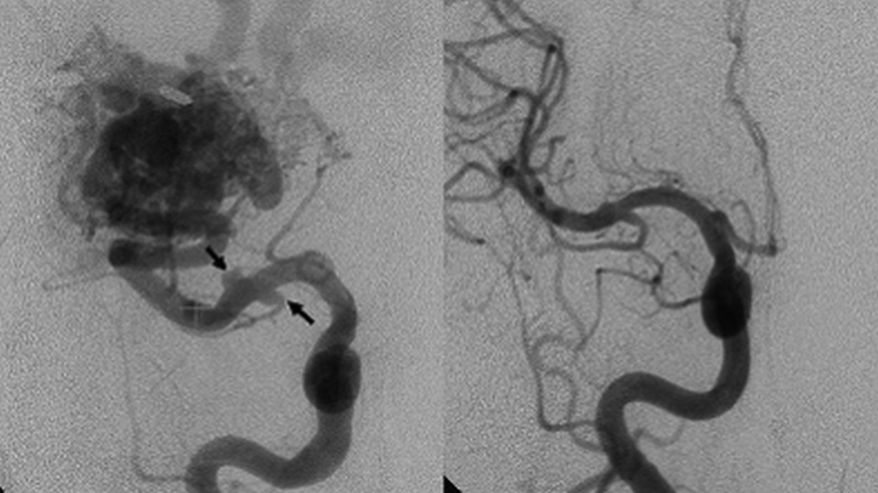
  • Fig 3.
    • Download figure
    • Open in new tab
    • Download powerpoint
    Fig 3.

    Left, The patient had a right inferior temporal BAVM and 2 distal FAs at the M2 segment of the right middle cerebral artery (arrows). Right, Conventional angiography at 37 months after GKS demonstrates total obliteration of the distal FAs with normalization of blood flow through the feeders to the BAVM and FAs.

Tables

  • Figures
    • View popup
    Table 1:

    Demographics of patients with BAVMs enrolled and characteristics of FAs

    Value
    No. of eligible patients936
    No. of excluded patients113
        No. of patients with <50% obliteration of BAVM39
        No. of patients without DSA follow-up57
        No. of interventions of FAs17
    No. of patients enrolled823
    No. of patients with FAs enrolled72 (8.8%)
    Mean age (yr)43 (range, 18–72)
    Sex (No.)
        Female29 (40%)
        Male43 (60%)
    No., location, and size of FAs (mm)
        Total No. of FAs (mean size)111 (4.1)
        Mean size of proximal FAs4.3
        Mean size of distal FA3.7
    No. of FAs per patient1.5
    No. of FAs in supratentorium98
        No. of FAs in ICA42
        No. of FAs in proximal/distal MCA16/14
        No. of FAs in proximal/distal ACA9/17
    No. of FAs in infratntorium13
        No. of proximal FAs5
        No. of distal FAs8
    • View popup
    Table 2.

    Demographics of 111 FAs in 72 patients with BAVMs after radiosurgerya

    ParametersStable FAs (n = 60)Regressive FAs (n = 51)P Value
    Sex.70
        Male (n = 43)35 (57)27 (44)
        Female (n = 29)25 (51)24 (49)
    Mean age of patients (yr)41 ± 1044 ± 15.21
    Mean volume of BAVM (mL)36 ± 940 ± 12.048b
    Mean size of FAs (mm)4.5 ± 0.93.6 ± 0.5.0001b
    No. of ruptured/unruptured BAVMs.85
        Ruptured (n = 31)26 (55)21 (45)
        Unruptured (n = 41)34 (53)30 (47)
    No. of complete/incomplete obliteration of BAVMs.29
        Complete (n = 52)41 (51)40 (49)
        Incomplete (n = 20)19 (63)11 (37)
    • ↵a Data are means. Numbers in parentheses do not add up to 100% due to rounding. Data in parentheses are percentages.

    • ↵b P < .05, indicates statistical significance.

    • View popup
    Table 3:

    Location and morphologic change of FAs of BAVMsa

    Location of FANo. of FAsNo. of Stable FAsNo. of Regressive FAs/Total RegressionP Value of Regression in Proximal and Distal FAs
    Supratentorium9856 (57)42 (43)/20 (20)
        ACA26
            Proximal (%)95 (56)4 (44)/1 (11).19
            Distal (%)174 (24)13 (76)/8 (47)
        MCA30
            Proximal (%)1610 (63)6 (38)/2 (13).01
            Distal (%)142 (14)12 (86)/9 (62)
        ICA (proximal)4235 (83)7 (17)/0 (0)NA
    Infratentorium13
        Proximal43 (75)1 (25)/1 (25).052
        Distal91 (11)8 (89)/5 (56)
    Overall11160 (54)51 (46)/26 (23)
            Proximal (%)7153 (75)18 (25)/4 (6)<.001b
            Distal (%)407 (18)33 (83)/22 (55)
    • Note:—NA indicates not available.

    • ↵a Data in parentheses are percentages. Numbers in parentheses do not add up to 100% due to rounding.

    • ↵b P < .05, indicates statistical significance.

    • View popup
    Table 4:

    Size and morphologic change of FAs of BAVMsa

    Location of FANo. of FAsNo. of Stable FAsNo. of Regressive FAs/Total RegressionP Value of Regression in Small FAs (<5 mm)
    Supratentorium9856 (57)42 (43)/20 (20)
        ACA26
            <5 mm (%)269 (35)17 (65)/13 (50)NA
            ≥5 mm (%)00 (0)0 (0)
        MCA30
            <5 mm (%)259 (36)16 (64)/9 (36).36
            ≥5 mm (%)53 (60)2 (40)/0 (0)
        ICA42
            <5 mm (%)3629 (80)7 (19)/0 (0).57
            ≥5 mm (%)66 (100)0 (0)
    Infratentorium133 (25)9 (75)/2 (17).31
         <5 mm (%)12
        ≥5 mm (%)11 (100)0 (0)
    Overall11160 (54)51 (46)/26 (23).036b
         <5 mm (%)9950 (51)49 (49)/26 (26)
        ≥5 mm (%)1210 (83)2 (17)/0 (0)
    • ↵a Data in parentheses are percentages. Numbers in parentheses do not add up to 100% due to rounding.

    • ↵b P < .05, indicates statistical significance.

PreviousNext
Back to top

In this issue

American Journal of Neuroradiology: 40 (4)
American Journal of Neuroradiology
Vol. 40, Issue 4
1 Apr 2019
  • Table of Contents
  • Index by author
  • Complete Issue (PDF)
Advertisement
Print
Download PDF
Email Article

Thank you for your interest in spreading the word on American Journal of Neuroradiology.

NOTE: We only request your email address so that the person you are recommending the page to knows that you wanted them to see it, and that it is not junk mail. We do not capture any email address.

Enter multiple addresses on separate lines or separate them with commas.
Morphologic Change of Flow-Related Aneurysms in Brain Arteriovenous Malformations after Stereotactic Radiosurgery
(Your Name) has sent you a message from American Journal of Neuroradiology
(Your Name) thought you would like to see the American Journal of Neuroradiology web site.
CAPTCHA
This question is for testing whether or not you are a human visitor and to prevent automated spam submissions.
Cite this article
Y.-S. Tsuei, C.-B. Luo, L.-Y. Fay, H.-C. Yang, W.-Y. Guo, H.-M. Wu, W.-Y. Chung, M.M.H. Teng
Morphologic Change of Flow-Related Aneurysms in Brain Arteriovenous Malformations after Stereotactic Radiosurgery
American Journal of Neuroradiology Apr 2019, 40 (4) 675-680; DOI: 10.3174/ajnr.A6018

Citation Manager Formats

  • BibTeX
  • Bookends
  • EasyBib
  • EndNote (tagged)
  • EndNote 8 (xml)
  • Medlars
  • Mendeley
  • Papers
  • RefWorks Tagged
  • Ref Manager
  • RIS
  • Zotero
0 Responses
Respond to this article
Share
Bookmark this article
Morphologic Change of Flow-Related Aneurysms in Brain Arteriovenous Malformations after Stereotactic Radiosurgery
Y.-S. Tsuei, C.-B. Luo, L.-Y. Fay, H.-C. Yang, W.-Y. Guo, H.-M. Wu, W.-Y. Chung, M.M.H. Teng
American Journal of Neuroradiology Apr 2019, 40 (4) 675-680; DOI: 10.3174/ajnr.A6018
del.icio.us logo Twitter logo Facebook logo Mendeley logo
  • Tweet Widget
  • Facebook Like
  • Google Plus One
Purchase

Jump to section

  • Article
    • Abstract
    • ABBREVIATIONS:
    • Materials and Methods
    • Results
    • Discussion
    • Conclusions
    • Footnotes
    • REFERENCES
  • Figures & Data
  • Info & Metrics
  • Responses
  • References
  • PDF

Related Articles

  • No related articles found.
  • PubMed
  • Google Scholar

Cited By...

  • No citing articles found.
  • Crossref
  • Google Scholar

This article has not yet been cited by articles in journals that are participating in Crossref Cited-by Linking.

More in this TOC Section

  • Prophylactic Endovascular Treatments for CBS
  • Smoking & Aneurysm Outcomes: A Meta-Analysis
  • “Buddy-wire anchoring” technique for TVE
Show more NEUROINTERVENTION

Similar Articles

Advertisement

Indexed Content

  • Current Issue
  • Accepted Manuscripts
  • Article Preview
  • Past Issues
  • Editorials
  • Editor's Choice
  • Fellows' Journal Club
  • Letters to the Editor
  • Video Articles

Cases

  • Case Collection
  • Archive - Case of the Week
  • Archive - Case of the Month
  • Archive - Classic Case

More from AJNR

  • Trainee Corner
  • Imaging Protocols
  • MRI Safety Corner
  • Book Reviews

Multimedia

  • AJNR Podcasts
  • AJNR Scantastics

Resources

  • Turnaround Time
  • Submit a Manuscript
  • Submit a Video Article
  • Submit an eLetter to the Editor/Response
  • Manuscript Submission Guidelines
  • Statistical Tips
  • Fast Publishing of Accepted Manuscripts
  • Graphical Abstract Preparation
  • Imaging Protocol Submission
  • Evidence-Based Medicine Level Guide
  • Publishing Checklists
  • Author Policies
  • Become a Reviewer/Academy of Reviewers
  • News and Updates

About Us

  • About AJNR
  • Editorial Board
  • Editorial Board Alumni
  • Alerts
  • Permissions
  • Not an AJNR Subscriber? Join Now
  • Advertise with Us
  • Librarian Resources
  • Feedback
  • Terms and Conditions
  • AJNR Editorial Board Alumni

American Society of Neuroradiology

  • Not an ASNR Member? Join Now

© 2025 by the American Society of Neuroradiology All rights, including for text and data mining, AI training, and similar technologies, are reserved.
Print ISSN: 0195-6108 Online ISSN: 1936-959X

Powered by HighWire