Advertisement

Main menu

  • Home
  • Content
    • Current Issue
    • Accepted Manuscripts
    • Article Preview
    • Past Issue Archive
    • Video Articles
    • AJNR Case Collection
    • Case of the Week Archive
    • Case of the Month Archive
    • Classic Case Archive
  • Special Collections
    • AJNR Awards
    • Low-Field MRI
    • Alzheimer Disease
    • ASNR Foundation Special Collection
    • Photon-Counting CT
    • View All
  • Multimedia
    • AJNR Podcasts
    • AJNR SCANtastic
    • Trainee Corner
    • MRI Safety Corner
    • Imaging Protocols
  • For Authors
    • Submit a Manuscript
    • Submit a Video Article
    • Submit an eLetter to the Editor/Response
    • Manuscript Submission Guidelines
    • Statistical Tips
    • Fast Publishing of Accepted Manuscripts
    • Graphical Abstract Preparation
    • Imaging Protocol Submission
    • Author Policies
  • About Us
    • About AJNR
    • Editorial Board
    • Editorial Board Alumni
  • More
    • Become a Reviewer/Academy of Reviewers
    • Subscribers
    • Permissions
    • Alerts
    • Feedback
    • Advertisers
    • ASNR Home

User menu

  • Alerts
  • Log in

Search

  • Advanced search
American Journal of Neuroradiology

American Journal of Neuroradiology

ASHNR American Society of Functional Neuroradiology ASHNR American Society of Pediatric Neuroradiology ASSR
  • Alerts
  • Log in

Advanced Search

  • Home
  • Content
    • Current Issue
    • Accepted Manuscripts
    • Article Preview
    • Past Issue Archive
    • Video Articles
    • AJNR Case Collection
    • Case of the Week Archive
    • Case of the Month Archive
    • Classic Case Archive
  • Special Collections
    • AJNR Awards
    • Low-Field MRI
    • Alzheimer Disease
    • ASNR Foundation Special Collection
    • Photon-Counting CT
    • View All
  • Multimedia
    • AJNR Podcasts
    • AJNR SCANtastic
    • Trainee Corner
    • MRI Safety Corner
    • Imaging Protocols
  • For Authors
    • Submit a Manuscript
    • Submit a Video Article
    • Submit an eLetter to the Editor/Response
    • Manuscript Submission Guidelines
    • Statistical Tips
    • Fast Publishing of Accepted Manuscripts
    • Graphical Abstract Preparation
    • Imaging Protocol Submission
    • Author Policies
  • About Us
    • About AJNR
    • Editorial Board
    • Editorial Board Alumni
  • More
    • Become a Reviewer/Academy of Reviewers
    • Subscribers
    • Permissions
    • Alerts
    • Feedback
    • Advertisers
    • ASNR Home
  • Follow AJNR on Twitter
  • Visit AJNR on Facebook
  • Follow AJNR on Instagram
  • Join AJNR on LinkedIn
  • RSS Feeds

AJNR Awards, New Junior Editors, and more. Read the latest AJNR updates

Research ArticleNeurointervention
Open Access

Clot Burden Score and Early Ischemia Predict Intracranial Hemorrhage following Endovascular Therapy

V. Yogendrakumar, F. Al-Ajlan, M. Najm, J. Puig, A. Calleja, S.-I. Sohn, S.H. Ahn, R. Mikulik, N. Asdaghi, T.S. Field, A. Jin, T. Asil, J.-M. Boulanger, M.D. Hill, A.M. Demchuk, B.K. Menon and D. Dowlatshahi on behalf of the INTERRSeCT Investigators
American Journal of Neuroradiology April 2019, 40 (4) 655-660; DOI: https://doi.org/10.3174/ajnr.A6009
V. Yogendrakumar
aFrom the Department of Medicine (Neurology) (V.Y., D.D.), University of Ottawa and Ottawa Hospital Research Institute, Ottawa, Canada
  • Find this author on Google Scholar
  • Find this author on PubMed
  • Search for this author on this site
  • ORCID record for V. Yogendrakumar
F. Al-Ajlan
bDepartment of Neurosciences (F.A.-A.), King Faisal Specialist Hospital and Research Center, Riyadh, Saudi Arabia
  • Find this author on Google Scholar
  • Find this author on PubMed
  • Search for this author on this site
  • ORCID record for F. Al-Ajlan
M. Najm
cCalgary Stroke Program (M.N., M.D.H., A.M.D., B.K.M.), Department of Clinical Neurosciences, University of Calgary, Calgary, Canada
  • Find this author on Google Scholar
  • Find this author on PubMed
  • Search for this author on this site
  • ORCID record for M. Najm
J. Puig
dInstitut de Diagnostic per la Imatge (J.P.), Girona Biomedical Research Institute, University Hospital Dr Josep Trueta, Girona, Spain
  • Find this author on Google Scholar
  • Find this author on PubMed
  • Search for this author on this site
  • ORCID record for J. Puig
A. Calleja
eDepartment of Neurology (A.C.), Stroke Unit, Hospital Clínico Universitario de Valladolid, Universidad de Valladolid, Valladolid, Spain
  • Find this author on Google Scholar
  • Find this author on PubMed
  • Search for this author on this site
  • ORCID record for A. Calleja
S.-I. Sohn
fDepartment of Neurology (S-.I.S.), Keimyung University, Daegu, Republic of Korea
  • Find this author on Google Scholar
  • Find this author on PubMed
  • Search for this author on this site
  • ORCID record for S.-I. Sohn
S.H. Ahn
gDepartment of Neurology (S.H.A.), Chosun University School of Medicine and Hospital, Gwangju, Republic of Korea
  • Find this author on Google Scholar
  • Find this author on PubMed
  • Search for this author on this site
  • ORCID record for S.H. Ahn
R. Mikulik
hInternational Clinical Research Center (R.M.), St. Anne's University Hospital, Brno, Czech Republic
  • Find this author on Google Scholar
  • Find this author on PubMed
  • Search for this author on this site
  • ORCID record for R. Mikulik
N. Asdaghi
iDepartment of Neurology (N.A.), Miller School of Medicine, University of Miami, Miami, Florida
  • Find this author on Google Scholar
  • Find this author on PubMed
  • Search for this author on this site
  • ORCID record for N. Asdaghi
T.S. Field
jDivision of Neurology (T.S.F.), Vancouver Stroke Program, University of British Columbia, Vancouver, Canada
  • Find this author on Google Scholar
  • Find this author on PubMed
  • Search for this author on this site
  • ORCID record for T.S. Field
A. Jin
kDivision of Neurology (A.J.), Queen's University, Kingston, Canada
  • Find this author on Google Scholar
  • Find this author on PubMed
  • Search for this author on this site
  • ORCID record for A. Jin
T. Asil
lBezmialem Vakif Üniversitesi Nöroloji (T.A., J.-M.B.), Istanbul, Turkey
  • Find this author on Google Scholar
  • Find this author on PubMed
  • Search for this author on this site
  • ORCID record for T. Asil
J.-M. Boulanger
lBezmialem Vakif Üniversitesi Nöroloji (T.A., J.-M.B.), Istanbul, Turkey
mDepartment of Medicine (J.-M.B.), Charles LeMoyne Hospital, Greenfield Park, Canada.
  • Find this author on Google Scholar
  • Find this author on PubMed
  • Search for this author on this site
  • ORCID record for J.-M. Boulanger
M.D. Hill
cCalgary Stroke Program (M.N., M.D.H., A.M.D., B.K.M.), Department of Clinical Neurosciences, University of Calgary, Calgary, Canada
  • Find this author on Google Scholar
  • Find this author on PubMed
  • Search for this author on this site
  • ORCID record for M.D. Hill
A.M. Demchuk
cCalgary Stroke Program (M.N., M.D.H., A.M.D., B.K.M.), Department of Clinical Neurosciences, University of Calgary, Calgary, Canada
  • Find this author on Google Scholar
  • Find this author on PubMed
  • Search for this author on this site
  • ORCID record for A.M. Demchuk
B.K. Menon
cCalgary Stroke Program (M.N., M.D.H., A.M.D., B.K.M.), Department of Clinical Neurosciences, University of Calgary, Calgary, Canada
  • Find this author on Google Scholar
  • Find this author on PubMed
  • Search for this author on this site
  • ORCID record for B.K. Menon
D. Dowlatshahi
aFrom the Department of Medicine (Neurology) (V.Y., D.D.), University of Ottawa and Ottawa Hospital Research Institute, Ottawa, Canada
  • Find this author on Google Scholar
  • Find this author on PubMed
  • Search for this author on this site
  • ORCID record for D. Dowlatshahi
  • Article
  • Figures & Data
  • Info & Metrics
  • Responses
  • References
  • PDF
Loading

Abstract

BACKGROUND AND PURPOSE: Intracranial hemorrhage is a known complication following endovascular thrombectomy. The radiologic characteristics of a CT scan may assist with hemorrhage risk stratification. We assessed the radiologic predictors of intracranial hemorrhage following endovascular therapy using data from the INTERRSeCT (Identifying New Approaches to Optimize Thrombus Characterization for Predicting Early Recanalization and Reperfusion With IV Alteplase and Other Treatments Using Serial CT Angiography) study.

MATERIALS AND METHODS: Patients undergoing endovascular therapy underwent baseline imaging, postprocedural angiography, and 24-hour follow-up imaging. The primary outcome was any intracranial hemorrhage observed on follow-up imaging. The secondary outcome was symptomatic hemorrhage. We assessed the relationship between hemorrhage occurrence and baseline patient characteristics, clinical course, and imaging factors: baseline ASPECTS, thrombus location, residual flow grade, collateralization, and clot burden score. Multivariable logistic regression with backward selection was used to adjust for relevant covariates.

RESULTS: Of the 199 enrolled patients who met the inclusion criteria, 46 (23%) had an intracranial hemorrhage at 24 hours. On multivariable analysis, postprocedural hemorrhage was associated with pretreatment ASPECTS (OR, 1.56 per point lost; 95% CI, 1.12–2.15), clot burden score (OR, 1.19 per point lost; 95% CI, 1.03–1.38), and ICA thrombus location (OR, 3.10; 95% CI, 1.07–8.91). In post hoc analysis, clot burden scores of ≤3 (sensitivity, 41%; specificity, 82%; OR, 3.12; 95% CI, 1.36–7.15) and pretreatment ASPECTS ≤ 7 (sensitivity, 48%; specificity, 82%; OR, 3.17; 95% CI, 1.35–7.45) robustly predicted hemorrhage. Residual flow grade and collateralization were not associated with hemorrhage occurrence. Symptomatic hemorrhage was observed in 4 patients.

CONCLUSIONS: Radiologic factors, early ischemia on CT, and increased CTA clot burden are associated with an increased risk of intracranial hemorrhage in patients undergoing endovascular therapy.

ABBREVIATIONS:

aOR
adjusted OR
CBS
clot burden score
EVT
endovascular therapy
ICH
intracranial hemorrhage

With the results of the Clinical Mismatch in the Triage of Wake-up and Late Presenting Strokes Undergoing Neurointervention with Trevo (DAWN) and Endovascular Therapy Following Imaging Evaluation for Ischemic Stroke 3 (DEFUSE-3) trials, the use of endovascular therapy (EVT) for large-vessel occlusion stroke will continue to increase as we transition from time-to tissue-based decision-making.1,2 While there is great potential to prevent disability and mortality, procedural risks still remain. Of note, post-EVT intracranial hemorrhage (ICH) continues to be observed consistently across studies.3 The incidence of intracranial hemorrhage varies from 9% to 25%.4 In recent randomized controlled trials, symptomatic hemorrhage was observed in 4%–8% of patients1,2,5; in nonrandomized studies, it was as high as 16%.4,6,7

Baseline imaging can help predict ICH following EVT.6,7 Low ASPECTS on presentation, low cerebral blood volume on perfusion, and thrombus length are associated with an increased risk of ICH following EVT.7⇓⇓–10 It remains to be seen whether emerging scores such as the clot burden score (CBS), residual flow grade, and collateral grade similarly predict post-EVT ICH because these scores may be more practical for clinicians in the acute setting.11 This knowledge may assist with pre-EVT prognostication and in the selection of borderline or “off-protocol”/higher risk individuals. The objective of our study was to investigate the predictors of ICH after EVT, with a focus on identifying imaging factors that may assist with risk stratification.

Materials and Methods

Local research ethics board approval was obtained at all enrolling sites as part of the Identifying New Approaches to Optimize Thrombus Characterization for Predicting Early Recanalization and Reperfusion with IV Alteplase and Other Treatments Using Serial CT Angiography (INTERRSeCT) study.12 Participants provided written consent. Data can be obtained from the senior author on reasonable request.

Patients

Patients were participants enrolled in the INTERRSeCT study.12 Briefly, INTERRSeCT was a prospective multicenter observational cohort study assessing recanalization rates with or without intravenous alteplase in patients with acute ischemic stroke. Patients enrolled in the study received treatment with intravenous tPA, EVT, both, or neither. Any patient presenting to the emergency department with symptoms consistent with ischemic stroke 12 hours from last known well and older than 40 years of age with a baseline CTA (before alteplase bolus if that drug was given) with evidence of a symptomatic intracranial thrombus was eligible. Patients with primary vertebrobasilar artery occlusions or a presenting ASPECTS of <6 were excluded from the study. Patients were also excluded if they had mRS > 2 at baseline, renal impairment (creatinine clearance level, <60 mL/min), contrast allergy, or hypoglycemia (serum glucose level, <2 mmol/L), or if they were unlikely to participate in follow-up. For this substudy, patients treated with IV tPA alone were excluded.

Imaging Evaluation and Treatment

Each patient was examined with noncontrast CT to exclude intracranial hemorrhage and to estimate signs of early ischemia via ASPECTS. Baseline CTA was used to identify acute occlusions in the anterior circulation and the following clot characteristics: thrombus location, CBS, residual flow grade, and collateralization grade. The CBS is a quantified assessment of the intracranial thrombus burden within the anterior circulation.13 Starting at 10, points are deducted on the basis of the loss of contrast opacification on CTA. Two points are deducted for contrast opacification within the supraclinoid ICA, proximal M1 MCA, or distal M1 MCA. One point is subtracted for opacification within the infraclinoid ICA, M2 branches of the MCA, or A1 segment of the anterior cerebral artery. A score of zero indicates occlusion of all major anterior circulation arteries. Residual flow was defined as the presence of attenuated contrast signal through the clot and was categorized as the following: Grade 0 is complete absence of contrast attenuation (no residual flow); grade 1, contrast attenuation denser than the surrounding brain parenchyma; and grade 2, a tiny lumen or streak of well-defined contrast is observed.14 Collaterals were graded as “good,” “intermediate,” or “poor” as per the Endovascular Treatment for Small Core and Proximal Occlusion Ischemic Stroke (ESCAPE) protocol.15 Clinical stroke severity was measured via the NIHSS at initial presentation, postprocedure, and at 24 hours. Follow-up at 24 hours was performed with MR imaging or CT. To maintain a pragmatic study design, participating sites were not required to use dual-energy CT. All imaging was sent to a core laboratory at the University of Calgary and reviewed by blinded study investigators.

Primary and Secondary Outcomes

The primary outcome of this study was any new intracranial hemorrhage diagnosed at 24-hour follow-up imaging. ICH was classified per the Heidelberg Bleeding system.16 Briefly, the Heidelberg system is based on the original European Cooperative Acute Stroke Study III definitions.17 To better reflect ICH in the context of intra-arterial therapy, hemorrhages are categorized into 5 major groups: 1a (hemorrhagic infarction [HI] 1), 1b (HI2), 1c (parenchymal hemorrhage [PH] 1), 2 (PH2), and 3 (other: remote ICH, intraventricular hemorrhage, subarachnoid, or subdural hemorrhage).18 Secondary outcome was symptomatic hemorrhage, defined as intracranial hemorrhage associated with an NIHSS ≥ 4 between the postprocedural assessment and 24-hour follow-up.

Statistical Analysis

Associations among patient characteristics (demographics, medical history, stroke severity, serum biomarkers), clinical course, imaging characteristics, and the occurrence of any intracranial hemorrhage were explored with the Fisher exact test, ANOVA, and Mann-Whitney U test, as appropriate. Factors with marginal significance (P < .1) were considered for inclusion in multivariable modeling. Continuous variables that did not meet the linearity assumption were categorized into quantiles, dichotomous variables or categoric variables. Collinearity was assessed using pair-wise correlations. Variables with a Spearman correlation of >0.25 were further assessed using 2-variable logistic regression models. Variables with confidence interval widening were assessed in separate multivariable models. Multivariable logistic regression with backward selection was performed to assess independent predictors of postprocedural intracranial hemorrhage. In post hoc analysis, radiologic score variables that were found to be statistically significant underwent further assessment via a receiver operating characteristic curve, and Youden's index was used to determine a clinically relevant threshold that predicts postprocedural hemorrhage. Statistical analysis was performed using SPSS, Version 25.0 (IBM, Armonk, New York) and SAS, Version 9.4 (SAS Institute, Cary, North Carolina).

Results

Of the 655 patients initially enrolled in the INTERRSeCT study, 199 were included in the primary analysis. Three hundred ninety-seven patients were not treated with mechanical thrombectomy. Forty-nine patients with vertebrobasilar occlusions, baseline modified Rankin Scale scores of > 2, and serum creatinine clearance levels of <60 mL/min were also excluded. Ten patients were excluded from analysis due to a presenting ASPECTS of 1–5, scores that would normally have excluded these patients from mechanical thrombectomy on the basis of selection criteria of the major treatment trials.2,15,19 The median age of the primary analysis population was 70 years (interquartile range, 59–78 years), and the median NIHSS score at initial presentation was 17 (interquartile range, 13–21). One hundred sixty of 199 (80%) patients received IV tPA along with mechanical thrombectomy. Forty-six patients (23%) had an ICH on 24-hour follow-up imaging. The breakdown of ICH subtypes is as follows: 1a (52.2%), 1b (17.4%), 1c (8.7%), 2 (19.6%), and 3 (2.2%). Only 4 of the 46 hemorrhage events were symptomatic (9%). Thirty patients with hemorrhage had a poor clinical outcome at 3 months (mRS 3–6). Patients with a postprocedural hemorrhage were associated with poor outcome (43% versus 12%, P < .001). The mean time to follow-up imaging was 24.4 ± 9.5 hours. There was no significant difference in mean follow-up time between patients positive for and negative for ICH (P = .86). MR imaging was used for follow-up imaging in 69 (35%) patients.

The associations between baseline patient characteristics and ICH occurrence are highlighted in Table 1. On univariate analysis, serum glucose and time to recanalization were higher in patients with ICH than in those without. Both ASPECTS and clot burden score were lower (indicating more early ischemia and increased clot burden, respectively) in patients with ICH. Occlusions located more proximally (eg, in the ICA) were associated with increased odds of post-EVT ICH. No significant relationship was observed among residual flow grade, collateralization, and hemorrhage incidence.

View this table:
  • View inline
  • View popup
Table 1:

Exploratory analysis of patients stratified by hemorrhage occurrence at 24-hour follow-upa

Collinearity between CBS and clot location was suspected on the basis of significant pair-wise correlation (Spearman rank correlation, 0.87; P < .001) and confidence interval widening in 2-variable logistic models. The median CBSs for the ICA and proximal M1 locations were 1 and 6, respectively. The CBS for the remaining locations (distal M1, M2 horizontal, M2 Sylvian) ranged from 6 to 9. Clot location was included in an alternate multivariable regression model in which the clot burden score was not included (Table 2). No collinearity between CBS and time to recanalization was observed, and there was no relationship between CBS and a successful final recanalization state.

View this table:
  • View inline
  • View popup
Table 2:

—Multivariable analysis—predictors of intracranial hemorrhage after endovascular therapy

If we adjusted for the relevant covariates, pretreatment ASPECTS (adjusted OR [aOR], 1.56 per point lost; 95% CI, 1.12–2.15) and clot burden score (aOR, 1.19 per point lost; 95% CI, 1.03–1.38) were associated with postprocedural hemorrhage. ICA occlusions were associated with a 3.10 times increased odds of postprocedure hemorrhage (95% CI, 1.07–8.91). Clinically, female sex (aOR, 2.34; 95% CI, 1.04–5.29), an elevated serum glucose level (aOR, 1.28 per 1 mmol/L increase; 95% CI, 1.09–1.51 mmol/L), and a prolonged time to recanalization (aOR, 1.33 per 60 minutes; 95% CI, 1.02–1.74 minutes) increased the risk of hemorrhage.

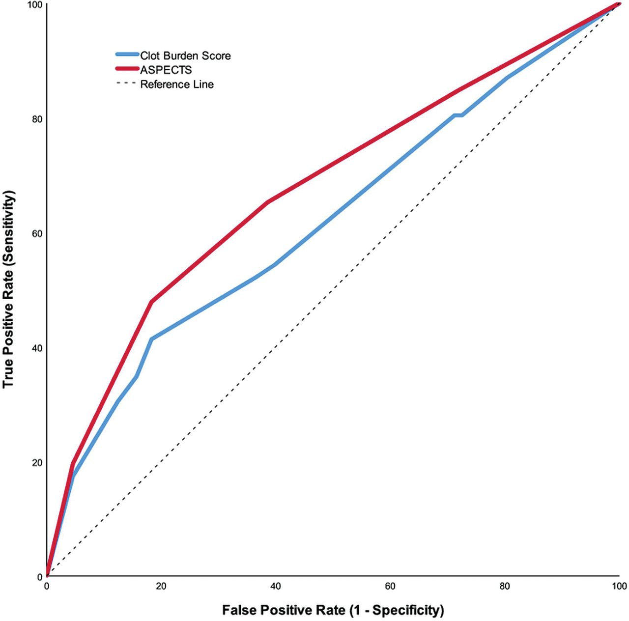
Baseline ASPECTS and CBS were selected for post hoc analysis. The receiver operating characteristic curves comparing these 2 radiologic scores with hemorrhage occurrence are shown in the Figure. According to Youden's index, the mathematically optimal cut-point for ASPECTS was ≤7 (sensitivity, 48%; specificity, 82%). Adjusted for relevant covariates, ASPECTS ≤7 was associated with a 3.17 times increased odds of ICH (95% CI, 1.35–7.45). Likewise, the ideal optimal cut-point for CBS was ≤3 (sensitivity, 41%; specificity, 82%; OR, 3.12; 95% CI, 1.36–7.15), indicative of extensive clot burden.

FIGURE.
  • Download figure
  • Open in new tab
  • Download powerpoint
FIGURE.

Receiver operating characteristic curves. Curves for baseline ASPECTS (area under the curve, 0.68; 95% confidence interval: 0.58–0.77) and CBS (area under the curve, 0.62; 95% confidence interval: 0.52–0.72).

Discussion

Our study assessed whether imaging features of large-vessel occlusion could help identify patients who are at high risk of post-EVT ICH. In a prospective cohort representative of the real-world application of EVT, we found that lower pretreatment ASPECTS, ICA clot location, and increased clot burden independently predicted ICH. Our study suggests that imaging features may be helpful for prognostication and risk stratification for EVT.

The findings of pretreatment ASPECTS affecting the risk of hemorrhage are in keeping with previous studies and provide further evidence that patients presenting with increasing amounts of early ischemia are at risk of developing postprocedural hemorrhage.7,20,21 States of relative hyperglycemia have been previously associated with postprocedural hemorrhage, and our findings are in line with previous work in this area.22,23 Prolonged symptom-to-recanalization times have also been previously associated with postprocedural hemorrhage and may aid in bedside prognostication.7 We hypothesize that a prolonged time to recanalization allows more time for the development of ischemia, ultimately increasing the risk of hemorrhage formation. Technical factors that could be associated with an increased time to recanalization, such as an increased number of stent-retriever passes, may play a contributing role as well.7,24

The CBS is a surrogate representation of both thrombus length and location.11 Accordingly, CBS was colinear with thrombus location. This idea is supported by the low clot burden scores observed in the ICA and proximal M1 locations and the increased risk of ICH associated with the ICA thrombus location in our alternate multivariable model. Our findings suggest that proximal occlusions in the ICA will often exhibit a high clot burden, thereby increasing the risk of hemorrhage in this particular patient subgroup. This finding, however, does not mean that clot burden equates to a difficult recanalization because we found no relationship among clot burden, time to recanalization, or successful recanalization.

Our findings complement safety data recently published from the Highly Effective Reperfusion evaluated in Multiple Endovascular Stroke Trials (HERMES) collaboration, a pooled patient-level analysis of the 7 major randomized controlled EVT trials.21 In this analysis, clot burden score, pooled into 3 groups (8–10, 5–7, 0–4), was not associated with symptomatic ICH. The symptomatic ICH event rate in INTERRSeCT was relatively low, and we therefore cannot comment on the associations between CBS and symptomatic ICH. However, asymptomatic hemorrhages are associated with poor outcome,25 and postprocedural ICH occurrence is still an important contributor to clinical outcome, symptomatic or not. While the risk of hemorrhage increases with certain locations and extensive clot burden (CBS ≤ 3), it is exactly these same patients who stand to benefit the most from EVT. Thus, our findings are important prognostic elements that can be discussed with patients and their families, even if they do not specifically influence the decision to offer EVT.

We expected to see an increased risk of hemorrhage in patients undergoing combined therapy; this was not observed. If IV tPA does not increase the risk of ICH, the size of the brain injury may ultimately have the largest influence on hemorrhagic transformation risk7,8,10; this notion is further supported by our results. Furthermore, our study supports the safety of combined therapy and provides additional context for the ongoing debate around whether EVT alone should be offered instead of combined therapy.26

Our study has several limitations. It can be difficult to differentiate contrast staining from reperfusion hemorrhage. Contrast staining is seen in approximately one-third of patients, and unlike reperfusion hemorrhage, it does not have a negative prognosis.27 For those 69 (35%) patients who underwent follow-up MR imaging, this concern is less because MR imaging can effectively differentiate blood from contrast staining.28 However, for the remaining scans, CT was the primary imaging technique of choice. Ideally, dual-energy CT would be used to effectively distinguish contrast from blood,29 but study sites were not required to do this. While the reported washout period of IV contrast is estimated to take place within 24–48 hours,29,30 studies seeking to differentiate contrast from hemorrhage have, in fact, used imaging at the 24-hour mark as a comparative end point.30 In addition, a recent study by Dekeyzer et al31 reported that follow-up CT scans performed at least 19 hours after the initial CT can differentiate contrast and hemorrhage with high specificity. The mean time to follow-up in our study was 24.4 ± 9.5 hours. Thus, while the risk of mistaking hemorrhage for contrast remains, we believe that we have reduced the risk of potential bias by strictly following our protocol timeline for follow-up imaging. Additional limitations include the use of an observational design, which may have introduced selection bias toward those patients offered EVT. Finally, given that EVT was not yet considered standard of care, technical data on the endovascular therapy procedure (number of passes, device used, aspiration used) were not collected.

Conclusions

Imaging characteristics, namely pretreatment ASPECTS, clot burden score, and clot location, are predictive of post-EVT ICH. These radiologic factors can be acquired with relative ease during an acute stroke and therefore can be practical tools to assist with EVT prognostication and can complement risk/benefit conversations with patients and their caregivers.

Footnotes

  • Disclosures: Robert Mikulik—RELATED: Grant: Project No. LQ1605 from the National Program of Sustainability II and the project of the International Clinical Research Center of St. Anne's University Hospital in Brno No. CZ.1.05/1.1.00/02.0123 (Operační program Výzkum a vývoj pro inovace).* Thalia S. Field—RELATED: Grant: Canadian Institutes of Health Research operating funds, Comments: The research program of the Vancouver Stroke Program received a per-patient payment to offset the cost of research nurse time and non-standard-of-care neuroimaging for the INTERRSeCT study*; UNRELATED: Consultancy: Bayer Canada, Pfizer-Bristol-Myers Squibb, Servier Laboratories, Comments: Advisory Board honoraria; Grants/Grants Pending: Canadian Institutes of Health Research, Heart and Stroke Foundation of Canada, Canadian Stroke Consortium, Bayer Canada, Boehringer-Ingelheim, Comments: Canadian Institutes of Health Research, Heart and Stroke Foundation of Canada, Canadian Stroke Consortium operating funds, Boehringer Canada operating funds, and Bayer Canada in-kind study medication*; Payment for Lectures Including Service on Speakers Bureaus: Bayer Canada, Pfizer-Bristol-Myers Squibb, Servier Laboratories, Boehringer-Ingelheim, Comments: Speakers Bureau honoraria from Bayer Canada, Pfizer-Bristol-Myers Squibb, Servier Laboratories, and Boehringer-Ingelheim Canada. Jean-Martin Boulanger—UNRELATED: Consultancy: Novartis, Bayer, Pfizer, Comments: conferences and Advisory Boards. Michael D. Hill—RELATED: Grant: Canadian Institutes of Health Research, Comments: grant to the University of Calgary. The principal Investigator was Dr A.M. Demchuk. I was a coinvestigator on the grant*; UNRELATED: Board Membership: Canadian Neuroscience Federation, Comments: not-for-profit entity; Consultancy: Boehringer-Ingelheim; Grants/Grants Pending: Medtronic, Stryker, Bayer Canada, Boehringer-Ingelheim, NoNO Inc, Canadian Institutes of Health Research, Alberta Innovates, British Heart Foundation, Comments: grants for ongoing studies; Stock/Stock Options: Calgary Scientific Inc, Comments: stock ownership. Andrew M. Demchuk—RELATED: Consulting Fee or Honorarium: Medtronic. Bijoy K. Menon—RELATED: Grant: Canadian Institutes of Health Research, Comments: operating grant.* Dar Dowlatshahi—UNRELATED: Consultancy: ApoPharma, Bayer; Payment for Lectures Including Service on Speakers Bureaus: Bristol-Myers Squibb. *Money paid to the institution.

  • INTERRSeCT was originally funded by an operating grant from the Canadian Institutes of Health Research. Dr Dowlatshahi was funded by a University of Ottawa Department of Medicine Clinician-Scientist Research Chair award and a Heart and Stroke Foundation of Canada New Investigator Award. Dr Yogendrakumar is supported by the Queen Elizabeth II Graduate Scholarship for Science and Technology and the Canadian Institutes of Health Research Banting and Best Graduate Scholarship. Dr Mikulik was supported by No. LQ1605 from the National Program of Sustainability II (MEYS CR) and by The International Clinical Research Center of St. Anne's University Hospital in Brno No. CZ.1.05/1.1.00/02.0123 (Operační program Výzkum a vývoj pro inovace).

Indicates open access to non-subscribers at www.ajnr.org

REFERENCES

  1. 1.↵
    1. Albers GW,
    2. Marks MP,
    3. Kemp S, et al
    ; DEFUSE 3 Investigators. Thrombectomy for stroke at 6 to 16 hours with selection by perfusion imaging. N Engl J Med 2018;378:708–18 doi:10.1056/NEJMoa1713973 pmid:29364767
    CrossRefPubMed
  2. 2.↵
    1. Nogueira RG,
    2. Jadhav AP,
    3. Haussen DC, et al
    ; DAWN Trial Investigators. Thrombectomy 6 to 24 hours after stroke with a mismatch between deficit and infarct. N Engl J Med 2018;378:11–21 doi:10.1056/NEJMoa1706442 pmid:29129157
    CrossRefPubMed
  3. 3.↵
    1. Yaghi S,
    2. Willey JZ,
    3. Cucchiara B, et al
    ; American Heart Association Stroke Council; Council on Cardiovascular and Stroke Nursing; Council on Clinical Cardiology; and Council on Quality of Care and Outcomes Research. Treatment and Outcome of Hemorrhagic Transformation After Intravenous Alteplase in Acute Ischemic Stroke: A Scientific Statement for Healthcare Professionals from the American Heart Association/American Stroke Association. Stroke 2017;48:e343–61 doi:10.1161/STR.0000000000000152 pmid:29097489
    Abstract/FREE Full Text
  4. 4.↵
    1. Balami JS,
    2. White PM,
    3. McMeekin PJ, et al
    . Complications of endovascular treatment for acute ischemic stroke: prevention and management. Int J Stroke 2018;13:348–61 doi:10.1177/1747493017743051 pmid:29171362
    CrossRefPubMed
  5. 5.↵
    1. Goyal M,
    2. Menon BK,
    3. van Zwam WH, et al
    ; HERMES collaborators. Endovascular thrombectomy after large-vessel ischaemic stroke: a meta-analysis of individual patient data from five randomised trials. Lancet 2016;387:1723–31 doi:10.1016/S0140-6736(16)00163-X pmid:26898852
    CrossRefPubMed
  6. 6.↵
    1. Enomoto Y,
    2. Yoshimura S,
    3. Egashira Y, et al
    ; Committee of Endovascular Salvage for Cerebral Ultra-acute Embolism (RESCUE)-Japan Study Group. The risk of intracranial hemorrhage in Japanese patients with acute large vessel occlusion; subanalysis of the RESCUE-Japan registry. J Stroke Cerebrovasc Dis 2016;25:1076–80 doi:10.1016/j.jstrokecerebrovasdis.2015.12.022 pmid:26923092
    CrossRefPubMed
  7. 7.↵
    1. Hao Y,
    2. Yang D,
    3. Wang H, et al
    . Predictors for symptomatic intracranial hemorrhage after endovascular treatment of acute ischemic stroke. Stroke 2017;48:1203–09 doi:10.1161/STROKEAHA.116.016368 pmid:28373302
    Abstract/FREE Full Text
  8. 8.↵
    1. Soize S,
    2. Barbe C,
    3. Kadziolka K, et al
    . Predictive factors of outcome and hemorrhage after acute ischemic stroke treated by mechanical thrombectomy with a stent-retriever. Neuroradiology 2013;55:977–87 doi:10.1007/s00234-013-1191-4 pmid:23644538
    CrossRefPubMed
  9. 9.↵
    1. Yoo AJ,
    2. Khatri P,
    3. Mocco J, et al
    ; THERAPY Trial Investigators. Impact of thrombus length on outcomes after intra-arterial aspiration thrombectomy in the THERAPY Trial. Stroke 2017;48:1895–900 doi:10.1161/STROKEAHA.116.016253 pmid:28596447
    Abstract/FREE Full Text
  10. 10.↵
    1. Mishra NK,
    2. Christensen S,
    3. Wouters A, et al
    ; DEFUSE 2 Investigators. Reperfusion of very low cerebral blood volume lesion predicts parenchymal hematoma after endovascular therapy. Stroke 2015;46:1245–49 doi:10.1161/STROKEAHA.114.008171 pmid:25828235
    Abstract/FREE Full Text
  11. 11.↵
    1. Tan IY,
    2. Demchuk AM,
    3. Hopyan J, et al
    . CT angiography clot burden score and collateral score: correlation with clinical and radiologic outcomes in acute middle cerebral artery infarct. AJNR Am J Neuroradiol 2009;30:525–31 doi:10.3174/ajnr.A1408 pmid:19147716
    Abstract/FREE Full Text
  12. 12.↵
    1. Menon BK,
    2. Al-Ajlan FS,
    3. Najm M, et al
    . Association of clinical, imaging, and thrombus characteristics with recanalization of visible intracranial occlusion in patients with acute ischemic stroke. JAMA 2018;320:1017–26 doi:10.1001/jama.2018.12498 pmid:30208455
    CrossRefPubMed
  13. 13.↵
    1. Puetz V,
    2. Dzialowski I,
    3. Hill MD, et al
    ; Calgary CTA Study Group. Intracranial thrombus extent predicts clinical outcome, final infarct size and hemorrhagic transformation in ischemic stroke: the clot burden score. Int J Stroke 2008;3:230–36 doi:10.1111/j.1747-4949.2008.00221.x pmid:18811738
    CrossRefPubMed
  14. 14.↵
    1. Mishra SM,
    2. Dykeman J,
    3. Sajobi TT, et al
    . Early reperfusion rates with IV tPA are determined by CTA clot characteristics. AJNR Am J Neuroradiol 2014;35:2265–72 doi:10.3174/ajnr.A4048 pmid:25059699
    Abstract/FREE Full Text
  15. 15.↵
    1. Goyal M,
    2. Demchuk AM,
    3. Menon BK, et al
    ; ESCAPE Trial Investigators. Randomized assessment of rapid endovascular treatment of ischemic stroke. N Engl J Med 2015;372:1019–30 doi:10.1056/NEJMoa1414905 pmid:25671798
    CrossRefPubMed
  16. 16.↵
    1. von Kummer R,
    2. Broderick JP,
    3. Campbell BC, et al
    . The Heidelberg Bleeding Classification: classification of bleeding events after ischemic stroke and reperfusion therapy. Stroke 2015;46:2981–86 doi:10.1161/STROKEAHA.115.010049 pmid:26330447
    FREE Full Text
  17. 17.↵
    1. Trouillas P,
    2. von Kummer R
    . Classification and pathogenesis of cerebral hemorrhages after thrombolysis in ischemic stroke. Stroke 2006;37:556–61 doi:10.1161/01.STR.0000196942.84707.71 pmid:16397182
    Abstract/FREE Full Text
  18. 18.↵
    1. Neuberger U,
    2. Möhlenbruch MA,
    3. Herweh C, et al
    . Classification of Bleeding Events: comparison of ECASS III (European Cooperative Acute Stroke Study) and the New Heidelberg Bleeding Classification. Stroke 2017;48:1983–85 doi:10.1161/STROKEAHA.117.016735 pmid:28455322
    Abstract/FREE Full Text
  19. 19.↵
    1. Berkhemer OA,
    2. Fransen PS,
    3. Beumer D, et al
    . A randomized trial of intraarterial treatment for acute ischemic stroke. N Engl J Med 2015;372:11–20 doi:10.1056/NEJMoa1411587 pmid:25517348
    CrossRefPubMed
  20. 20.↵
    1. Lin K,
    2. Zink WE,
    3. Tsiouris AJ, et al
    . Risk assessment of hemorrhagic transformation of acute middle cerebral artery stroke using multimodal CT. J Neuroimaging 2012;22:160–66 doi:10.1111/j.1552-6569.2010.00562.x pmid:21143549
    CrossRefPubMed
  21. 21.↵
    1. Román LS,
    2. Menon BK,
    3. Blasco J, et al
    . Imaging features and safety and efficacy of endovascular stroke treatment: a meta-analysis of individual patient-level data. Lancet Neurol 2018;17:895–904 doi:10.1016/S1474-4422(18)30242-4 pmid:30264728
    CrossRefPubMed
  22. 22.↵
    1. Osei E,
    2. den Hertog HM,
    3. Berkhemer OA, et al
    ; MR CLEAN Investigators. Admission glucose and effect of intra-arterial treatment in patients with acute ischemic stroke. Stroke 2017;48:1299–305 doi:10.1161/STROKEAHA.116.016071 pmid:28389610
    Abstract/FREE Full Text
  23. 23.↵
    1. Poppe AY,
    2. Majumdar SR,
    3. Jeerakathil T, et al
    ; Canadian Alteplase for Stroke Effectiveness Study Investigators. Admission hyperglycemia predicts a worse outcome in stroke patients treated with intravenous thrombolysis. Diabetes Care 2009;32:617–22 doi:10.2337/dc08-1754 pmid:19131465
    Abstract/FREE Full Text
  24. 24.↵
    1. Bourcier R,
    2. Saleme S,
    3. Labreuche J, et al
    ; ASTER Trial Investigators. More than three passes of stent retriever is an independent predictor of parenchymal hematoma in acute ischemic stroke. J Neurointerv Surg 2018 Nov 2. [Epub ahead of print] doi:10.1136/neurintsurg-2018-014380 pmid:30389897
    Abstract/FREE Full Text
  25. 25.↵
    1. Dzialowski I,
    2. Pexman JH,
    3. Barber PA, et al
    ; CASES Investigators. Asymptomatic hemorrhage after thrombolysis may not be benign: prognosis by hemorrhage type in the Canadian alteplase for stroke effectiveness study registry. Stroke 2007;38:75–79 doi:10.1161/01.STR.0000251644.76546.62 pmid:17122437
    Abstract/FREE Full Text
  26. 26.↵
    1. Fischer U,
    2. Kaesmacher J,
    3. Mendes Pereira V, et al
    . Direct mechanical thrombectomy versus combined intravenous and mechanical thrombectomy in large-artery anterior circulation stroke: a topical review. Stroke 2017;48:2912–18 doi:10.1161/STROKEAHA.117.017208 pmid:28887391
    FREE Full Text
  27. 27.↵
    1. Parrilla G,
    2. García-Villalba B,
    3. Espinosa de Rueda M, et al
    . Hemorrhage/contrast staining areas after mechanical intra-arterial thrombectomy in acute ischemic stroke: imaging findings and clinical significance. AJNR Am J Neuroradiol 2012;33:1791–96 doi:10.3174/ajnr.A3044 pmid:22538076
    Abstract/FREE Full Text
  28. 28.↵
    1. Greer DM,
    2. Koroshetz WJ,
    3. Cullen S, et al
    . Magnetic resonance imaging improves detection of intracerebral hemorrhage over computed tomography after intra-arterial thrombolysis. Stroke 2004;35:491–95 doi:10.1161/01.STR.0000114201.11353.C5 pmid:14739411
    Abstract/FREE Full Text
  29. 29.↵
    1. Phan CM,
    2. Yoo AJ,
    3. Hirsch JA, et al
    . Differentiation of hemorrhage from iodinated contrast in different intracranial compartments using dual-energy head CT. AJNR Am J Neuroradiol 2012;33:1088–94 doi:10.3174/ajnr.A2909 pmid:22268092
    Abstract/FREE Full Text
  30. 30.↵
    1. Payabvash S,
    2. Qureshi MH,
    3. Khan SM, et al
    . Differentiating intraparenchymal hemorrhage from contrast extravasation on post-procedural noncontrast CT scan in acute ischemic stroke patients undergoing endovascular treatment. Neuroradiology 2014;56:737–44 doi:10.1007/s00234-014-1381-8 pmid:24925217
    CrossRefPubMed
  31. 31.↵
    1. Dekeyzer S,
    2. Nikoubashman O,
    3. Lutin B, et al
    . Distinction between contrast staining and hemorrhage after endovascular stroke treatment: one CT is not enough. J Neurointerv Surg 2017;9:394–98 doi:10.1136/neurintsurg-2016-012290 pmid:27036980
    Abstract/FREE Full Text
  • Received November 23, 2018.
  • Accepted after revision February 11, 2019.
  • © 2019 by American Journal of Neuroradiology
View Abstract
PreviousNext
Back to top

In this issue

American Journal of Neuroradiology: 40 (4)
American Journal of Neuroradiology
Vol. 40, Issue 4
1 Apr 2019
  • Table of Contents
  • Index by author
  • Complete Issue (PDF)
Advertisement
Print
Download PDF
Email Article

Thank you for your interest in spreading the word on American Journal of Neuroradiology.

NOTE: We only request your email address so that the person you are recommending the page to knows that you wanted them to see it, and that it is not junk mail. We do not capture any email address.

Enter multiple addresses on separate lines or separate them with commas.
Clot Burden Score and Early Ischemia Predict Intracranial Hemorrhage following Endovascular Therapy
(Your Name) has sent you a message from American Journal of Neuroradiology
(Your Name) thought you would like to see the American Journal of Neuroradiology web site.
CAPTCHA
This question is for testing whether or not you are a human visitor and to prevent automated spam submissions.
Cite this article
V. Yogendrakumar, F. Al-Ajlan, M. Najm, J. Puig, A. Calleja, S.-I. Sohn, S.H. Ahn, R. Mikulik, N. Asdaghi, T.S. Field, A. Jin, T. Asil, J.-M. Boulanger, M.D. Hill, A.M. Demchuk, B.K. Menon, D. Dowlatshahi
Clot Burden Score and Early Ischemia Predict Intracranial Hemorrhage following Endovascular Therapy
American Journal of Neuroradiology Apr 2019, 40 (4) 655-660; DOI: 10.3174/ajnr.A6009

Citation Manager Formats

  • BibTeX
  • Bookends
  • EasyBib
  • EndNote (tagged)
  • EndNote 8 (xml)
  • Medlars
  • Mendeley
  • Papers
  • RefWorks Tagged
  • Ref Manager
  • RIS
  • Zotero
0 Responses
Respond to this article
Share
Bookmark this article
Clot Burden Score and Early Ischemia Predict Intracranial Hemorrhage following Endovascular Therapy
V. Yogendrakumar, F. Al-Ajlan, M. Najm, J. Puig, A. Calleja, S.-I. Sohn, S.H. Ahn, R. Mikulik, N. Asdaghi, T.S. Field, A. Jin, T. Asil, J.-M. Boulanger, M.D. Hill, A.M. Demchuk, B.K. Menon, D. Dowlatshahi
American Journal of Neuroradiology Apr 2019, 40 (4) 655-660; DOI: 10.3174/ajnr.A6009
del.icio.us logo Twitter logo Facebook logo Mendeley logo
  • Tweet Widget
  • Facebook Like
  • Google Plus One
Purchase

Jump to section

  • Article
    • Abstract
    • ABBREVIATIONS:
    • Materials and Methods
    • Results
    • Discussion
    • Conclusions
    • Footnotes
    • REFERENCES
  • Figures & Data
  • Info & Metrics
  • Responses
  • References
  • PDF

Related Articles

  • No related articles found.
  • PubMed
  • Google Scholar

Cited By...

  • Effect of Clot Burden Score on Safety and Efficacy of Intravenous Alteplase Prior to Mechanical Thrombectomy in Acute Ischemic Stroke: A Subgroup Analysis of a Randomized Phase 3 Trial
  • Crossref (6)
  • Google Scholar

This article has been cited by the following articles in journals that are participating in Crossref Cited-by Linking.

  • Frequency, Determinants, and Outcomes of Emboli to Distal and New Territories Related to Mechanical Thrombectomy for Acute Ischemic Stroke
    Gregory J. Wong, Bryan Yoo, David Liebeskind, Humain Baharvahdat, Jeffrey Gornbein, Reza Jahan, Viktor Szeder, Gary Duckwiler, Satoshi Tateshima, Geoffrey Colby, May Nour, Latisha Sharma, Neal Rao, Jason Hinman, Sidney Starkman, Jeffrey L. Saver, Rodel Alfonso, Allison Arch, Gilda Avila, Mersedeh Bahr Hosseini, Fionna Chatfield, Arun S. Chhabra, Bruce Dobkin, Lindsey K. Frischmann, Ileana Grunberg, Judy Guzy, Josephine F. Huang, Doojin Kim, Konark Malhotra, Michael McManus, Lucas Restrepo, Parampreet Singh, Xiannan (Xander) Tang, Jason W. Tarpley, Anita Tipirneni
    Stroke 2021 52 7
  • Predictive value of clot imaging in acute ischemic stroke: A systematic review of artificial intelligence and conventional studies
    Daniela Dumitriu LaGrange, Jeremy Hofmeister, Andrea Rosi, Maria Isabel Vargas, Isabel Wanke, Paolo Machi, Karl-Olof Lövblad
    Neuroscience Informatics 2023 3 1
  • What You Always Wanted to Know about Endovascular Therapy in Acute Ischemic Stroke but Never Dared to Ask: A Comprehensive Review
    Philipp Bücke, Jose E. Cohen, Thomas Horvath, Alexandru Cimpoca, Pervinder Bhogal, Hansjörg Bäzner, Hans Henkes
    Reviews in Cardiovascular Medicine 2022 23 10
  • The value of radiomics-based hyperdense middle cerebral artery sign in predicting hemorrhagic transformation in acute ischemic stroke patients undergoing endovascular treatment
    Chundan Gong, Yun Liu, Wei Ma, Yang Jing, Li Liu, Yan Huang, Jinlin Yang, Chen Feng, Yuan Fang, Weidong Fang
    Frontiers in Neurology 2024 15
  • Can Computed Tomographic Angiography Be Used to Predict Who Will Not Benefit from Endovascular Treatment in Patients with Acute Ischemic Stroke? The CTA-ABC Score
    Hyo-Sung Kwak, Jung-Soo Park
    Journal of Korean Neurosurgical Society 2020 63 4
  • Effect of Clot Burden Score on Safety and Efficacy of Intravenous Alteplase Prior to Mechanical Thrombectomy in Acute Ischemic Stroke: A Subgroup Analysis of a Randomized Phase 3 Trial
    Zifu Li, Yu Zhou, Xiaoxi Zhang, Lei Zhang, Yongwei Zhang, Pengfei Xing, Yongxin Zhang, Qinghai Huang, Qiang Li, Qiao Zuo, Xiaofei Ye, Jianmin Liu, Pengfei Yang
    American Journal of Neuroradiology 2024 45 3

More in this TOC Section

  • Rescue Reentry in Carotid Near-Occlusion
  • Contour Neurovascular System: Five Year Follow Up
  • Effect of SARS-CoV2 on Endovascular Thrombectomy
Show more NEUROINTERVENTION

Similar Articles

Advertisement

Indexed Content

  • Current Issue
  • Accepted Manuscripts
  • Article Preview
  • Past Issues
  • Editorials
  • Editor's Choice
  • Fellows' Journal Club
  • Letters to the Editor
  • Video Articles

Cases

  • Case Collection
  • Archive - Case of the Week
  • Archive - Case of the Month
  • Archive - Classic Case

More from AJNR

  • Trainee Corner
  • Imaging Protocols
  • MRI Safety Corner
  • Book Reviews

Multimedia

  • AJNR Podcasts
  • AJNR Scantastics

Resources

  • Turnaround Time
  • Submit a Manuscript
  • Submit a Video Article
  • Submit an eLetter to the Editor/Response
  • Manuscript Submission Guidelines
  • Statistical Tips
  • Fast Publishing of Accepted Manuscripts
  • Graphical Abstract Preparation
  • Imaging Protocol Submission
  • Evidence-Based Medicine Level Guide
  • Publishing Checklists
  • Author Policies
  • Become a Reviewer/Academy of Reviewers
  • News and Updates

About Us

  • About AJNR
  • Editorial Board
  • Editorial Board Alumni
  • Alerts
  • Permissions
  • Not an AJNR Subscriber? Join Now
  • Advertise with Us
  • Librarian Resources
  • Feedback
  • Terms and Conditions
  • AJNR Editorial Board Alumni

American Society of Neuroradiology

  • Not an ASNR Member? Join Now

© 2025 by the American Society of Neuroradiology All rights, including for text and data mining, AI training, and similar technologies, are reserved.
Print ISSN: 0195-6108 Online ISSN: 1936-959X

Powered by HighWire