
Article Figures & Data
Figures
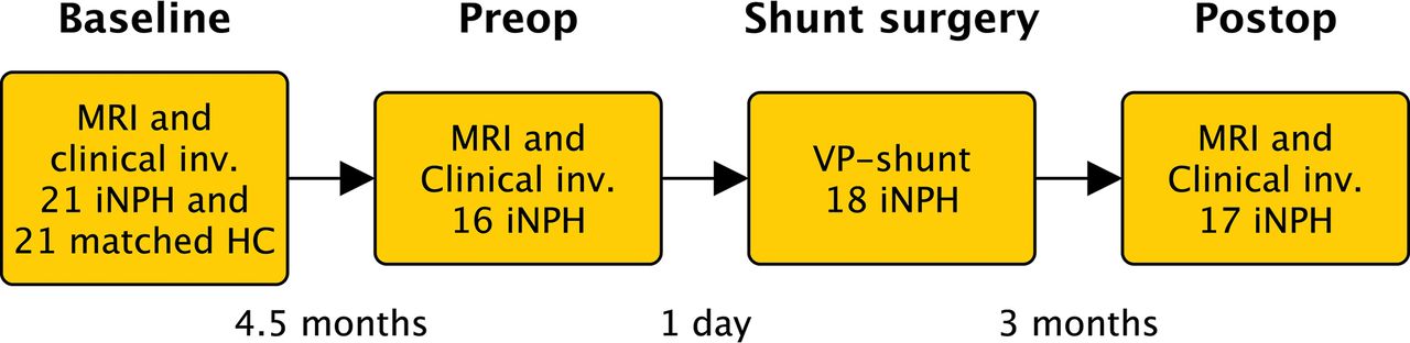
- Fig 1.
Timeline. MRI indicates MR imaging with the phase-contrast MR imaging sequence; Clinical inv., clinical investigation with tests of gait function, cognition, and urinary symptoms; HC, age- and sex-matched healthy controls. Preop = preoperative investigations; Postop, postoperative follow-up.
- Fig 2.
A, Aqueductal cerebral stroke volume in patients with iNPH and healthy controls. The lines connect each patient with a matched control. B, Aqueductal cerebral stroke volume in patients with iNPH at baseline, the day before shunt surgery (preop), and at 3 months after the operation (postop). Error bars represent 10th and 90th percentiles. The asterisk indicates P < .05; double asterisks, P < .01; NS, not significant.
- Fig 3.
A, Sagittal T2-weighted turbo spin-echo image (without flow compensation) with the red line illustrating the location of the phase-contrast MR imaging scan plane. B, Transverse magnitude image through the aqueduct. The red circle illustrates the ROI drawn for flow measurements. C, Corresponding velocity (phase) image.
- Fig 4.
Aqueductal CSF flow during 1 cardiac cycle. Positive values are in the craniocaudal direction.


Tables
Patients (n = 21) Controls (n = 21) P Value Age (median) (range) (yr) 74 (65–81) 74 (65–82) NSb Sex (No. of men) (%) 11 (52%) 11 (52%) NSc MMSE 25 (22–27) 30 (29–30) <.001d Urgency scale 3 (1–4) 1 (1–1) <.001d mRS 2 (2–3) 0 (0–0) <.001d TUG (sec) 20 (14–31) 9 (8–11) <.001d TUG (No. of steps) 22 (18–34) 12 (11–14) <.001d 10 Meter Walk test (sec) 12 (8–17) 5 (5–6) <.001d 10 Meter Walk Test (No. of steps) 22 (16–30) 12 (11–13) <.001d Evans index 0.35 (0.34–0.39) 0.28 (0.24–0.30) <.001d DWMH 1 (1–3) 1 (1–2) NSd DESH (No.) (%) 14 (67%) 0 (0%) <.001c Callosal angle 66° (60°–73°) 113° (104°–121°) <.001d Flow void 3 (2–3) 2 (2–2) NSd - Table 2:
Aqueductal stroke volume, peak velocity, and aqueductal area in controls and patients at baselinea
Healthy Controls (n = 21) Patient Baseline (n = 21) P Valueb ACSV (μL) 62.5 (58.3–73.8) 103.5 (69.8–142.8) <.01 Peak velocity (mm/s) 103 (79.5–113.5) 103 (68.5–166.5) NS Net flow (μL) −2.9 (−5.65–2.55) −1.6 (−19–14) NS Aq area (mm2) 18 (15.5–19) 22 (19–25) <.001 - Table 3:
Aqueductal stroke volume, peak velocity, and aqueductal area in patients at all assessment timesa
Patient Baseline (n = 21) Patient Preop (n = 16) Patient Postop (n = 17) P Value ACSV (μL) 103.5 (69.8–142.8) 94.8 (81–241) 88 (51.8–173.3) <.05b Peak velocity (mm/s) 103 (68.5–166.5) 127 (72.5–154.8) 108 (71.5–146.5) NS Net flow (μL) −1.6 (−19–14) −1.8 (−14.5–10.8) 0 (−12.5–2.1) NS Aq area (mm2) 22 (19–25) 22.5 (19.3–25.8) 24 (20.5–25.5) NS Note:—Preop indicates preoperative; Postop, postoperative; Aq, aqueductal; NS, not signifiicant.
↵a Data are median with interquartile range in parentheses.
↵b Wilcoxon signed rank test. Significant difference between baseline and postoperative investigation and between preoperative and postoperative investigation. Comparisons between baseline and preoperative measurements were all nonsignificant.


Jump to section
Related Articles
Cited By...
- Characterization of oscillations in the brain and cerebrospinal fluid using ultra-high field magnetic resonance imaging
- Decreased Craniocervical CSF Flow in Patients with Normal Pressure Hydrocephalus: A Pilot Study
- Can Shunt Response in Patients with Idiopathic Normal Pressure Hydrocephalus Be Predicted from Preoperative Brain Imaging? A Retrospective Study of the Diagnostic Use of the Normal Pressure Hydrocephalus Radscale in 119 Patients