Advertisement

Main menu

  • Home
  • Content
    • Current Issue
    • Accepted Manuscripts
    • Article Preview
    • Past Issue Archive
    • Video Articles
    • AJNR Case Collection
    • Case of the Week Archive
    • Case of the Month Archive
    • Classic Case Archive
  • Special Collections
    • AJNR Awards
    • Low-Field MRI
    • Alzheimer Disease
    • ASNR Foundation Special Collection
    • Photon-Counting CT
    • View All
  • Multimedia
    • AJNR Podcasts
    • AJNR SCANtastic
    • Trainee Corner
    • MRI Safety Corner
    • Imaging Protocols
  • For Authors
    • Submit a Manuscript
    • Submit a Video Article
    • Submit an eLetter to the Editor/Response
    • Manuscript Submission Guidelines
    • Statistical Tips
    • Fast Publishing of Accepted Manuscripts
    • Graphical Abstract Preparation
    • Imaging Protocol Submission
    • Author Policies
  • About Us
    • About AJNR
    • Editorial Board
    • Editorial Board Alumni
  • More
    • Become a Reviewer/Academy of Reviewers
    • Subscribers
    • Permissions
    • Alerts
    • Feedback
    • Advertisers
    • ASNR Home

User menu

  • Alerts
  • Log in

Search

  • Advanced search
American Journal of Neuroradiology

American Journal of Neuroradiology

ASHNR American Society of Functional Neuroradiology ASHNR American Society of Pediatric Neuroradiology ASSR
  • Alerts
  • Log in

Advanced Search

  • Home
  • Content
    • Current Issue
    • Accepted Manuscripts
    • Article Preview
    • Past Issue Archive
    • Video Articles
    • AJNR Case Collection
    • Case of the Week Archive
    • Case of the Month Archive
    • Classic Case Archive
  • Special Collections
    • AJNR Awards
    • Low-Field MRI
    • Alzheimer Disease
    • ASNR Foundation Special Collection
    • Photon-Counting CT
    • View All
  • Multimedia
    • AJNR Podcasts
    • AJNR SCANtastic
    • Trainee Corner
    • MRI Safety Corner
    • Imaging Protocols
  • For Authors
    • Submit a Manuscript
    • Submit a Video Article
    • Submit an eLetter to the Editor/Response
    • Manuscript Submission Guidelines
    • Statistical Tips
    • Fast Publishing of Accepted Manuscripts
    • Graphical Abstract Preparation
    • Imaging Protocol Submission
    • Author Policies
  • About Us
    • About AJNR
    • Editorial Board
    • Editorial Board Alumni
  • More
    • Become a Reviewer/Academy of Reviewers
    • Subscribers
    • Permissions
    • Alerts
    • Feedback
    • Advertisers
    • ASNR Home
  • Follow AJNR on Twitter
  • Visit AJNR on Facebook
  • Follow AJNR on Instagram
  • Join AJNR on LinkedIn
  • RSS Feeds

AJNR Awards, New Junior Editors, and more. Read the latest AJNR updates

Research ArticleAdult Brain
Open Access

3T MRI Whole-Brain Microscopy Discrimination of Subcortical Anatomy, Part 1: Brain Stem

M.J. Hoch, M.T. Bruno, A. Faustin, N. Cruz, L. Crandall, T. Wisniewski, O. Devinsky and T.M. Shepherd
American Journal of Neuroradiology March 2019, 40 (3) 401-407; DOI: https://doi.org/10.3174/ajnr.A5956
M.J. Hoch
aFrom the Department of Radiology and Imaging Sciences (M.J.H.), Emory University, Atlanta, Georgia
  • Find this author on Google Scholar
  • Find this author on PubMed
  • Search for this author on this site
  • ORCID record for M.J. Hoch
M.T. Bruno
bDepartments of Radiology (M.T.B., N.C., T.M.S.)
  • Find this author on Google Scholar
  • Find this author on PubMed
  • Search for this author on this site
  • ORCID record for M.T. Bruno
A. Faustin
cPathology (A.F.)
  • Find this author on Google Scholar
  • Find this author on PubMed
  • Search for this author on this site
  • ORCID record for A. Faustin
N. Cruz
bDepartments of Radiology (M.T.B., N.C., T.M.S.)
  • Find this author on Google Scholar
  • Find this author on PubMed
  • Search for this author on this site
  • ORCID record for N. Cruz
L. Crandall
dNeurology (L.C., T.W., O.D.)
fSUDC Registry and Research Collaborative (L.C., O.D.), New York, New York
  • Find this author on Google Scholar
  • Find this author on PubMed
  • Search for this author on this site
  • ORCID record for L. Crandall
T. Wisniewski
dNeurology (L.C., T.W., O.D.)
ePsychiatry (T.W.), New York University, New York, New York
  • Find this author on Google Scholar
  • Find this author on PubMed
  • Search for this author on this site
  • ORCID record for T. Wisniewski
O. Devinsky
dNeurology (L.C., T.W., O.D.)
fSUDC Registry and Research Collaborative (L.C., O.D.), New York, New York
  • Find this author on Google Scholar
  • Find this author on PubMed
  • Search for this author on this site
  • ORCID record for O. Devinsky
T.M. Shepherd
bDepartments of Radiology (M.T.B., N.C., T.M.S.)
gCenter for Advanced Imaging Innovation and Research (T.M.S.), New York, New York.
  • Find this author on Google Scholar
  • Find this author on PubMed
  • Search for this author on this site
  • ORCID record for T.M. Shepherd
  • Article
  • Figures & Data
  • Supplemental
  • Info & Metrics
  • Responses
  • References
  • PDF
Loading

Article Figures & Data

Figures

  • Tables
  • Fig 1.
    • Download figure
    • Open in new tab
    • Download powerpoint
    Fig 1.

    Parasagittal and coronal T2-weighted MR images of the postmortem human brain stem. A, Canonical axial brain stem levels parallel to the anterior/posterior commissure plane that are found in Fig 2 are represented with the solid lines and On-line Fig 1 with the dashed line. Only selected brain stem substructures are labeled in coaligned sagittal and coronal images to orient the reader relative to the craniocaudal axial slice positions. The On-line Table provides a complete list of labeled anatomy for all figures, indicated by the numbers in parentheses in the legends. Note the trochlear nerve (asterisk, C) only seen in some brains.
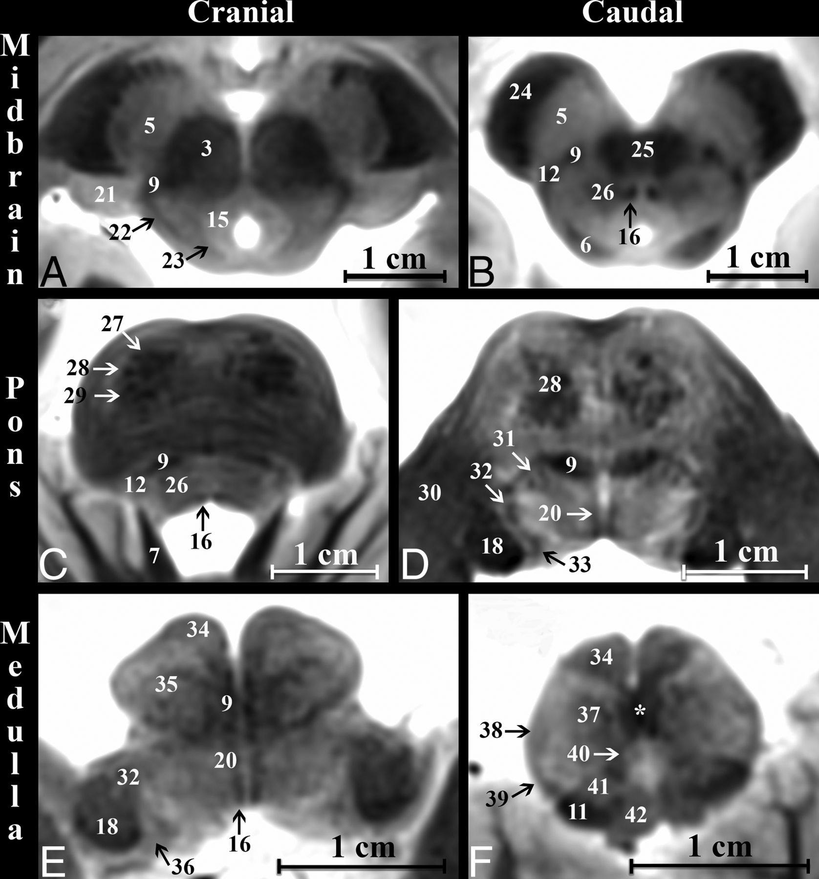
  • Fig 2.
    • Download figure
    • Open in new tab
    • Download powerpoint
    Fig 2.

    Axial modified T2-weighted TSE images at 6 canonical levels of the postmortem brain stem orientated parallel to the anterior/posterior commissure plane. Upper row: A, cranial midbrain; B, caudal midbrain. Middle row: C, cranial pons; D, caudal pons. Lower row: E, cranial medulla, F, caudal medulla. Improved image contrast from the modified TSE sequence directly demonstrates even small structures like the medial longitudinal fasciculus (16). Note the sensory decussation of the medial lemniscus in the caudal medulla (asterisk, F). The motor decussation is demonstrated in Fig 3.
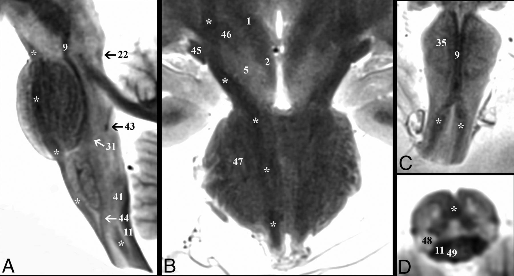
  • Fig 3.
    • Download figure
    • Open in new tab
    • Download powerpoint
    Fig 3.

    Demonstration of the corticospinal tract (asterisk) throughout the brain stem. A, Parasagittal image depicts the corticospinal tract descending within the brain stem from the cerebral peduncle to the upper cervical cord. B, Coronal image shows the course of the corticospinal tract from the posterior limb of the internal capsule to the most superior aspect of the medullary pyramids. Note in the diencephalic junction, the close relationship of the corticospinal tract to the optic tract (45) laterally and the subthalamic nucleus (46) medially. Oblique coronal (C) and oblique axial (D) images highlight the decussation of the corticospinal tracts at the cervicomedullary junction. C, The paramedian dark lines are the medial lemniscus (9), which is superficial to the corticospinal tract on this oblique axial image.
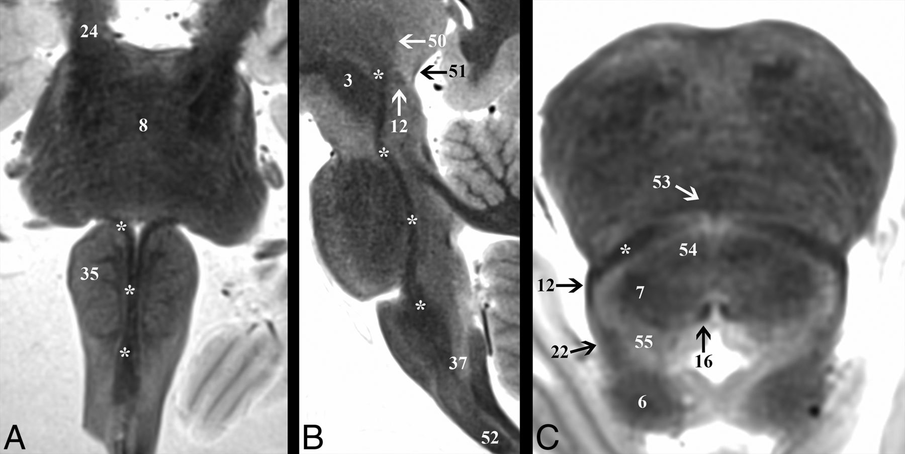
  • Fig 4.
    • Download figure
    • Open in new tab
    • Download powerpoint
    Fig 4.

    Demonstration of the medial lemniscus (asterisk) throughout the brain stem. A, Coronal image shows the change in the long-axis orientation of the medial lemniscus from anteroposterior to transverse as it ascends the medulla to the pontomedullary junction. B, Parasagittal image highlights the terminations of the medial lemniscus in the ventral posterolateral thalamic nucleus (50). C, Axial image angled anteroinferior to posterosuperior 20° relative to the ACPC plane through the inferior colliculus (6) shows the relationship of the medial lemniscus to the spinothalamic tract (12) and lateral lemniscus (22) at the lateral tegmentum.
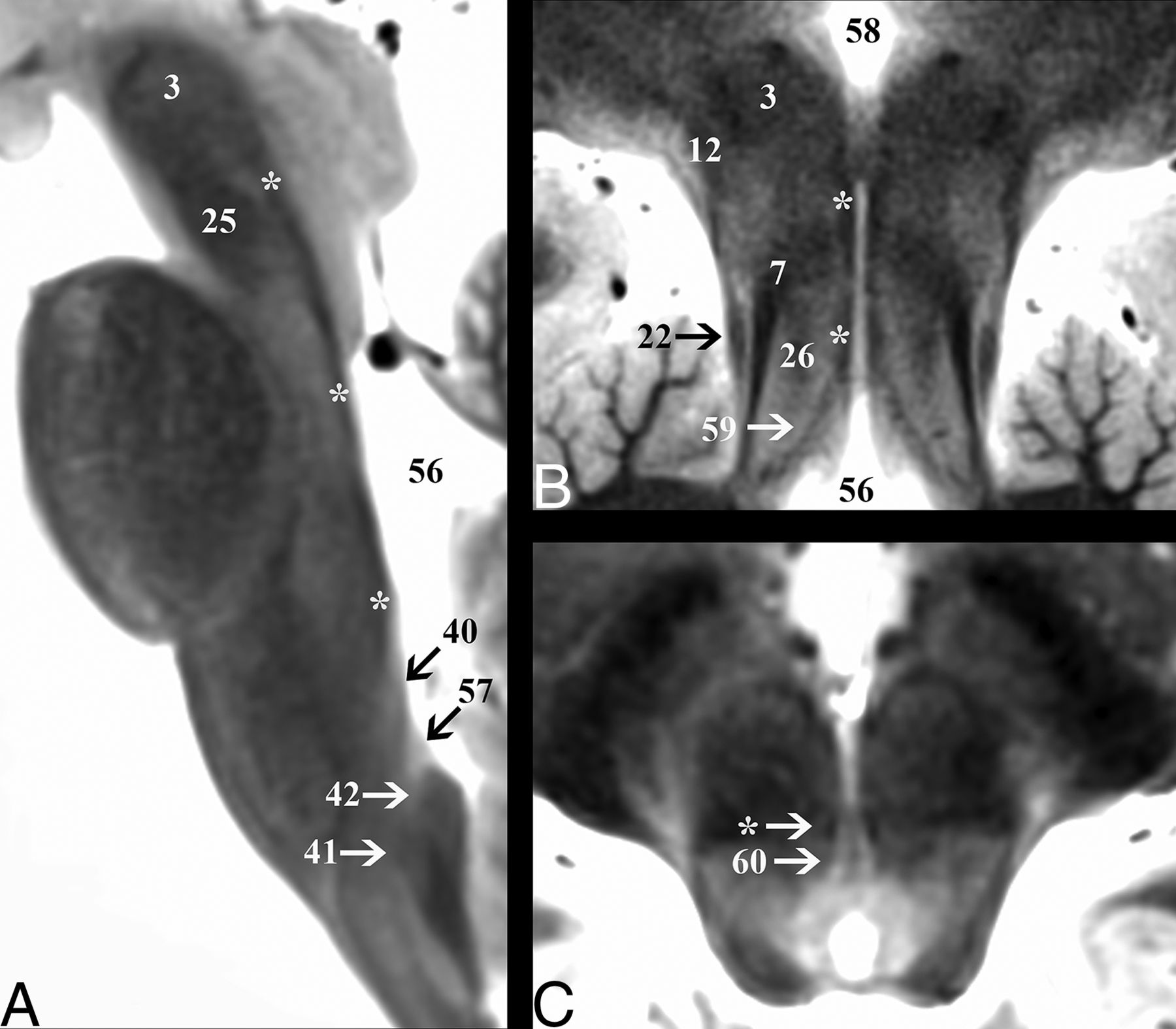
  • Fig 5.
    • Download figure
    • Open in new tab
    • Download powerpoint
    Fig 5.

    Demonstration of the medial longitudinal fasciculus (asterisk) throughout the brain stem. A, Sagittal image depicts the dorsal course of the medial longitudinal fasciculus from its origin in the cranial medulla just superior to the hypoglossal nucleus (40) to the level of the red nucleus (3). B, Coronal oblique image that is perpendicular to the long axis of the hippocampus (structure not shown) at the level of the posterior commissure shows the terminations of the tract in the inferior walls of the third ventricle (58). This coronal image also highlights vertical columns of the central midbrain from lateral to medial: lateral lemniscus (22), superior cerebellar peduncle (7), central tegmental tract (26), and medial longitudinal fasciculus (asterisk). C, Axial cranial midbrain image angled anterosuperior to posteroinferior 20° relative to the ACPC plane highlights the close relationship of the medial longitudinal fasciculus to the oculomotor nucleus (60).

Tables

  • Figures
  • Selected measurements for 5 major brain stem white matter tracts at 3 canonical axial planesa

    TractFigCCCranial MedullaMid PonsCaudal Midbrain
    APTVAreaAPTVAreaAPTVArea
    CST (L)351.7 ± 4.82.9 ± 0.63.7 ± 0.58.8 ± 2.6b6.6 ± 1.06.5 ± 0.733.9 ± 6.8b6.1 ± 0.63.5 ± 0.517.1 ± 3.0b
    CST (R)351.7 ± 4.82.7 ± 0.33.3 ± 0.47.3 ± 1.4b5.7 ± 0.96.2 ± 0.828.2 ± 8.1b6.1 ± 0.83.2 ± 0.815.5 ± 5.2b
    ML446.9 ± 3.55.6 ± 0.80.6 ± 0.12.9 ± 0.61.2 ± 0.44.6 ± 0.74.7 ± 1.92.0 ± 0.43.0 ± 0.44.9 ± 1.4
    MLF539.6 ± 3.41.0 ± 0.30.5 ± .060.5 ± 0.21.4 ± 0.30.8 ± 0.10.9 ± 0.23.2 ± 0.40.7 ± 0.11.9 ± 0.5
    CTT637.7 ± 4.02.6 ± 0.41.6 ± 0.33.4 ± 0.81.7 ± 0.22.1 ± 0.42.9 ± 0.83.2 ± 0.33.6 ± 0.49.1 ± 1.5
    • Note:—CC indicates craniocaudal; AP, anteroposterior; TV, transverse; Fig, figure; L, left; R, right.

    • ↵a Units are millimeters or square millimeters, and data are mean ± SD, with 13 SUDC samples.

    • ↵b All measurements of the right and left corticospinal tracts were compared separately. Cross-sectional areas trended toward small statistical differences in the medulla (P = .099) and pons (P = .063), but not the midbrain (P = .361).

PreviousNext
Back to top

In this issue

American Journal of Neuroradiology: 40 (3)
American Journal of Neuroradiology
Vol. 40, Issue 3
1 Mar 2019
  • Table of Contents
  • Index by author
  • Complete Issue (PDF)
Advertisement
Print
Download PDF
Email Article

Thank you for your interest in spreading the word on American Journal of Neuroradiology.

NOTE: We only request your email address so that the person you are recommending the page to knows that you wanted them to see it, and that it is not junk mail. We do not capture any email address.

Enter multiple addresses on separate lines or separate them with commas.
3T MRI Whole-Brain Microscopy Discrimination of Subcortical Anatomy, Part 1: Brain Stem
(Your Name) has sent you a message from American Journal of Neuroradiology
(Your Name) thought you would like to see the American Journal of Neuroradiology web site.
CAPTCHA
This question is for testing whether or not you are a human visitor and to prevent automated spam submissions.
Cite this article
M.J. Hoch, M.T. Bruno, A. Faustin, N. Cruz, L. Crandall, T. Wisniewski, O. Devinsky, T.M. Shepherd
3T MRI Whole-Brain Microscopy Discrimination of Subcortical Anatomy, Part 1: Brain Stem
American Journal of Neuroradiology Mar 2019, 40 (3) 401-407; DOI: 10.3174/ajnr.A5956

Citation Manager Formats

  • BibTeX
  • Bookends
  • EasyBib
  • EndNote (tagged)
  • EndNote 8 (xml)
  • Medlars
  • Mendeley
  • Papers
  • RefWorks Tagged
  • Ref Manager
  • RIS
  • Zotero
0 Responses
Respond to this article
Share
Bookmark this article
3T MRI Whole-Brain Microscopy Discrimination of Subcortical Anatomy, Part 1: Brain Stem
M.J. Hoch, M.T. Bruno, A. Faustin, N. Cruz, L. Crandall, T. Wisniewski, O. Devinsky, T.M. Shepherd
American Journal of Neuroradiology Mar 2019, 40 (3) 401-407; DOI: 10.3174/ajnr.A5956
del.icio.us logo Twitter logo Facebook logo Mendeley logo
  • Tweet Widget
  • Facebook Like
  • Google Plus One
Purchase

Jump to section

  • Article
    • Abstract
    • ABBREVIATIONS:
    • Materials and Methods
    • Results
    • Discussion
    • Conclusions
    • Acknowledgments
    • Footnotes
    • References
  • Figures & Data
  • Supplemental
  • Info & Metrics
  • Responses
  • References
  • PDF

Related Articles

  • PubMed
  • Google Scholar

Cited By...

  • The Subcortical Atlas of the Marmoset ("SAM") monkey based on high-resolution MRI and histology
  • Multimodal anatomical mapping of subcortical regions in Marmoset monkeys using high-resolution MRI and matched histology with multiple stains
  • High-resolution mapping and digital atlas of subcortical regions in the macaque monkey based on matched MAP-MRI and histology
  • Direct In Vivo MRI Discrimination of Brain Stem Nuclei and Pathways
  • 3T MRI Whole-Brain Microscopy Discrimination of Subcortical Anatomy, Part 2: Basal Forebrain
  • Crossref (5)
  • Google Scholar

This article has been cited by the following articles in journals that are participating in Crossref Cited-by Linking.

  • 3T MRI Whole-Brain Microscopy Discrimination of Subcortical Anatomy, Part 2: Basal Forebrain
    M.J. Hoch, M.T. Bruno, A. Faustin, N. Cruz, A.Y. Mogilner, L. Crandall, T. Wisniewski, O. Devinsky, T.M. Shepherd
    American Journal of Neuroradiology 2019 40 7
  • Direct In Vivo MRI Discrimination of Brain Stem Nuclei and Pathways
    T.M. Shepherd, B. Ades-Aron, M. Bruno, H.M. Schambra, M.J. Hoch
    American Journal of Neuroradiology 2020 41 5
  • Inner SPACE: 400-Micron Isotropic Resolution MRI of the Human Brain
    Timothy M. Shepherd, Michael J. Hoch, Mary Bruno, Arline Faustin, Antonios Papaioannou, Stephen E. Jones, Orrin Devinsky, Thomas Wisniewski
    Frontiers in Neuroanatomy 2020 14
  • Brainstem anatomy with 7-T MRI: in vivo assessment and ex vivo comparison
    Graziella Donatelli, Aron Emmi, Mauro Costagli, Paolo Cecchi, Veronica Macchi, Laura Biagi, Marta Lancione, Michela Tosetti, Andrea Porzionato, Raffaele De Caro, Mirco Cosottini
    European Radiology Experimental 2023 7 1
  • The influence of gender on the morphometric variability of the medulla oblongata and pons in adults, taking into account the type of skull base according to MRI data
    O.Yu. Aleshkina, N.H. Kuliev, T.S. Bikbaeva, I.A. Polkovova, O.B. Bogomyakova, M.A. Guseva, A.A. Tulupov, Yu.L. Vasil’ev
    Russian Journal of Operative Surgery and Clinical Anatomy 2025 9 1

More in this TOC Section

  • Diagnostic Neuroradiology of Monoclonal Antibodies
  • Cerebral ADC Changes in Fabry Disease
  • ML for Glioma Molecular Subtype Prediction
Show more ADULT BRAIN

Similar Articles

Advertisement

Indexed Content

  • Current Issue
  • Accepted Manuscripts
  • Article Preview
  • Past Issues
  • Editorials
  • Editor's Choice
  • Fellows' Journal Club
  • Letters to the Editor
  • Video Articles

Cases

  • Case Collection
  • Archive - Case of the Week
  • Archive - Case of the Month
  • Archive - Classic Case

More from AJNR

  • Trainee Corner
  • Imaging Protocols
  • MRI Safety Corner

Multimedia

  • AJNR Podcasts
  • AJNR Scantastics

Resources

  • Turnaround Time
  • Submit a Manuscript
  • Submit a Video Article
  • Submit an eLetter to the Editor/Response
  • Manuscript Submission Guidelines
  • Statistical Tips
  • Fast Publishing of Accepted Manuscripts
  • Graphical Abstract Preparation
  • Imaging Protocol Submission
  • Evidence-Based Medicine Level Guide
  • Publishing Checklists
  • Author Policies
  • Become a Reviewer/Academy of Reviewers
  • News and Updates

About Us

  • About AJNR
  • Editorial Board
  • Editorial Board Alumni
  • Alerts
  • Permissions
  • Not an AJNR Subscriber? Join Now
  • Advertise with Us
  • Librarian Resources
  • Feedback
  • Terms and Conditions
  • AJNR Editorial Board Alumni

American Society of Neuroradiology

  • Not an ASNR Member? Join Now

© 2025 by the American Society of Neuroradiology All rights, including for text and data mining, AI training, and similar technologies, are reserved.
Print ISSN: 0195-6108 Online ISSN: 1936-959X

Powered by HighWire