Advertisement

Main menu

  • Home
  • Content
    • Current Issue
    • Accepted Manuscripts
    • Article Preview
    • Past Issue Archive
    • Video Articles
    • AJNR Case Collection
    • Case of the Week Archive
    • Case of the Month Archive
    • Classic Case Archive
  • Special Collections
    • AJNR Awards
    • Low-Field MRI
    • Alzheimer Disease
    • ASNR Foundation Special Collection
    • Photon-Counting CT
    • View All
  • Multimedia
    • AJNR Podcasts
    • AJNR SCANtastic
    • Trainee Corner
    • MRI Safety Corner
    • Imaging Protocols
  • For Authors
    • Submit a Manuscript
    • Submit a Video Article
    • Submit an eLetter to the Editor/Response
    • Manuscript Submission Guidelines
    • Statistical Tips
    • Fast Publishing of Accepted Manuscripts
    • Graphical Abstract Preparation
    • Imaging Protocol Submission
    • Author Policies
  • About Us
    • About AJNR
    • Editorial Board
    • Editorial Board Alumni
  • More
    • Become a Reviewer/Academy of Reviewers
    • Subscribers
    • Permissions
    • Alerts
    • Feedback
    • Advertisers
    • ASNR Home

User menu

  • Alerts
  • Log in

Search

  • Advanced search
American Journal of Neuroradiology

American Journal of Neuroradiology

ASHNR American Society of Functional Neuroradiology ASHNR American Society of Pediatric Neuroradiology ASSR
  • Alerts
  • Log in

Advanced Search

  • Home
  • Content
    • Current Issue
    • Accepted Manuscripts
    • Article Preview
    • Past Issue Archive
    • Video Articles
    • AJNR Case Collection
    • Case of the Week Archive
    • Case of the Month Archive
    • Classic Case Archive
  • Special Collections
    • AJNR Awards
    • Low-Field MRI
    • Alzheimer Disease
    • ASNR Foundation Special Collection
    • Photon-Counting CT
    • View All
  • Multimedia
    • AJNR Podcasts
    • AJNR SCANtastic
    • Trainee Corner
    • MRI Safety Corner
    • Imaging Protocols
  • For Authors
    • Submit a Manuscript
    • Submit a Video Article
    • Submit an eLetter to the Editor/Response
    • Manuscript Submission Guidelines
    • Statistical Tips
    • Fast Publishing of Accepted Manuscripts
    • Graphical Abstract Preparation
    • Imaging Protocol Submission
    • Author Policies
  • About Us
    • About AJNR
    • Editorial Board
    • Editorial Board Alumni
  • More
    • Become a Reviewer/Academy of Reviewers
    • Subscribers
    • Permissions
    • Alerts
    • Feedback
    • Advertisers
    • ASNR Home
  • Follow AJNR on Twitter
  • Visit AJNR on Facebook
  • Follow AJNR on Instagram
  • Join AJNR on LinkedIn
  • RSS Feeds

AJNR Awards, New Junior Editors, and more. Read the latest AJNR updates

Research ArticleAdult Brain
Open Access

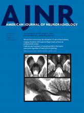
3T MRI Whole-Brain Microscopy Discrimination of Subcortical Anatomy, Part 1: Brain Stem

M.J. Hoch, M.T. Bruno, A. Faustin, N. Cruz, L. Crandall, T. Wisniewski, O. Devinsky and T.M. Shepherd
American Journal of Neuroradiology March 2019, 40 (3) 401-407; DOI: https://doi.org/10.3174/ajnr.A5956
M.J. Hoch
aFrom the Department of Radiology and Imaging Sciences (M.J.H.), Emory University, Atlanta, Georgia
  • Find this author on Google Scholar
  • Find this author on PubMed
  • Search for this author on this site
  • ORCID record for M.J. Hoch
M.T. Bruno
bDepartments of Radiology (M.T.B., N.C., T.M.S.)
  • Find this author on Google Scholar
  • Find this author on PubMed
  • Search for this author on this site
  • ORCID record for M.T. Bruno
A. Faustin
cPathology (A.F.)
  • Find this author on Google Scholar
  • Find this author on PubMed
  • Search for this author on this site
  • ORCID record for A. Faustin
N. Cruz
bDepartments of Radiology (M.T.B., N.C., T.M.S.)
  • Find this author on Google Scholar
  • Find this author on PubMed
  • Search for this author on this site
  • ORCID record for N. Cruz
L. Crandall
dNeurology (L.C., T.W., O.D.)
fSUDC Registry and Research Collaborative (L.C., O.D.), New York, New York
  • Find this author on Google Scholar
  • Find this author on PubMed
  • Search for this author on this site
  • ORCID record for L. Crandall
T. Wisniewski
dNeurology (L.C., T.W., O.D.)
ePsychiatry (T.W.), New York University, New York, New York
  • Find this author on Google Scholar
  • Find this author on PubMed
  • Search for this author on this site
  • ORCID record for T. Wisniewski
O. Devinsky
dNeurology (L.C., T.W., O.D.)
fSUDC Registry and Research Collaborative (L.C., O.D.), New York, New York
  • Find this author on Google Scholar
  • Find this author on PubMed
  • Search for this author on this site
  • ORCID record for O. Devinsky
T.M. Shepherd
bDepartments of Radiology (M.T.B., N.C., T.M.S.)
gCenter for Advanced Imaging Innovation and Research (T.M.S.), New York, New York.
  • Find this author on Google Scholar
  • Find this author on PubMed
  • Search for this author on this site
  • ORCID record for T.M. Shepherd
  • Article
  • Figures & Data
  • Supplemental
  • Info & Metrics
  • Responses
  • References
  • PDF
Loading

Abstract

BACKGROUND AND PURPOSE: The brain stem is compactly organized with life-sustaining sensorimotor and autonomic structures that can be affected by numerous pathologies but can be difficult to resolve on conventional MR imaging.

MATERIALS AND METHODS: We applied an optimized TSE T2 sequence to washed postmortem brain samples to reveal exquisite and reproducible brain stem anatomic MR imaging contrast comparable with histologic atlases. This resource-efficient approach can be performed across multiple whole-brain samples with relatively short acquisition times (2 hours per imaging plane) using clinical 3T MR imaging systems.

RESULTS: We identified most brain stem structures at 7 canonical axial levels. Multiplanar or oblique planes illustrate the 3D course and spatial relationships of major brain stem white matter pathways. Measurements of the relative position, course, and cross-sectional area of these pathways across multiple samples allow estimation of pathway location in other samples or clinical subjects. Possible structure-function asymmetries in these pathways will require further study—that is, the cross-sectional area of the left corticospinal tract in the midpons appeared 20% larger (n = 13 brains, P < .10).

CONCLUSIONS: Compared with traditional atlases, multiplanar MR imaging contrast has advantages for learning and retaining brain stem anatomy for clinicians and trainees. Direct TSE MR imaging sequence discrimination of brain stem anatomy can help validate other MR imaging contrasts, such as diffusion tractography, or serve as a structural template for extracting quantitative MR imaging data in future postmortem investigations.

ABBREVIATIONS:

ACPC
anterior/posterior commissure
CST
corticospinal tract
CTT
central tegmental tract
ML
medial lemniscus
MLF
medial longitudinal fasciculus
SUDC
sudden unexplained death of childhood
  • © 2019 by American Journal of Neuroradiology

Indicates open access to non-subscribers at www.ajnr.org

View Full Text
PreviousNext
Back to top

In this issue

American Journal of Neuroradiology: 40 (3)
American Journal of Neuroradiology
Vol. 40, Issue 3
1 Mar 2019
  • Table of Contents
  • Index by author
  • Complete Issue (PDF)
Advertisement
Print
Download PDF
Email Article

Thank you for your interest in spreading the word on American Journal of Neuroradiology.

NOTE: We only request your email address so that the person you are recommending the page to knows that you wanted them to see it, and that it is not junk mail. We do not capture any email address.

Enter multiple addresses on separate lines or separate them with commas.
3T MRI Whole-Brain Microscopy Discrimination of Subcortical Anatomy, Part 1: Brain Stem
(Your Name) has sent you a message from American Journal of Neuroradiology
(Your Name) thought you would like to see the American Journal of Neuroradiology web site.
CAPTCHA
This question is for testing whether or not you are a human visitor and to prevent automated spam submissions.
Cite this article
M.J. Hoch, M.T. Bruno, A. Faustin, N. Cruz, L. Crandall, T. Wisniewski, O. Devinsky, T.M. Shepherd
3T MRI Whole-Brain Microscopy Discrimination of Subcortical Anatomy, Part 1: Brain Stem
American Journal of Neuroradiology Mar 2019, 40 (3) 401-407; DOI: 10.3174/ajnr.A5956

Citation Manager Formats

  • BibTeX
  • Bookends
  • EasyBib
  • EndNote (tagged)
  • EndNote 8 (xml)
  • Medlars
  • Mendeley
  • Papers
  • RefWorks Tagged
  • Ref Manager
  • RIS
  • Zotero
0 Responses
Respond to this article
Share
Bookmark this article
3T MRI Whole-Brain Microscopy Discrimination of Subcortical Anatomy, Part 1: Brain Stem
M.J. Hoch, M.T. Bruno, A. Faustin, N. Cruz, L. Crandall, T. Wisniewski, O. Devinsky, T.M. Shepherd
American Journal of Neuroradiology Mar 2019, 40 (3) 401-407; DOI: 10.3174/ajnr.A5956
del.icio.us logo Twitter logo Facebook logo Mendeley logo
  • Tweet Widget
  • Facebook Like
  • Google Plus One
Purchase

Jump to section

  • Article
    • Abstract
    • ABBREVIATIONS:
    • Materials and Methods
    • Results
    • Discussion
    • Conclusions
    • Acknowledgments
    • Footnotes
    • References
  • Figures & Data
  • Supplemental
  • Info & Metrics
  • Responses
  • References
  • PDF

Related Articles

  • PubMed
  • Google Scholar

Cited By...

  • The Subcortical Atlas of the Marmoset ("SAM") monkey based on high-resolution MRI and histology
  • Multimodal anatomical mapping of subcortical regions in Marmoset monkeys using high-resolution MRI and matched histology with multiple stains
  • High-resolution mapping and digital atlas of subcortical regions in the macaque monkey based on matched MAP-MRI and histology
  • Direct In Vivo MRI Discrimination of Brain Stem Nuclei and Pathways
  • 3T MRI Whole-Brain Microscopy Discrimination of Subcortical Anatomy, Part 2: Basal Forebrain
  • Crossref (5)
  • Google Scholar

This article has been cited by the following articles in journals that are participating in Crossref Cited-by Linking.

  • 3T MRI Whole-Brain Microscopy Discrimination of Subcortical Anatomy, Part 2: Basal Forebrain
    M.J. Hoch, M.T. Bruno, A. Faustin, N. Cruz, A.Y. Mogilner, L. Crandall, T. Wisniewski, O. Devinsky, T.M. Shepherd
    American Journal of Neuroradiology 2019 40 7
  • Direct In Vivo MRI Discrimination of Brain Stem Nuclei and Pathways
    T.M. Shepherd, B. Ades-Aron, M. Bruno, H.M. Schambra, M.J. Hoch
    American Journal of Neuroradiology 2020 41 5
  • Inner SPACE: 400-Micron Isotropic Resolution MRI of the Human Brain
    Timothy M. Shepherd, Michael J. Hoch, Mary Bruno, Arline Faustin, Antonios Papaioannou, Stephen E. Jones, Orrin Devinsky, Thomas Wisniewski
    Frontiers in Neuroanatomy 2020 14
  • Brainstem anatomy with 7-T MRI: in vivo assessment and ex vivo comparison
    Graziella Donatelli, Aron Emmi, Mauro Costagli, Paolo Cecchi, Veronica Macchi, Laura Biagi, Marta Lancione, Michela Tosetti, Andrea Porzionato, Raffaele De Caro, Mirco Cosottini
    European Radiology Experimental 2023 7 1
  • The influence of gender on the morphometric variability of the medulla oblongata and pons in adults, taking into account the type of skull base according to MRI data
    O.Yu. Aleshkina, N.H. Kuliev, T.S. Bikbaeva, I.A. Polkovova, O.B. Bogomyakova, M.A. Guseva, A.A. Tulupov, Yu.L. Vasil’ev
    Russian Journal of Operative Surgery and Clinical Anatomy 2025 9 1

More in this TOC Section

  • Diagnostic Neuroradiology of Monoclonal Antibodies
  • NCCT vs. MRI for Brain Atrophy in Acute Stroke
  • Clinical Outcomes After Chiari I Decompression
Show more ADULT BRAIN

Similar Articles

Advertisement

Indexed Content

  • Current Issue
  • Accepted Manuscripts
  • Article Preview
  • Past Issues
  • Editorials
  • Editor's Choice
  • Fellows' Journal Club
  • Letters to the Editor
  • Video Articles

Cases

  • Case Collection
  • Archive - Case of the Week
  • Archive - Case of the Month
  • Archive - Classic Case

More from AJNR

  • Trainee Corner
  • Imaging Protocols
  • MRI Safety Corner

Multimedia

  • AJNR Podcasts
  • AJNR Scantastics

Resources

  • Turnaround Time
  • Submit a Manuscript
  • Submit a Video Article
  • Submit an eLetter to the Editor/Response
  • Manuscript Submission Guidelines
  • Statistical Tips
  • Fast Publishing of Accepted Manuscripts
  • Graphical Abstract Preparation
  • Imaging Protocol Submission
  • Evidence-Based Medicine Level Guide
  • Publishing Checklists
  • Author Policies
  • Become a Reviewer/Academy of Reviewers
  • News and Updates

About Us

  • About AJNR
  • Editorial Board
  • Editorial Board Alumni
  • Alerts
  • Permissions
  • Not an AJNR Subscriber? Join Now
  • Advertise with Us
  • Librarian Resources
  • Feedback
  • Terms and Conditions
  • AJNR Editorial Board Alumni

American Society of Neuroradiology

  • Not an ASNR Member? Join Now

© 2025 by the American Society of Neuroradiology All rights, including for text and data mining, AI training, and similar technologies, are reserved.
Print ISSN: 0195-6108 Online ISSN: 1936-959X

Powered by HighWire