Advertisement

Main menu

  • Home
  • Content
    • Current Issue
    • Accepted Manuscripts
    • Article Preview
    • Past Issue Archive
    • Video Articles
    • AJNR Case Collection
    • Case of the Week Archive
    • Case of the Month Archive
    • Classic Case Archive
  • Special Collections
    • AJNR Awards
    • Low-Field MRI
    • Alzheimer Disease
    • ASNR Foundation Special Collection
    • Photon-Counting CT
    • View All
  • Multimedia
    • AJNR Podcasts
    • AJNR SCANtastic
    • Trainee Corner
    • MRI Safety Corner
    • Imaging Protocols
  • For Authors
    • Submit a Manuscript
    • Submit a Video Article
    • Submit an eLetter to the Editor/Response
    • Manuscript Submission Guidelines
    • Statistical Tips
    • Fast Publishing of Accepted Manuscripts
    • Graphical Abstract Preparation
    • Imaging Protocol Submission
    • Author Policies
  • About Us
    • About AJNR
    • Editorial Board
    • Editorial Board Alumni
  • More
    • Become a Reviewer/Academy of Reviewers
    • Subscribers
    • Permissions
    • Alerts
    • Feedback
    • Advertisers
    • ASNR Home

User menu

  • Alerts
  • Log in

Search

  • Advanced search
American Journal of Neuroradiology

American Journal of Neuroradiology

ASHNR American Society of Functional Neuroradiology ASHNR American Society of Pediatric Neuroradiology ASSR
  • Alerts
  • Log in

Advanced Search

  • Home
  • Content
    • Current Issue
    • Accepted Manuscripts
    • Article Preview
    • Past Issue Archive
    • Video Articles
    • AJNR Case Collection
    • Case of the Week Archive
    • Case of the Month Archive
    • Classic Case Archive
  • Special Collections
    • AJNR Awards
    • Low-Field MRI
    • Alzheimer Disease
    • ASNR Foundation Special Collection
    • Photon-Counting CT
    • View All
  • Multimedia
    • AJNR Podcasts
    • AJNR SCANtastic
    • Trainee Corner
    • MRI Safety Corner
    • Imaging Protocols
  • For Authors
    • Submit a Manuscript
    • Submit a Video Article
    • Submit an eLetter to the Editor/Response
    • Manuscript Submission Guidelines
    • Statistical Tips
    • Fast Publishing of Accepted Manuscripts
    • Graphical Abstract Preparation
    • Imaging Protocol Submission
    • Author Policies
  • About Us
    • About AJNR
    • Editorial Board
    • Editorial Board Alumni
  • More
    • Become a Reviewer/Academy of Reviewers
    • Subscribers
    • Permissions
    • Alerts
    • Feedback
    • Advertisers
    • ASNR Home
  • Follow AJNR on Twitter
  • Visit AJNR on Facebook
  • Follow AJNR on Instagram
  • Join AJNR on LinkedIn
  • RSS Feeds

AJNR Awards, New Junior Editors, and more. Read the latest AJNR updates

Research ArticleAdult Brain

A Deep Learning–Based Approach to Reduce Rescan and Recall Rates in Clinical MRI Examinations

A. Sreekumari, D. Shanbhag, D. Yeo, T. Foo, J. Pilitsis, J. Polzin, U. Patil, A. Coblentz, A. Kapadia, J. Khinda, A. Boutet, J. Port and I. Hancu
American Journal of Neuroradiology February 2019, 40 (2) 217-223; DOI: https://doi.org/10.3174/ajnr.A5926
A. Sreekumari
aFrom the GE Global Research Center (A.S., D.S., U.P.), Bangalore, India
  • Find this author on Google Scholar
  • Find this author on PubMed
  • Search for this author on this site
  • ORCID record for A. Sreekumari
D. Shanbhag
aFrom the GE Global Research Center (A.S., D.S., U.P.), Bangalore, India
  • Find this author on Google Scholar
  • Find this author on PubMed
  • Search for this author on this site
  • ORCID record for D. Shanbhag
D. Yeo
bGE Global Research Center (D.Y., T.F., I.H.), Niskayuna, New York
  • Find this author on Google Scholar
  • Find this author on PubMed
  • Search for this author on this site
  • ORCID record for D. Yeo
T. Foo
bGE Global Research Center (D.Y., T.F., I.H.), Niskayuna, New York
  • Find this author on Google Scholar
  • Find this author on PubMed
  • Search for this author on this site
  • ORCID record for T. Foo
J. Pilitsis
cAlbany Medical College (J.Pilitsis), Albany, New York
  • Find this author on Google Scholar
  • Find this author on PubMed
  • Search for this author on this site
  • ORCID record for J. Pilitsis
J. Polzin
dGE Healthcare (J.Polzin), Milwaukee, Wisconsin
  • Find this author on Google Scholar
  • Find this author on PubMed
  • Search for this author on this site
  • ORCID record for J. Polzin
U. Patil
aFrom the GE Global Research Center (A.S., D.S., U.P.), Bangalore, India
  • Find this author on Google Scholar
  • Find this author on PubMed
  • Search for this author on this site
  • ORCID record for U. Patil
A. Coblentz
eUniversity Hospital Network (A.C., A.K., J.K., A.B.), Toronto, Ontario, Canada
  • Find this author on Google Scholar
  • Find this author on PubMed
  • Search for this author on this site
  • ORCID record for A. Coblentz
A. Kapadia
eUniversity Hospital Network (A.C., A.K., J.K., A.B.), Toronto, Ontario, Canada
  • Find this author on Google Scholar
  • Find this author on PubMed
  • Search for this author on this site
  • ORCID record for A. Kapadia
J. Khinda
eUniversity Hospital Network (A.C., A.K., J.K., A.B.), Toronto, Ontario, Canada
  • Find this author on Google Scholar
  • Find this author on PubMed
  • Search for this author on this site
  • ORCID record for J. Khinda
A. Boutet
eUniversity Hospital Network (A.C., A.K., J.K., A.B.), Toronto, Ontario, Canada
  • Find this author on Google Scholar
  • Find this author on PubMed
  • Search for this author on this site
  • ORCID record for A. Boutet
J. Port
fMayo Clinic (J.Port), Rochester, Minnesota.
  • Find this author on Google Scholar
  • Find this author on PubMed
  • Search for this author on this site
  • ORCID record for J. Port
I. Hancu
bGE Global Research Center (D.Y., T.F., I.H.), Niskayuna, New York
  • Find this author on Google Scholar
  • Find this author on PubMed
  • Search for this author on this site
  • ORCID record for I. Hancu
  • Article
  • Figures & Data
  • Supplemental
  • Info & Metrics
  • Responses
  • References
  • PDF
Loading

Article Figures & Data

Figures

  • Tables
  • Fig 1.
    • Download figure
    • Open in new tab
    • Download powerpoint
    Fig 1.

    Workflow for image rating and usage.

  • Fig 2.
    • Download figure
    • Open in new tab
    • Download powerpoint
    Fig 2.

    CNN architecture used in the experiment. Here NF represents number of filters and FS represents filter size.
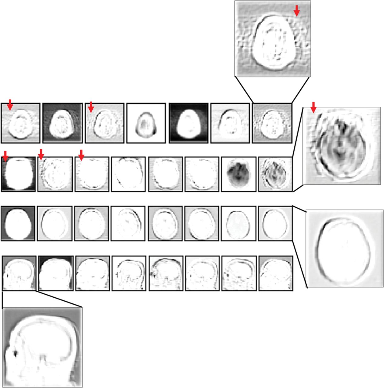
  • Fig 3.
    • Download figure
    • Open in new tab
    • Download powerpoint
    Fig 3.

    Representative filter responses from the fourth convolution layer of the CNN (Conv2D_4). Rows 1 and 2, Filter responses for motion-corrupted axial FLAIR/T2* input images, respectively. Rows 3 and 4, Filter responses from axial/sagittal T1 input images without motion, respectively. Filter responses are independent of image contrast and highlight the recognizable motion artifacts in the motion-corrupted images (arrows).

  • Fig 4.
    • Download figure
    • Open in new tab
    • Download powerpoint
    Fig 4.

    Examples of classification performance for 3 series. A few slices are displayed from each series (left), together with the slice ratings for the entire series (right). The numbers at the top left corner of each image represent the slice number.

Tables

  • Figures
    • View popup
    Table 1:

    Results of the survey—rule out MS clinical scan indicationa

    Doctor IDNo. Series of Insufficient QualityTechnician ID
    T1 (n = 26)T2 (n = 31)T3 (n = 12)T4 (n = 13)
    Unneeded RescansUnneeded RecallsUnneeded RescansUnneeded RecallsUnneeded RescansUnneeded RecallsUnneeded RescansUnneeded Recalls
    D0247570214011
    D1131411914522
    D23541348023022
    D324118135213010
    D4287862116014
    D5304843119017
    Mean ± SD25.7 ± 7.47.8 ± 47.2 ± 48.8 ± 63.2 ± 2.91.7 ± 1.415 ± 6.10.3 ± 0.812.7 ± 6.8
    • Note:—ID indicates identification. The numbers in parenthesis next to the technician identification numbers represent the total numbers of insufficient quality series identified by each rater.

    • ↵a All numbers reported are from the 49 series of the survey. Each series was evaluated twice, assuming that the scan indication was MS and stroke.

    • View popup
    Table 2:

    Results of the survey—rule out stroke clinical scan indicationa

    Doctor IDNo. Series of Insufficient QualityTechnician ID
    T1 (n = 12)T2 (n = 28)T3 (n = 7)T4 (n = 13)
    Unneeded RescansUnneeded RecallsUnneeded RescansUnneeded RecallsUnneeded RescansUnneeded RecallsUnneeded RescansUnneeded Recalls
    D1210026050110
    D213231500622
    D38612213371
    D411431812604
    D57502103360
    Mean ± SD8.2 ± 4.25.4 ± 31.4 ± 0.720.4 ± 4.20.4 ± 0.22.6 ± 0.83.6 ± 1.16 ± 3.41 ± 0.4
    • Note:—ID indicates identification. The numbers in parenthesis next to the technician identification numbers represent the total numbers of insufficient quality series identified by each rater.

    • ↵a All numbers reported are from the 49 series of the survey. Each series was evaluated twice, assuming that the scan indication was multiple sclerosis and stroke.

    • View popup
    Table 3:

    Matrix documenting the number of unneeded rescans and recalls created by the DL approach with different thresholds, assuming that series were scanned to rule out MSa

    DL (T = 0.1)DL (T = 0.5)DL (T = 0.8)
    RescansRecallsRescansRecallsRescansRecalls
    D0286390
    D183151211
    D211831146
    D3510106144
    D42115571
    D51133663
    Mean ± SD3.2 ± 2.810.5 ± 2.17 ± 4.75.3 ± 1.410.2 ± 2.62.5 ± 0.9
    • ↵a All numbers are from the 49 test series. Here D0–D5 represent the same individuals as in Tables 1 and 2.

    • View popup
    Table 4:

    Matrix documenting the number of unneeded rescans and recalls created by the DL approach with different thresholds, assuming that the series were scanned to rule out strokea

    DL (T = 0.5)DL (T = 0.1)DL (T = 5e–4)DL (T = 1e–6)
    RescansRecallsRescansRecallsRescansRecallsRescansRecalls
    D125016011040
    D2151833307
    D32001328234
    D41711036427
    D52011217123
    Mean ± SD19.4 ± 3.80.4 ± 0.211.8 ± 1.41.8 ± 1.37 ± 2.92 ± 1.62.2 ± 1.54.2 ± 3
    • ↵a All numbers are from the 49 test series. Here D1–D5 represent the same individuals as in Tables 1 and 2. Physician D0, whose ratings were used to train the DL algorithm, is now absent (as in Table 2) because no “stroke” ratings were available for this reader.

PreviousNext
Back to top

In this issue

American Journal of Neuroradiology: 40 (2)
American Journal of Neuroradiology
Vol. 40, Issue 2
1 Feb 2019
  • Table of Contents
  • Index by author
  • Complete Issue (PDF)
Advertisement
Print
Download PDF
Email Article

Thank you for your interest in spreading the word on American Journal of Neuroradiology.

NOTE: We only request your email address so that the person you are recommending the page to knows that you wanted them to see it, and that it is not junk mail. We do not capture any email address.

Enter multiple addresses on separate lines or separate them with commas.
A Deep Learning–Based Approach to Reduce Rescan and Recall Rates in Clinical MRI Examinations
(Your Name) has sent you a message from American Journal of Neuroradiology
(Your Name) thought you would like to see the American Journal of Neuroradiology web site.
CAPTCHA
This question is for testing whether or not you are a human visitor and to prevent automated spam submissions.
Cite this article
A. Sreekumari, D. Shanbhag, D. Yeo, T. Foo, J. Pilitsis, J. Polzin, U. Patil, A. Coblentz, A. Kapadia, J. Khinda, A. Boutet, J. Port, I. Hancu
A Deep Learning–Based Approach to Reduce Rescan and Recall Rates in Clinical MRI Examinations
American Journal of Neuroradiology Feb 2019, 40 (2) 217-223; DOI: 10.3174/ajnr.A5926

Citation Manager Formats

  • BibTeX
  • Bookends
  • EasyBib
  • EndNote (tagged)
  • EndNote 8 (xml)
  • Medlars
  • Mendeley
  • Papers
  • RefWorks Tagged
  • Ref Manager
  • RIS
  • Zotero
0 Responses
Respond to this article
Share
Bookmark this article
A Deep Learning–Based Approach to Reduce Rescan and Recall Rates in Clinical MRI Examinations
A. Sreekumari, D. Shanbhag, D. Yeo, T. Foo, J. Pilitsis, J. Polzin, U. Patil, A. Coblentz, A. Kapadia, J. Khinda, A. Boutet, J. Port, I. Hancu
American Journal of Neuroradiology Feb 2019, 40 (2) 217-223; DOI: 10.3174/ajnr.A5926
del.icio.us logo Twitter logo Facebook logo Mendeley logo
  • Tweet Widget
  • Facebook Like
  • Google Plus One
Purchase

Jump to section

  • Article
    • Abstract
    • ABBREVIATIONS:
    • Materials and Methods
    • Results
    • Discussion
    • Conclusions
    • Footnotes
    • References
  • Figures & Data
  • Supplemental
  • Info & Metrics
  • Responses
  • References
  • PDF

Related Articles

  • PubMed
  • Google Scholar

Cited By...

  • Opportunities for Understanding MS Mechanisms and Progression With MRI Using Large-Scale Data Sharing and Artificial Intelligence
  • Artificial Intelligence in Neuroradiology: Current Status and Future Directions
  • Crossref (29)
  • Google Scholar

This article has been cited by the following articles in journals that are participating in Crossref Cited-by Linking.

  • Convergence of Artificial Intelligence and Neuroscience towards the Diagnosis of Neurological Disorders—A Scoping Review
    Chellammal Surianarayanan, John Jeyasekaran Lawrence, Pethuru Raj Chelliah, Edmond Prakash, Chaminda Hewage
    Sensors 2023 23 6
  • Enhancing head and neck tumor management with artificial intelligence: Integration and perspectives
    Nian-Nian Zhong, Han-Qi Wang, Xin-Yue Huang, Zi-Zhan Li, Lei-Ming Cao, Fang-Yi Huo, Bing Liu, Lin-Lin Bu
    Seminars in Cancer Biology 2023 95
  • MR Imaging in the 21st Century: Technical Innovation over the First Two Decades
    Hiroyuki Kabasawa
    Magnetic Resonance in Medical Sciences 2022 21 1
  • Diverse Applications of Artificial Intelligence in Neuroradiology
    Michael Tran Duong, Andreas M. Rauschecker, Suyash Mohan
    Neuroimaging Clinics of North America 2020 30 4
  • Applications of Artificial Intelligence in the Radiology Roundtrip: Process Streamlining, Workflow Optimization, and Beyond
    Kevin Pierre, Adam G. Haneberg, Sean Kwak, Keith R. Peters, Bruno Hochhegger, Thiparom Sananmuang, Padcha Tunlayadechanont, Patrick J. Tighe, Anthony Mancuso, Reza Forghani
    Seminars in Roentgenology 2023 58 2
  • Improved Multiclass Brain Tumor Detection via Customized Pretrained EfficientNetB7 Model
    Hafiz Muhammad Tayyab Khushi, Tehreem Masood, Arfan Jaffar, Muhammad Rashid, Sheeraz Akram
    IEEE Access 2023 11
  • Overview of Noninterpretive Artificial Intelligence Models for Safety, Quality, Workflow, and Education Applications in Radiology Practice
    Yasasvi Tadavarthi, Valeria Makeeva, William Wagstaff, Henry Zhan, Anna Podlasek, Neil Bhatia, Marta Heilbrun, Elizabeth Krupinski, Nabile Safdar, Imon Banerjee, Judy Gichoya, Hari Trivedi
    Radiology: Artificial Intelligence 2022 4 2
  • Lung Nodule Malignancy Prediction in Sequential CT Scans: Summary of ISBI 2018 Challenge
    Yoganand Balagurunathan, Andrew Beers, Michael Mcnitt-Gray, Lubomir Hadjiiski, Sandy Napel, Dmitry Goldgof, Gustavo Perez, Pablo Arbelaez, Alireza Mehrtash, Tina Kapur, Ehwa Yang, Jung Won Moon, Gabriel Bernardino Perez, Ricard Delgado-Gonzalo, M. Mehdi Farhangi, Amir A. Amini, Renkun Ni, Xue Feng, Aditya Bagari, Kiran Vaidhya, Benjamin Veasey, Wiem Safta, Hichem Frigui, Joseph Enguehard, Ali Gholipour, Laura Silvana Castillo, Laura Alexandra Daza, Paul Pinsky, Jayashree Kalpathy-Cramer, Keyvan Farahani
    IEEE Transactions on Medical Imaging 2021 40 12
  • Economic evaluations of big data analytics for clinical decision-making: a scoping review
    Lytske Bakker, Jos Aarts, Carin Uyl-de Groot, William Redekop
    Journal of the American Medical Informatics Association 2020 27 9
  • Motion artifact reduction for magnetic resonance imaging with deep learning and k-space analysis
    Long Cui, Yang Song, Yida Wang, Rui Wang, Dongmei Wu, Haibin Xie, Jianqi Li, Guang Yang, Zhifan Gao
    PLOS ONE 2023 18 1

More in this TOC Section

  • Diagnostic Neuroradiology of Monoclonal Antibodies
  • ML for Glioma Molecular Subtype Prediction
  • NCCT vs. MRI for Brain Atrophy in Acute Stroke
Show more ADULT BRAIN

Similar Articles

Advertisement

Indexed Content

  • Current Issue
  • Accepted Manuscripts
  • Article Preview
  • Past Issues
  • Editorials
  • Editor's Choice
  • Fellows' Journal Club
  • Letters to the Editor
  • Video Articles

Cases

  • Case Collection
  • Archive - Case of the Week
  • Archive - Case of the Month
  • Archive - Classic Case

More from AJNR

  • Trainee Corner
  • Imaging Protocols
  • MRI Safety Corner
  • Book Reviews

Multimedia

  • AJNR Podcasts
  • AJNR Scantastics

Resources

  • Turnaround Time
  • Submit a Manuscript
  • Submit a Video Article
  • Submit an eLetter to the Editor/Response
  • Manuscript Submission Guidelines
  • Statistical Tips
  • Fast Publishing of Accepted Manuscripts
  • Graphical Abstract Preparation
  • Imaging Protocol Submission
  • Evidence-Based Medicine Level Guide
  • Publishing Checklists
  • Author Policies
  • Become a Reviewer/Academy of Reviewers
  • News and Updates

About Us

  • About AJNR
  • Editorial Board
  • Editorial Board Alumni
  • Alerts
  • Permissions
  • Not an AJNR Subscriber? Join Now
  • Advertise with Us
  • Librarian Resources
  • Feedback
  • Terms and Conditions
  • AJNR Editorial Board Alumni

American Society of Neuroradiology

  • Not an ASNR Member? Join Now

© 2025 by the American Society of Neuroradiology All rights, including for text and data mining, AI training, and similar technologies, are reserved.
Print ISSN: 0195-6108 Online ISSN: 1936-959X

Powered by HighWire