
Article Figures & Data
Figures
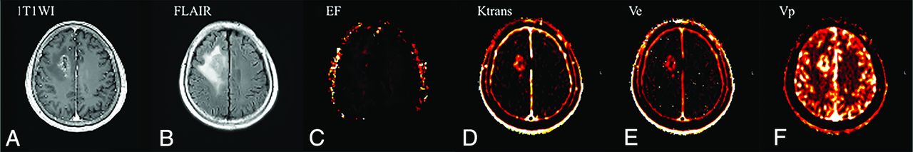
- Fig 1.
A 64-year-old patient with glioblastoma who had early disease progression (PFS = 14 months) after standard treatment. The preoperative axial contrast-enhanced T1WI (A) and FLAIR (B) images represent an enhancing area and nonenhancing FLAIR high-signal-intensity lesions, respectively. The preoperative parametric maps of contrast leakage information (EF, Ktrans, Ve, and Vp) are shown (C, D, E, and F, respectively). A low EF value on the nonenhancing FLAIR high-signal-intensity lesion was observed, with an EF 95th percentile value of 5.67.
- Fig 2.
A 60-year-old patient with glioblastoma who had nonprogression (PFS = 31 months) after standard treatment. The preoperative axial contrast-enhanced T1WI (A) and FLAIR (B) images represent an enhancing area and a nonenhancing FLAIR high-signal-intensity lesion, respectively. The preoperative parametric maps of contrast leakage information (EF, Ktrans, Ve, and Vp) are shown (C, D, E, and F, respectively). A high EF value on a nonenhancing FLAIR high-signal-intensity lesion was noted, with an EF 95th percentile value of 16.69.


Tables
Characteristics Total (n = 102) Progression (n = 87) Nonprogression (n = 15) P Value Mean age (yr) 56.9 59.2 ± 13.5 51.3 ± 14.0 .04b Mean radiation dose (Gy) 51.9 52.5 ± 18.4 48.6 ± 0.6 .43b Sex .40c Male 59 52 7 Female 43 35 8 Methylated MGMT promoter .09c Positive 59 47 12 Negative 42 39 3 IDH1/2 mutation .28c Positive 7 5 2 Negative 94 81 13 - Table 2:
Comparison of the parametric values of the progression and nonprogression groupsa
Parameters Progression (n = 71) Nonprogression (n = 15) P Valueb Mean tumor volume (mL) 119.84 ± 85.65 161.81 ± 102.59 .10 EF 95th PV (%) 11.053 ± 7.651 15.790 ± 8.693 .04 EF mean (%) 2.473 ± 1.552 3.140 ± 1.463 .13 Ktrans 95th PV (min–1) 0.140 ± 0.118 0.177 ± 0.132 .28 Ktrans mean (min–1) 0.022 ± 0.024 0.0260 ± 0.021 .59 Ve 95th PV 51.604 ± 71.965 44.480 ± 29.773 .71 Ve mean 7.750 ± 16.050 6.209 ± 4.917 .71 Vp 95th PV 5.334 ± 5.488 6.594 ± 4.807 .41 Vp mean 1.264 ± 1.352 1.384 ± 0.100 .75


Jump to section
Related Articles
Cited By...
- No citing articles found.