Advertisement

Main menu

  • Home
  • Content
    • Current Issue
    • Accepted Manuscripts
    • Article Preview
    • Past Issue Archive
    • Video Articles
    • AJNR Case Collection
    • Case of the Week Archive
    • Case of the Month Archive
    • Classic Case Archive
  • Special Collections
    • AJNR Awards
    • Low-Field MRI
    • Alzheimer Disease
    • ASNR Foundation Special Collection
    • Photon-Counting CT
    • View All
  • Multimedia
    • AJNR Podcasts
    • AJNR SCANtastic
    • Trainee Corner
    • MRI Safety Corner
    • Imaging Protocols
  • For Authors
    • Submit a Manuscript
    • Submit a Video Article
    • Submit an eLetter to the Editor/Response
    • Manuscript Submission Guidelines
    • Statistical Tips
    • Fast Publishing of Accepted Manuscripts
    • Graphical Abstract Preparation
    • Imaging Protocol Submission
    • Author Policies
  • About Us
    • About AJNR
    • Editorial Board
    • Editorial Board Alumni
  • More
    • Become a Reviewer/Academy of Reviewers
    • Subscribers
    • Permissions
    • Alerts
    • Feedback
    • Advertisers
    • ASNR Home

User menu

  • Alerts
  • Log in

Search

  • Advanced search
American Journal of Neuroradiology

American Journal of Neuroradiology

ASHNR American Society of Functional Neuroradiology ASHNR American Society of Pediatric Neuroradiology ASSR
  • Alerts
  • Log in

Advanced Search

  • Home
  • Content
    • Current Issue
    • Accepted Manuscripts
    • Article Preview
    • Past Issue Archive
    • Video Articles
    • AJNR Case Collection
    • Case of the Week Archive
    • Case of the Month Archive
    • Classic Case Archive
  • Special Collections
    • AJNR Awards
    • Low-Field MRI
    • Alzheimer Disease
    • ASNR Foundation Special Collection
    • Photon-Counting CT
    • View All
  • Multimedia
    • AJNR Podcasts
    • AJNR SCANtastic
    • Trainee Corner
    • MRI Safety Corner
    • Imaging Protocols
  • For Authors
    • Submit a Manuscript
    • Submit a Video Article
    • Submit an eLetter to the Editor/Response
    • Manuscript Submission Guidelines
    • Statistical Tips
    • Fast Publishing of Accepted Manuscripts
    • Graphical Abstract Preparation
    • Imaging Protocol Submission
    • Author Policies
  • About Us
    • About AJNR
    • Editorial Board
    • Editorial Board Alumni
  • More
    • Become a Reviewer/Academy of Reviewers
    • Subscribers
    • Permissions
    • Alerts
    • Feedback
    • Advertisers
    • ASNR Home
  • Follow AJNR on Twitter
  • Visit AJNR on Facebook
  • Follow AJNR on Instagram
  • Join AJNR on LinkedIn
  • RSS Feeds

AJNR Awards, New Junior Editors, and more. Read the latest AJNR updates

LetterLetter

An Important Pitfall in Diagnosing Intracanalicular Vestibular Schwannoma

A. Hakim and F. Wagner
American Journal of Neuroradiology October 2019, 40 (10) E58-E59; DOI: https://doi.org/10.3174/ajnr.A6192
A. Hakim
aUniversity Institute of Diagnostic and Interventional Neuroradiology Bern University Hospital, Inselspital University of Bern, Bern, Switzerland
  • Find this author on Google Scholar
  • Find this author on PubMed
  • Search for this author on this site
  • ORCID record for A. Hakim
F. Wagner
aUniversity Institute of Diagnostic and Interventional Neuroradiology Bern University Hospital, Inselspital University of Bern, Bern, Switzerland
  • Find this author on Google Scholar
  • Find this author on PubMed
  • Search for this author on this site
  • ORCID record for F. Wagner
  • Article
  • Figures & Data
  • Info & Metrics
  • Responses
  • References
  • PDF
Loading

Technical advances in MR imaging enable scanning of millimeter- and submillimeter-thick slices in a relatively short acquisition time, so sequences such as contrast-enhanced 3D T1 (eg, MPRAGE) are included in standard MR imaging protocols in many institutions. The MPRAGE sequence is especially important for the detection of small lesions such as a small intracanalicular vestibular schwannoma (Koos grade I).

The members of our interdisciplinary schwannoma board have encountered several patients with varying degrees of enhancement in the fundus of the internal auditory canal (IAC) who were referred to us with suspected schwannomas (Fig 1A–D). However, when we reviewed the patients' initial MR images and the follow-up images, that was a false-positive finding.

Fig 1.
  • Download figure
  • Open in new tab
  • Download powerpoint
Fig 1.

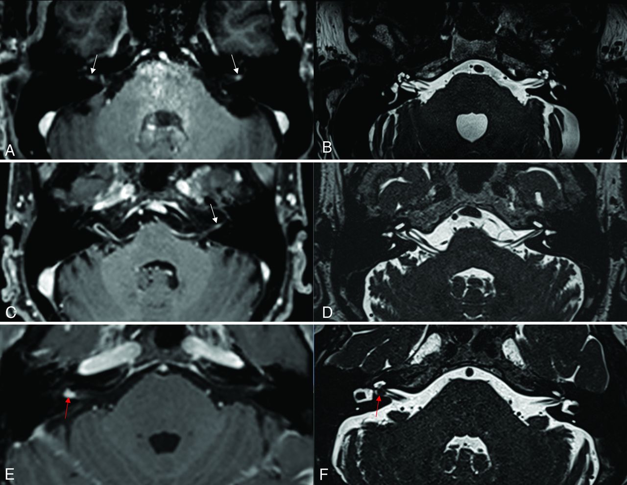
MPRAGE (A, C, and E) and CISS (B, D, and F) in 3 different patients (patient 1: A and B; patient 2: C and D; and patient 3: E and F). MPRAGE shows different degrees of enhancement in the fundus of the internal auditory canal bilaterally (left more than right) in patient 1 and on the left side in patient 2 (white arrows in A and C). However, in both patients, no analogous lesion was detected in the CISS sequence. Therefore, the enhancement was not due to the presence of a schwannoma but was most likely due to a vascular structure. In contrast, in patient 3, there was nodular enhancement in the fundus of the internal auditory canal on the right side (red arrow in E), which was accompanied by a corresponding lesion “filling defect” on the CISS sequence (red arrow in F) due to the presence of a small intracanalicular vestibular schwannoma (Koos I). Correlating findings in MPRAGE with the CISS sequence is crucial to avoiding misinterpretation of this enhancement.

Enhancement in the fundus of the IAC is sometimes detected in subjects without pathologic conditions, and it can be unilateral or bilateral and present in varying degrees (Fig 1A, -C). This is most likely due to the enhancement of vascular structures such as the venous plexus surrounding the nerve sheath (eg, around the Scarpa ganglion) in the acoustic facial cistern1 or due to the presence of capillaries in the meningeal layers covering the fundus.

In a recently published study performed using synchrotron phase-contrast imaging, Mei et al2 demonstrated the presence of previously unknown pillars or villi that support the cranial nerves in the IAC and are occasionally associated with blood vessels, mostly capillaries. These vascular structures within the villi may play a key role in the enhancement in this region, and variation in the amount of blood vessels between different subjects may be one cause for the different degrees of enhancement observed on postcontrast MR images (Fig 1A, -C).

Therefore, on the basis of our experience, the presence of enhancement in this region should be always correlated with the CISS sequence to confirm or exclude the presence of a nodular lesion, which represents the schwannoma (Fig 1F).

Knowledge of this pitfall is important so that CISS can be added if such an incidental finding is present, to reduce the number of false-positive cases and thereby reduce the unnecessary follow-up scans and consequentially to lower health care costs. This precaution will also avoid instilling unnecessary fear in patients who incorrectly believe they have a tumor that may affect their hearing and lifestyle.

Footnotes

  • Disclosures: Arsany Hakim—UNRELATED: Grants/Grants Pending: Swiss Heart Association.* Franca Wagner—UNRELATED: Grants/Grants Pending: Swiss MS Society.* *Money paid to the institution.

References

  1. 1.↵
    1. Lescanne E,
    2. Velut S,
    3. Lefrancq T, et al
    . The internal acoustic meatus and its meningeal layers: a microanatomical study. J Neurosurg 2002;97:1191–97 doi:10.3171/jns.2002.97.5.1191 pmid:12450043
    CrossRefPubMed
  2. 2.↵
    1. Mei X,
    2. Schart‐Morén N,
    3. Li H, et al
    . Three‐dimensional imaging of the human internal acoustic canal and arachnoid cistern: a synchrotron study with clinical implications. J Anat 2019;234:316–26 doi:10.1111/joa.12926 pmid:30565214
    CrossRefPubMed
  • © 2019 by American Journal of Neuroradiology
PreviousNext
Back to top

In this issue

American Journal of Neuroradiology: 40 (10)
American Journal of Neuroradiology
Vol. 40, Issue 10
1 Oct 2019
  • Table of Contents
  • Index by author
  • Complete Issue (PDF)
Advertisement
Print
Download PDF
Email Article

Thank you for your interest in spreading the word on American Journal of Neuroradiology.

NOTE: We only request your email address so that the person you are recommending the page to knows that you wanted them to see it, and that it is not junk mail. We do not capture any email address.

Enter multiple addresses on separate lines or separate them with commas.
An Important Pitfall in Diagnosing Intracanalicular Vestibular Schwannoma
(Your Name) has sent you a message from American Journal of Neuroradiology
(Your Name) thought you would like to see the American Journal of Neuroradiology web site.
CAPTCHA
This question is for testing whether or not you are a human visitor and to prevent automated spam submissions.
Cite this article
A. Hakim, F. Wagner
An Important Pitfall in Diagnosing Intracanalicular Vestibular Schwannoma
American Journal of Neuroradiology Oct 2019, 40 (10) E58-E59; DOI: 10.3174/ajnr.A6192

Citation Manager Formats

  • BibTeX
  • Bookends
  • EasyBib
  • EndNote (tagged)
  • EndNote 8 (xml)
  • Medlars
  • Mendeley
  • Papers
  • RefWorks Tagged
  • Ref Manager
  • RIS
  • Zotero
0 Responses
Respond to this article
Share
Bookmark this article
An Important Pitfall in Diagnosing Intracanalicular Vestibular Schwannoma
A. Hakim, F. Wagner
American Journal of Neuroradiology Oct 2019, 40 (10) E58-E59; DOI: 10.3174/ajnr.A6192
del.icio.us logo Twitter logo Facebook logo Mendeley logo
  • Tweet Widget
  • Facebook Like
  • Google Plus One
Purchase

Jump to section

  • Article
    • Footnotes
    • References
  • Figures & Data
  • Info & Metrics
  • Responses
  • References
  • PDF

Related Articles

  • No related articles found.
  • PubMed
  • Google Scholar

Cited By...

  • No citing articles found.
  • Crossref (2)
  • Google Scholar

This article has been cited by the following articles in journals that are participating in Crossref Cited-by Linking.

  • Radiopaedia.org
    Frank Gaillard, Rohit Sharma, Carlos Peñafiel Salgado
    2008
  • What Radiology Residents Need to Know: Neuroradiology
    Yuh-Shin Chang, Gul Moonis
    2024

More in this TOC Section

  • Brain AVM’s Nidus: What if We Hadn’t Understood Anything?
  • Letter to the Editor regarding “Automated Volumetric Software in Dementia: Help or Hindrance to the Neuroradiologist?”
  • Reply:
Show more LETTERS

Similar Articles

Advertisement

Indexed Content

  • Current Issue
  • Accepted Manuscripts
  • Article Preview
  • Past Issues
  • Editorials
  • Editor's Choice
  • Fellows' Journal Club
  • Letters to the Editor
  • Video Articles

Cases

  • Case Collection
  • Archive - Case of the Week
  • Archive - Case of the Month
  • Archive - Classic Case

More from AJNR

  • Trainee Corner
  • Imaging Protocols
  • MRI Safety Corner
  • Book Reviews

Multimedia

  • AJNR Podcasts
  • AJNR Scantastics

Resources

  • Turnaround Time
  • Submit a Manuscript
  • Submit a Video Article
  • Submit an eLetter to the Editor/Response
  • Manuscript Submission Guidelines
  • Statistical Tips
  • Fast Publishing of Accepted Manuscripts
  • Graphical Abstract Preparation
  • Imaging Protocol Submission
  • Evidence-Based Medicine Level Guide
  • Publishing Checklists
  • Author Policies
  • Become a Reviewer/Academy of Reviewers
  • News and Updates

About Us

  • About AJNR
  • Editorial Board
  • Editorial Board Alumni
  • Alerts
  • Permissions
  • Not an AJNR Subscriber? Join Now
  • Advertise with Us
  • Librarian Resources
  • Feedback
  • Terms and Conditions
  • AJNR Editorial Board Alumni

American Society of Neuroradiology

  • Not an ASNR Member? Join Now

© 2025 by the American Society of Neuroradiology All rights, including for text and data mining, AI training, and similar technologies, are reserved.
Print ISSN: 0195-6108 Online ISSN: 1936-959X

Powered by HighWire