Advertisement

Main menu

  • Home
  • Content
    • Current Issue
    • Accepted Manuscripts
    • Article Preview
    • Past Issue Archive
    • Video Articles
    • AJNR Case Collection
    • Case of the Week Archive
    • Case of the Month Archive
    • Classic Case Archive
  • Special Collections
    • Advancing NeuroMRI with High-Relaxivity Contrast Agents
    • AJNR Awards
    • Low-Field MRI
    • Alzheimer Disease
    • ASNR Foundation Special Collection
    • Photon-Counting CT
    • View All
  • Multimedia
    • AJNR Podcasts
    • AJNR SCANtastic
    • Trainee Corner
    • MRI Safety Corner
    • Imaging Protocols
  • For Authors
    • Submit a Manuscript
    • Submit a Video Article
    • Submit an eLetter to the Editor/Response
    • Manuscript Submission Guidelines
    • Statistical Tips
    • Fast Publishing of Accepted Manuscripts
    • Graphical Abstract Preparation
    • Imaging Protocol Submission
    • Author Policies
  • About Us
    • About AJNR
    • Editorial Board
    • Editorial Board Alumni
  • More
    • Become a Reviewer/Academy of Reviewers
    • Subscribers
    • Permissions
    • Alerts
    • Feedback
    • Advertisers
    • ASNR Home

User menu

  • Alerts
  • Log in

Search

  • Advanced search
American Journal of Neuroradiology

American Journal of Neuroradiology

ASHNR American Society of Functional Neuroradiology ASHNR American Society of Pediatric Neuroradiology ASSR
  • Alerts
  • Log in

Advanced Search

  • Home
  • Content
    • Current Issue
    • Accepted Manuscripts
    • Article Preview
    • Past Issue Archive
    • Video Articles
    • AJNR Case Collection
    • Case of the Week Archive
    • Case of the Month Archive
    • Classic Case Archive
  • Special Collections
    • Advancing NeuroMRI with High-Relaxivity Contrast Agents
    • AJNR Awards
    • Low-Field MRI
    • Alzheimer Disease
    • ASNR Foundation Special Collection
    • Photon-Counting CT
    • View All
  • Multimedia
    • AJNR Podcasts
    • AJNR SCANtastic
    • Trainee Corner
    • MRI Safety Corner
    • Imaging Protocols
  • For Authors
    • Submit a Manuscript
    • Submit a Video Article
    • Submit an eLetter to the Editor/Response
    • Manuscript Submission Guidelines
    • Statistical Tips
    • Fast Publishing of Accepted Manuscripts
    • Graphical Abstract Preparation
    • Imaging Protocol Submission
    • Author Policies
  • About Us
    • About AJNR
    • Editorial Board
    • Editorial Board Alumni
  • More
    • Become a Reviewer/Academy of Reviewers
    • Subscribers
    • Permissions
    • Alerts
    • Feedback
    • Advertisers
    • ASNR Home
  • Follow AJNR on Twitter
  • Visit AJNR on Facebook
  • Follow AJNR on Instagram
  • Join AJNR on LinkedIn
  • RSS Feeds

AJNR Awards, New Junior Editors, and more. Read the latest AJNR updates


Improved Turnaround Times | Median time to first decision: 12 days

Research ArticleHead and Neck Imaging

Solitary Parathyroid Adenoma Localization in Technetium Tc99m Sestamibi SPECT and Multiphase Multidetector 4D CT

T.H. Vu, D. Schellingerhout, N. Guha-Thakurta, J. Sun, W. Wei, S.C. Kappadth, N. Perrier, E.E. Kim, E. Rohren, H.H. Chuang and F.C. Wong
American Journal of Neuroradiology January 2019, 40 (1) 142-149; DOI: https://doi.org/10.3174/ajnr.A5901
T.H. Vu
aFrom the Departments of Diagnostic Radiology (T.H.V., D.S., N.G.-T.)
  • Find this author on Google Scholar
  • Find this author on PubMed
  • Search for this author on this site
  • ORCID record for T.H. Vu
D. Schellingerhout
aFrom the Departments of Diagnostic Radiology (T.H.V., D.S., N.G.-T.)
  • Find this author on Google Scholar
  • Find this author on PubMed
  • Search for this author on this site
  • ORCID record for D. Schellingerhout
N. Guha-Thakurta
aFrom the Departments of Diagnostic Radiology (T.H.V., D.S., N.G.-T.)
  • Find this author on Google Scholar
  • Find this author on PubMed
  • Search for this author on this site
  • ORCID record for N. Guha-Thakurta
J. Sun
bBiostatistics (J.S., W.W.)
  • Find this author on Google Scholar
  • Find this author on PubMed
  • Search for this author on this site
  • ORCID record for J. Sun
W. Wei
bBiostatistics (J.S., W.W.)
  • Find this author on Google Scholar
  • Find this author on PubMed
  • Search for this author on this site
  • ORCID record for W. Wei
S.C. Kappadth
cImaging Physics (S.C.K.)
  • Find this author on Google Scholar
  • Find this author on PubMed
  • Search for this author on this site
  • ORCID record for S.C. Kappadth
N. Perrier
dSurgical Oncology (N.P.)
  • Find this author on Google Scholar
  • Find this author on PubMed
  • Search for this author on this site
  • ORCID record for N. Perrier
E.E. Kim
fDepartment of Radiological Sciences (E.E.K.), University of California at Irvine, Orange, California
  • Find this author on Google Scholar
  • Find this author on PubMed
  • Search for this author on this site
  • ORCID record for E.E. Kim
E. Rohren
gBaylor College of Medicine (E.R.), Houston, Texas.
  • Find this author on Google Scholar
  • Find this author on PubMed
  • Search for this author on this site
  • ORCID record for E. Rohren
H.H. Chuang
eNuclear Medicine (H.H.C., F.C.W.), The University of Texas MD Anderson Cancer Center, Houston, Texas
  • Find this author on Google Scholar
  • Find this author on PubMed
  • Search for this author on this site
  • ORCID record for H.H. Chuang
F.C. Wong
eNuclear Medicine (H.H.C., F.C.W.), The University of Texas MD Anderson Cancer Center, Houston, Texas
  • Find this author on Google Scholar
  • Find this author on PubMed
  • Search for this author on this site
  • ORCID record for F.C. Wong
  • Article
  • Figures & Data
  • Info & Metrics
  • Responses
  • References
  • PDF
Loading

Article Figures & Data

Figures

  • Tables
  • Fig 1.
    • Download figure
    • Open in new tab
    • Download powerpoint
    Fig 1.

    Surgical classification of parathyroid adenoma locations, anterior view. A, Superior, in proximity to the posterior surface of the thyroid parenchyma. B, Superior, fallen posteriorly into the tracheoesophageal groove and no longer in contact with the posterior surface of the thyroid tissue. C, Superior, fallen posteriorly into the tracheoesophageal groove and no longer in contact with the posterior surface of the thyroid tissue at the inferior pole close to the clavicles. D, Superior or inferior, in the midregion of the posterior surface of the thyroid parenchyma near the junction of the recurrent laryngeal nerve and the inferior thyroidal artery. E, Inferior, in the region inferior to the thyroid gland, lying in the anteroposterior plane of the thyroid and anterior to the trachea. F, Inferior, descended into the thyrothymic ligament or superior thymus and possibly appearing to be “ectopic” or in the mediastinum. G, Intrathyroidal.
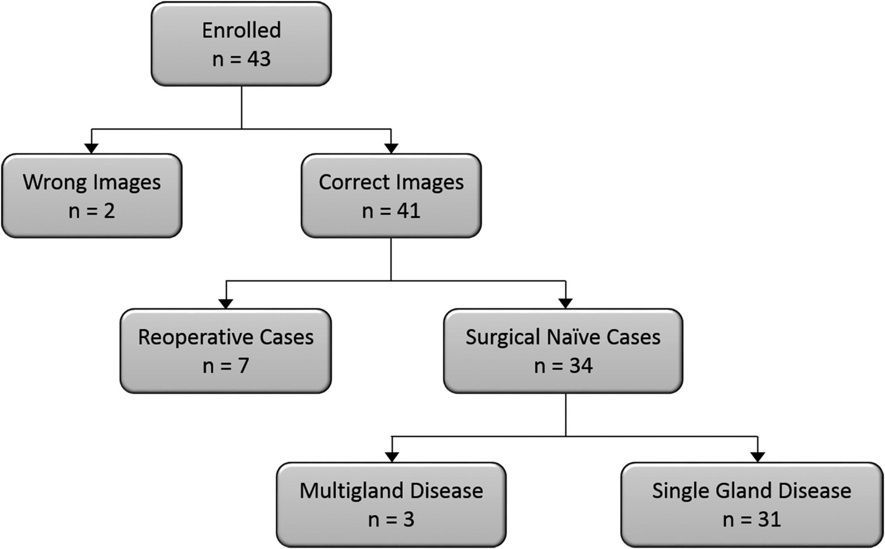
  • Fig 2.
    • Download figure
    • Open in new tab
    • Download powerpoint
    Fig 2.

    Patient selection flowchart. Thirty-one patients were included in the final analysis.

  • Fig 3.
    • Download figure
    • Open in new tab
    • Download powerpoint
    Fig 3.

    A, The axial arterial phase of a multiphase multidetector 4D CT image shows a right tracheoesophageal parathyroid adenoma (type C; Fig 1). B, Anterior and posterior delay planar scintigraphy shows retention of the radiotracer on the right side. C, An axial SPECT image shows retention of the radiotracer in the lower neck. D, An axial SPECT image fused to a noncontrast CT image localizes the retention of the radiotracer to the right tracheoesophageal groove. E, An axial SPECT image fused to the axial arterial phase of a multiphase multidetector 4D CT image localizes the retention of the radiotracer with concomitant early enhancement to the right tracheoesophageal groove. Red and white arrows show the parathyroid adenoma.

  • Fig 4.
    • Download figure
    • Open in new tab
    • Download powerpoint
    Fig 4.

    A case in which 4DCT could diagnose a parathyroid adenoma in the face of a sestamibi study with negative findings. A, The axial arterial phase of a multiphase multidetector 4D CT image shows a small early-enhancing left paraesophageal parathyroid adenoma (arrow). B, An axial MIBI SPECT image fused to a noncontrast CT image reveals no retention of the radiotracer in the left central compartment of a surgically proven parathyroid adenoma, which was effectively demonstrated on 4DCT (arrow). C, An axial SPECT image fused to the axial arterial phase of a multiphase multidetector 4D CT image shows a small early-enhancing left paraesophageal parathyroid adenoma (arrow).

Tables

  • Figures
    • View popup
    Table 1:

    Diagnostic accuracy of left/right localization of parathyroid adenomas in the 31 patients in our study group

    Imaging ModalityLeft No. (%)Right No. (%)Total No. (%)Accuracy (95% CI)
    MIBI SPECT93.5 (78.6–99.2)
        Left13 (41.9)1 (3.2)14 (45.2)
        Right1 (3.2)16 (51.6)17 (54.8)
    4DCT96.8 (83.3–99.9)
        Left14 (45.2)1 (3.2)15 (48.4)
        Right0 (0.0)16 (51.6)16 (51.6)
    MIBI SPECT + 4DCT96.8 (83.3–99.9)
        Left14 (45.2)1 (3.2)15 (48.4)
        Right0 (0.0)16 (51.6)16 (51.6)
        Total14 (45.2)17 (54.8)
    • View popup
    Table 2:

    Diagnostic accuracy of embryologic origin of the abnormal parathyroid gland in the 31 patients in our study group

    Imaging ModalitySuperior No. (%)Inferior No. (%)Total No. (%)Accuracy (95% CI)
    MIBI SPECT74.2 (55.4–88.1)
        Superior gland10 (32.3)0 (0)10 (32.3)
        Inferior gland8 (25.8)13 (41.9)21 (67.7)
        Total18 (58.1)13 (41.9)
    4DCT90.3 (74.3–98.0)
        Superior gland15 (48.4)0 (0)15 (48.6)
        Inferior gland3 (9.7)13 (41.9)16 (51.6)
        Total18 (58.1)13 (41.9)
    MIBI SPECT + 4DCT96.8 (83.3–99.9)
        Superior gland17 (54.8)0 (0)17 (54.8)
        Inferior gland1 (3.2)13 (41.9)14 (45.2)
        Total18 (58.1)13 (41.9)
    • View popup
    Table 3:

    Diagnostic accuracy of quadrant localization of parathyroid adenomas in the 31 patients in our study group

    Imaging ModalityLI No. (%)LS No. (%)RI No. (%)RS No. (%)Total No. (%)Accuracy (95% CI)
    MIBI SPECT67.7 (48.6–83.3)
        LI2 (6.5)2 (6.5)1 (3.2)0 (0)5 (16.1)
        LS0 (0)9 (29.0)0 (0)0 (0)9 (29.0)
        RI0 (0)0 (0)10 (32.3)6 (19.4)16 (51.6)
        RS0 (0)1 (3.2)0 (0)0 (0)1 (3.2)
    4DCT87.1 (70.2–96.4)
        LI2 (6.5)1 (3.2)1 (3.2)0 (0)4 (12.9)
        LS0 (0)11 (35.5)0 (0)0 (0)11 (35.5)
        RI0 (0)0 (0)10 (32.3)2 (6.5)12 (38.7)
        RS0 (0)0 (0)0 (0)4 (12.9)4 (12.9)
    MIBI SPECT + 4DCT93.5 (78.6–99.2)
        LI2 (6.5)0 (0)1 (3.2)0 (0)3 (9.7)
        LS0 (0)12 (38.7)0 (0)0 (0)12 (38.7)
        RI0 (0)0 (0)10 (32.3)1 (3.2)11 (35.5)
        RS0 (0)0 (0)0 (0)5 (16.1)5 (16.1)
        Total2 (6.5)12 (38.7)11 (35.5)6 (19.4)
    • Note:—LI indicates left inferior; LS, left superior; RI, right inferior; RS, right superior.

    • View popup
    Table 4:

    Diagnostic accuracy of surgical classification (Fig 1) of parathyroid adenomas in the 31 patients in our study group

    Imaging ModalityA No. (%)B No. (%)C No. (%)D No. (%)E No. (%)Total No. (%)Accuracy (95% CI)
    MIBI SPECT54.8 (36.0–72.7)
        A3 (9.7)1 (3.2)0 (0)0 (0)0 (0)4 (12.9)
        B2 (6.5)3 (9.7)0 (0)0 (0)0 (0)5 (16.1)
        C2 (6.5)2 (6.5)3 (9.7)1 (3.2)0 (0)8 (25.8)
        D0 (0)1 (3.2)0 (0)0 (0)0 (0)1 (3.2)
        E2 (6.5)2 (6.5)0 (0)0 (0)8 (25.8)12 (38.7)
        F0 (0)0 (0)0 (0)0 (0)1 (3.2)1 (3.2)
    4DCT61.3 (42.2–78.2)
        A7 (22.6)4 (12.9)0 (0)0 (0)0 (0)11 (35.5)
        B1 (3.2)2 (6.5)0 (0)0 (0)0 (0)3 (9.7)
        C0 (0)1 (3.2)3 (9.7)0 (0)0 (0)4 (12.9)
        D0 (0)0 (0)0 (0)1 (3.2)3 (9.7)4 (12.9)
        E1 (3.2)2 (6.5)0 (0)0 (0)6 (19.4)9 (29.0)
        F0 (0)0 (0)0 (0)0 (0)0 (0)0 (0)
    MIBI SPECT + 4DCT74.2 (55.4–88.1)
        A7 (22.6)3 (9.7)0 (0)0 (0)0 (0)10 (32.3)
        B0 (0)4 (12.9)0 (0)0 (0)0 (0)4 (12.9)
        C0 (0)2 (6.5)3 (9.7)1 (3.2)0 (0)6 (19.4)
        D2 (6.5)0 (0)0 (0)0 (0)0 (0)2 (6.5)
        E0 (0)0 (0)0 (0)0 (0)9 (29)9 (29.0)
        F0 (0)0 (0)0 (0)0 (0)0 (0)0 (0)
        Total9 (29)9 (29)3 (9.7)1 (3.2)9 (29.0)
    • View popup
    Table 5:

    Level of confidence response from 3 reading groups for lateralization, upper/lower quadrant localization, and localization by surgical classification

    Response/Imaging ModalityCertain (%)Equivocal (%)Uncertain (%)
    Lateralization
        MIBI SPECT27 (87.1)1 (3.2)3 (9.7)
        4DCT30 (96.8)1 (3.2)0 (0.0)
        MIBI SPECT + 4DCT29 (93.5)2 (6.5)0 (0.0)
    Upper/lower
        MIBI SPECT22 (71.0)6 (19.4)3 (9.7)
        4DCT30 (96.8)1 (3.2)0 (0.0)
        MIBI SPECT + 4DCT31 (100.0)0 (0.0)0 (0.0)
    Surgical localization
        MIBI SPECT15 (48.4)15 (48.4)1 (3.2)
        4DCT28 (90.3)3 (9.7)0 (0.0)
        MIBI SPECT + 4DCT29 (93.5)1 (3.2)1 (3.2)
    • View popup
    Table 6:

    McNemar test of paired imaging modalities for diagnostic accuracy of left/right localization of parathyroid adenomas in the 31 patients in our study group

    Imaging ModalityCorrectWrongTotalP Value
    4DCT vs
    MIBI SPECT
        Correct28129.56
        Wrong202
        Total30131
    MIBI SPECT + 4DCT vs
    4DCT
        Correct291301.00
        Wrong101
        Total30131
    MIBI SPECT + 4DCT vs
    MIBI SPECT
        Correct28129.56
        Wrong202
        Total30131
    • View popup
    Table 7:

    McNemar test of paired imaging modalities for diagnostic accuracy of embryologic origin of the abnormal parathyroid gland in the 31 patients in our study group

    Imaging ModalityCorrectWrongTotalP Value
    4DCT vs
    MIBI SPECT
        Correct22123.06
        Wrong628
        Total28331
    MIBI SPECT + 4DCT vs
    4DCT
        Correct28028.16
        Wrong213
        Total30131
    MIBI SPECT + 4DCT vs
    MIBI SPECT
        Correct23023.008
        Wrong718
        Total30131
    • View popup
    Table 8:

    McNemar test of paired imaging modalities for diagnostic accuracy of quadrant localization of the abnormal parathyroid gland in the 31 patients in our study group

    Imaging ModalityCorrectWrongTotalP Value
    4DCT vs
    MIBI SPECT
        Correct19221.06
        Wrong8210
        Total27431
    MIBI SPECT + 4DCT vs
    4DCT
        Correct26127.32
        Wrong314
        Total29231
    MIBI SPECT + 4DCT vs
    MIBI SPECT
        Correct20121.01
        Wrong9110
        Total29231
    • View popup
    Table 9:

    McNemar test of paired imaging modalities for diagnostic accuracy of surgical localization of the abnormal parathyroid gland in the 31 patients in our study group

    Imaging ModalityCorrectWrongTotalP Value
    4DCT vs
    MIBI SPECT
        Correct11617.06
        Wrong8614
        Total191231
    MIBI SPECT + 4DCT vs
    4DCT
        Correct163190.21
        Wrong7512
        Total23831
    MIBI SPECT + 4DCT vs
    MIBI SPECT
        Correct15217.06
        Wrong8614
        Total23831
PreviousNext
Back to top

In this issue

American Journal of Neuroradiology: 40 (1)
American Journal of Neuroradiology
Vol. 40, Issue 1
1 Jan 2019
  • Table of Contents
  • Index by author
  • Complete Issue (PDF)
Advertisement
Print
Download PDF
Email Article

Thank you for your interest in spreading the word on American Journal of Neuroradiology.

NOTE: We only request your email address so that the person you are recommending the page to knows that you wanted them to see it, and that it is not junk mail. We do not capture any email address.

Enter multiple addresses on separate lines or separate them with commas.
Solitary Parathyroid Adenoma Localization in Technetium Tc99m Sestamibi SPECT and Multiphase Multidetector 4D CT
(Your Name) has sent you a message from American Journal of Neuroradiology
(Your Name) thought you would like to see the American Journal of Neuroradiology web site.
CAPTCHA
This question is for testing whether or not you are a human visitor and to prevent automated spam submissions.
Cite this article
T.H. Vu, D. Schellingerhout, N. Guha-Thakurta, J. Sun, W. Wei, S.C. Kappadth, N. Perrier, E.E. Kim, E. Rohren, H.H. Chuang, F.C. Wong
Solitary Parathyroid Adenoma Localization in Technetium Tc99m Sestamibi SPECT and Multiphase Multidetector 4D CT
American Journal of Neuroradiology Jan 2019, 40 (1) 142-149; DOI: 10.3174/ajnr.A5901

Citation Manager Formats

  • BibTeX
  • Bookends
  • EasyBib
  • EndNote (tagged)
  • EndNote 8 (xml)
  • Medlars
  • Mendeley
  • Papers
  • RefWorks Tagged
  • Ref Manager
  • RIS
  • Zotero
0 Responses
Respond to this article
Share
Bookmark this article
Solitary Parathyroid Adenoma Localization in Technetium Tc99m Sestamibi SPECT and Multiphase Multidetector 4D CT
T.H. Vu, D. Schellingerhout, N. Guha-Thakurta, J. Sun, W. Wei, S.C. Kappadth, N. Perrier, E.E. Kim, E. Rohren, H.H. Chuang, F.C. Wong
American Journal of Neuroradiology Jan 2019, 40 (1) 142-149; DOI: 10.3174/ajnr.A5901
del.icio.us logo Twitter logo Facebook logo Mendeley logo
  • Tweet Widget
  • Facebook Like
  • Google Plus One
Purchase

Jump to section

  • Article
    • Abstract
    • ABBREVIATIONS:
    • Materials and Methods
    • Results
    • Discussion
    • Conclusions
    • Acknowledgments
    • Footnotes
    • References
  • Figures & Data
  • Info & Metrics
  • Responses
  • References
  • PDF

Related Articles

  • No related articles found.
  • PubMed
  • Google Scholar

Cited By...

  • No citing articles found.
  • Crossref
  • Google Scholar

This article has not yet been cited by articles in journals that are participating in Crossref Cited-by Linking.

More in this TOC Section

  • Diagnostic Performance of US in Neck Node NIRADS 2
  • Hydrops Herniation into the Semicircular Canals
  • ASL Sensitivity for Head and Neck Paraganglioma
Show more HEAD AND NECK IMAGING

Similar Articles

Advertisement

Indexed Content

  • Current Issue
  • Accepted Manuscripts
  • Article Preview
  • Past Issues
  • Editorials
  • Editor's Choice
  • Fellows' Journal Club
  • Letters to the Editor
  • Video Articles

Cases

  • Case Collection
  • Archive - Case of the Week
  • Archive - Case of the Month
  • Archive - Classic Case

More from AJNR

  • Trainee Corner
  • Imaging Protocols
  • MRI Safety Corner
  • Book Reviews

Multimedia

  • AJNR Podcasts
  • AJNR Scantastics

Resources

  • Turnaround Time
  • Submit a Manuscript
  • Submit a Video Article
  • Submit an eLetter to the Editor/Response
  • Manuscript Submission Guidelines
  • Statistical Tips
  • Fast Publishing of Accepted Manuscripts
  • Graphical Abstract Preparation
  • Imaging Protocol Submission
  • Evidence-Based Medicine Level Guide
  • Publishing Checklists
  • Author Policies
  • Become a Reviewer/Academy of Reviewers
  • News and Updates

About Us

  • About AJNR
  • Editorial Board
  • Editorial Board Alumni
  • Alerts
  • Permissions
  • Not an AJNR Subscriber? Join Now
  • Advertise with Us
  • Librarian Resources
  • Feedback
  • Terms and Conditions
  • AJNR Editorial Board Alumni

American Society of Neuroradiology

  • Not an ASNR Member? Join Now

© 2025 by the American Society of Neuroradiology All rights, including for text and data mining, AI training, and similar technologies, are reserved.
Print ISSN: 0195-6108 Online ISSN: 1936-959X

Powered by HighWire