Advertisement

Main menu

  • Home
  • Content
    • Current Issue
    • Accepted Manuscripts
    • Article Preview
    • Past Issue Archive
    • Video Articles
    • AJNR Case Collection
    • Case of the Week Archive
    • Case of the Month Archive
    • Classic Case Archive
  • Special Collections
    • AJNR Awards
    • Low-Field MRI
    • Alzheimer Disease
    • ASNR Foundation Special Collection
    • Photon-Counting CT
    • View All
  • Multimedia
    • AJNR Podcasts
    • AJNR SCANtastic
    • Trainee Corner
    • MRI Safety Corner
    • Imaging Protocols
  • For Authors
    • Submit a Manuscript
    • Submit a Video Article
    • Submit an eLetter to the Editor/Response
    • Manuscript Submission Guidelines
    • Statistical Tips
    • Fast Publishing of Accepted Manuscripts
    • Graphical Abstract Preparation
    • Imaging Protocol Submission
    • Author Policies
  • About Us
    • About AJNR
    • Editorial Board
    • Editorial Board Alumni
  • More
    • Become a Reviewer/Academy of Reviewers
    • Subscribers
    • Permissions
    • Alerts
    • Feedback
    • Advertisers
    • ASNR Home

User menu

  • Alerts
  • Log in

Search

  • Advanced search
American Journal of Neuroradiology

American Journal of Neuroradiology

ASHNR American Society of Functional Neuroradiology ASHNR American Society of Pediatric Neuroradiology ASSR
  • Alerts
  • Log in

Advanced Search

  • Home
  • Content
    • Current Issue
    • Accepted Manuscripts
    • Article Preview
    • Past Issue Archive
    • Video Articles
    • AJNR Case Collection
    • Case of the Week Archive
    • Case of the Month Archive
    • Classic Case Archive
  • Special Collections
    • AJNR Awards
    • Low-Field MRI
    • Alzheimer Disease
    • ASNR Foundation Special Collection
    • Photon-Counting CT
    • View All
  • Multimedia
    • AJNR Podcasts
    • AJNR SCANtastic
    • Trainee Corner
    • MRI Safety Corner
    • Imaging Protocols
  • For Authors
    • Submit a Manuscript
    • Submit a Video Article
    • Submit an eLetter to the Editor/Response
    • Manuscript Submission Guidelines
    • Statistical Tips
    • Fast Publishing of Accepted Manuscripts
    • Graphical Abstract Preparation
    • Imaging Protocol Submission
    • Author Policies
  • About Us
    • About AJNR
    • Editorial Board
    • Editorial Board Alumni
  • More
    • Become a Reviewer/Academy of Reviewers
    • Subscribers
    • Permissions
    • Alerts
    • Feedback
    • Advertisers
    • ASNR Home
  • Follow AJNR on Twitter
  • Visit AJNR on Facebook
  • Follow AJNR on Instagram
  • Join AJNR on LinkedIn
  • RSS Feeds

AJNR Awards, New Junior Editors, and more. Read the latest AJNR updates


Improved Turnaround Times | Median time to first decision: 12 days

Letter

MR Imaging Features of Adult-Onset Neuronal Intranuclear Inclusion Disease May Be Indistinguishable from Fragile X–Associated Tremor/Ataxia Syndrome

I.G. Padilha, R.H. Nunes, F.A. Scortegagna, J.L. Pedroso, V.H. Marussi, M.R. Rodrigues Gonçalves, O.G.P. Barsottini and A.J. da Rocha
American Journal of Neuroradiology September 2018, 39 (9) E100-E101; DOI: https://doi.org/10.3174/ajnr.A5729
I.G. Padilha
aDivision of Neuroradiology Santa Casa de Sao Paulo School of Medical Sciences Sao Paulo, Brazil
bDivision of Neuroradiology Diagnosticos da America SA Sao Paulo, Brazil
  • Find this author on Google Scholar
  • Find this author on PubMed
  • Search for this author on this site
  • ORCID record for I.G. Padilha
R.H. Nunes
aDivision of Neuroradiology Santa Casa de Sao Paulo School of Medical Sciences Sao Paulo, Brazil
bDivision of Neuroradiology Diagnosticos da America SA Sao Paulo, Brazil
  • Find this author on Google Scholar
  • Find this author on PubMed
  • Search for this author on this site
  • ORCID record for R.H. Nunes
F.A. Scortegagna
aDivision of Neuroradiology Santa Casa de Sao Paulo School of Medical Sciences Sao Paulo, Brazil
bDivision of Neuroradiology Diagnosticos da America SA Sao Paulo, Brazil
  • Find this author on Google Scholar
  • Find this author on PubMed
  • Search for this author on this site
  • ORCID record for F.A. Scortegagna
J.L. Pedroso
cAtaxia Unit, Department of Neurology Universidade Federal de São Paulo Sao Paulo, Brazil
  • Find this author on Google Scholar
  • Find this author on PubMed
  • Search for this author on this site
  • ORCID record for J.L. Pedroso
V.H. Marussi
dDivision of Neuroradiology Med Imagem, Hospital da Beneficencia Portuguesa de Sao Paulo Sao Paulo, Brazil
  • Find this author on Google Scholar
  • Find this author on PubMed
  • Search for this author on this site
  • ORCID record for V.H. Marussi
M.R. Rodrigues Gonçalves
eDepartment of Neurology Universidade de São Paulo Sao Paulo, Brazil
  • Find this author on Google Scholar
  • Find this author on PubMed
  • Search for this author on this site
  • ORCID record for M.R. Rodrigues Gonçalves
O.G.P. Barsottini
fDivision of Neuroradiology Med Imagem, Hospital da Beneficencia Portuguesa de Sao Paulo Sao Paulo, Brazil
  • Find this author on Google Scholar
  • Find this author on PubMed
  • Search for this author on this site
  • ORCID record for O.G.P. Barsottini
A.J. da Rocha
gDivision of Neuroradiology Santa Casa de Sao Paulo School of Medical Sciences Sao Paulo, Brazil Division of Neuroradiology Diagnosticos da America SA Sao Paulo, Brazil
  • Find this author on Google Scholar
  • Find this author on PubMed
  • Search for this author on this site
  • ORCID record for A.J. da Rocha
  • Article
  • Figures & Data
  • Info & Metrics
  • Responses
  • References
  • PDF
Loading

We have read with great interest the article by Sugiyama et al1 published in the November 2017 edition of the American Journal of Neuroradiology, which described typical brain MR imaging findings in adult-onset neuronal intranuclear inclusion disease (NIID). The authors highlighted the paravermal signal changes on FLAIR sequences as well as the high-intensity signal on DWI along the corticomedullary junction as typical neuroimaging findings in NIID.

According to Sone et al,2 who described clinical features, MR imaging findings, and pathologic features in a larger series of 57 patients with adult-onset NIID, the pathophysiology of fragile X–associated tremor/ataxia syndrome (FXTAS) and NIID overlaps, and it is not reliable for distinguishing both disorders based on either clinical presentation, imaging findings, or family history. Moreover, pathologic changes are also similar because FXTAS and NIID usually present with intranuclear eosinophilic inclusions. The study of Sugiyama et al1 did not evaluate the CGG repeat length of the FMR1 gene in their patients, which is a crucial step to support their conclusions. When we reviewed our institutional records, 3 recent patients were clinically evaluated and genetic studies confirmed FXTAS. Most interesting, our 3 patients with FXTAS presented with imaging findings (Figs 1 and 2) very similar to the those in patients described by Sugiyama et al, whose diagnoses were NIID.

Fig 1.
  • Download figure
  • Open in new tab
  • Download powerpoint
Fig 1.

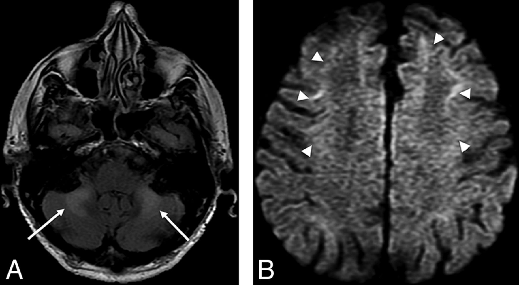
Representative case (patient 1). A 49-year-old male patient with a genetically confirmed diagnosis of FXTAS (CGG 115). Coronal (A) and sagittal (B) FLAIR images demonstrate cerebellar atrophy and a high signal intensity in the paravermal area (arrows). C, DWI shows abnormal high-intensity signal along the corticomedullary junction (arrowheads).

Fig 2.
  • Download figure
  • Open in new tab
  • Download powerpoint
Fig 2.

Representative case (patient 2). A 69-year-old male patient with slowly progressive ataxia, tremor, and paraparesis. Axial FLAIR image (A) shows bilateral abnormal high signal intensity in the middle cerebellar peduncles (arrows). Axial DWI (b = 1000 s/mm2) (B) image depicts similar signal changes along the corticomedullary junction (arrowheads). Genetic investigation showed 100 CGG repetition (premutation) of the FMR1 gene, which confirmed FXTAS.

Research in neuroradiology must concentrate on predicting specific neurologic disorders, identifying either radiophenotypes and/or useful algorithms for clinical practice. Considering that FXTAS seems to be more common than adult-onset NIID, it is reasonable that the algorithm using genetic studies (FMR1 gene premutation) proposed by Sone et al2 remains unpredictable because the pathologic changes and the imaging findings reported by Sugiyama et al1 may occur in both disorders.

Footnotes

  • Disclosures: Orlando G.P. Barsottini—UNRELATED: Employment: Federal University of Sao Paulo, Brazil, Comments: Professor of Neurology.

References

  1. 1.↵
    1. Sugiyama A,
    2. Sato N,
    3. Kimura Y, et al
    . MR imaging features of the cerebellum in adult-onset neuronal intranuclear inclusion disease: 8 cases. AJNR Am J Neuroradiol 2017;38:2100–04 doi:10.3174/ajnr.A5336 pmid:28818825
    Abstract/FREE Full Text
  2. 2.↵
    1. Sone J,
    2. Mori K,
    3. Inagaki T, et al
    . Clinicopathological features of adult-onset neuronal intranuclear inclusion disease. Brain 2016;139:3170–86 doi:10.1093/brain/aww249 pmid:27797808
    CrossRefPubMed
  • © 2018 by American Journal of Neuroradiology
PreviousNext
Back to top

In this issue

American Journal of Neuroradiology: 39 (9)
American Journal of Neuroradiology
Vol. 39, Issue 9
1 Sep 2018
  • Table of Contents
  • Index by author
  • Complete Issue (PDF)
Advertisement
Print
Download PDF
Email Article

Thank you for your interest in spreading the word on American Journal of Neuroradiology.

NOTE: We only request your email address so that the person you are recommending the page to knows that you wanted them to see it, and that it is not junk mail. We do not capture any email address.

Enter multiple addresses on separate lines or separate them with commas.
MR Imaging Features of Adult-Onset Neuronal Intranuclear Inclusion Disease May Be Indistinguishable from Fragile X–Associated Tremor/Ataxia Syndrome
(Your Name) has sent you a message from American Journal of Neuroradiology
(Your Name) thought you would like to see the American Journal of Neuroradiology web site.
CAPTCHA
This question is for testing whether or not you are a human visitor and to prevent automated spam submissions.
Cite this article
I.G. Padilha, R.H. Nunes, F.A. Scortegagna, J.L. Pedroso, V.H. Marussi, M.R. Rodrigues Gonçalves, O.G.P. Barsottini, A.J. da Rocha
MR Imaging Features of Adult-Onset Neuronal Intranuclear Inclusion Disease May Be Indistinguishable from Fragile X–Associated Tremor/Ataxia Syndrome
American Journal of Neuroradiology Sep 2018, 39 (9) E100-E101; DOI: 10.3174/ajnr.A5729

Citation Manager Formats

  • BibTeX
  • Bookends
  • EasyBib
  • EndNote (tagged)
  • EndNote 8 (xml)
  • Medlars
  • Mendeley
  • Papers
  • RefWorks Tagged
  • Ref Manager
  • RIS
  • Zotero
0 Responses
Respond to this article
Share
Bookmark this article
MR Imaging Features of Adult-Onset Neuronal Intranuclear Inclusion Disease May Be Indistinguishable from Fragile X–Associated Tremor/Ataxia Syndrome
I.G. Padilha, R.H. Nunes, F.A. Scortegagna, J.L. Pedroso, V.H. Marussi, M.R. Rodrigues Gonçalves, O.G.P. Barsottini, A.J. da Rocha
American Journal of Neuroradiology Sep 2018, 39 (9) E100-E101; DOI: 10.3174/ajnr.A5729
del.icio.us logo Twitter logo Facebook logo Mendeley logo
  • Tweet Widget
  • Facebook Like
  • Google Plus One
Purchase

Jump to section

  • Article
    • Footnotes
    • References
  • Figures & Data
  • Info & Metrics
  • Responses
  • References
  • PDF

Related Articles

  • Reply:
  • PubMed
  • Google Scholar

Cited By...

  • MRI diagnosis of neuronal intranuclear inclusion disease leukoencephalopathy
  • Reader response: Neuronal intranuclear inclusion disease showing intranuclear inclusions in renal biopsy 12 years earlier
  • Crossref
  • Google Scholar

This article has not yet been cited by articles in journals that are participating in Crossref Cited-by Linking.

Similar Articles

Advertisement

Indexed Content

  • Current Issue
  • Accepted Manuscripts
  • Article Preview
  • Past Issues
  • Editorials
  • Editor's Choice
  • Fellows' Journal Club
  • Letters to the Editor
  • Video Articles

Cases

  • Case Collection
  • Archive - Case of the Week
  • Archive - Case of the Month
  • Archive - Classic Case

More from AJNR

  • Trainee Corner
  • Imaging Protocols
  • MRI Safety Corner
  • Book Reviews

Multimedia

  • AJNR Podcasts
  • AJNR Scantastics

Resources

  • Turnaround Time
  • Submit a Manuscript
  • Submit a Video Article
  • Submit an eLetter to the Editor/Response
  • Manuscript Submission Guidelines
  • Statistical Tips
  • Fast Publishing of Accepted Manuscripts
  • Graphical Abstract Preparation
  • Imaging Protocol Submission
  • Evidence-Based Medicine Level Guide
  • Publishing Checklists
  • Author Policies
  • Become a Reviewer/Academy of Reviewers
  • News and Updates

About Us

  • About AJNR
  • Editorial Board
  • Editorial Board Alumni
  • Alerts
  • Permissions
  • Not an AJNR Subscriber? Join Now
  • Advertise with Us
  • Librarian Resources
  • Feedback
  • Terms and Conditions
  • AJNR Editorial Board Alumni

American Society of Neuroradiology

  • Not an ASNR Member? Join Now

© 2025 by the American Society of Neuroradiology All rights, including for text and data mining, AI training, and similar technologies, are reserved.
Print ISSN: 0195-6108 Online ISSN: 1936-959X

Powered by HighWire