Advertisement

Main menu

  • Home
  • Content
    • Current Issue
    • Accepted Manuscripts
    • Article Preview
    • Past Issue Archive
    • Video Articles
    • AJNR Case Collection
    • Case of the Week Archive
    • Case of the Month Archive
    • Classic Case Archive
  • Special Collections
    • AJNR Awards
    • Low-Field MRI
    • Alzheimer Disease
    • ASNR Foundation Special Collection
    • Photon-Counting CT
    • View All
  • Multimedia
    • AJNR Podcasts
    • AJNR SCANtastic
    • Trainee Corner
    • MRI Safety Corner
    • Imaging Protocols
  • For Authors
    • Submit a Manuscript
    • Submit a Video Article
    • Submit an eLetter to the Editor/Response
    • Manuscript Submission Guidelines
    • Statistical Tips
    • Fast Publishing of Accepted Manuscripts
    • Graphical Abstract Preparation
    • Imaging Protocol Submission
    • Author Policies
  • About Us
    • About AJNR
    • Editorial Board
    • Editorial Board Alumni
  • More
    • Become a Reviewer/Academy of Reviewers
    • Subscribers
    • Permissions
    • Alerts
    • Feedback
    • Advertisers
    • ASNR Home

User menu

  • Alerts
  • Log in

Search

  • Advanced search
American Journal of Neuroradiology

American Journal of Neuroradiology

ASHNR American Society of Functional Neuroradiology ASHNR American Society of Pediatric Neuroradiology ASSR
  • Alerts
  • Log in

Advanced Search

  • Home
  • Content
    • Current Issue
    • Accepted Manuscripts
    • Article Preview
    • Past Issue Archive
    • Video Articles
    • AJNR Case Collection
    • Case of the Week Archive
    • Case of the Month Archive
    • Classic Case Archive
  • Special Collections
    • AJNR Awards
    • Low-Field MRI
    • Alzheimer Disease
    • ASNR Foundation Special Collection
    • Photon-Counting CT
    • View All
  • Multimedia
    • AJNR Podcasts
    • AJNR SCANtastic
    • Trainee Corner
    • MRI Safety Corner
    • Imaging Protocols
  • For Authors
    • Submit a Manuscript
    • Submit a Video Article
    • Submit an eLetter to the Editor/Response
    • Manuscript Submission Guidelines
    • Statistical Tips
    • Fast Publishing of Accepted Manuscripts
    • Graphical Abstract Preparation
    • Imaging Protocol Submission
    • Author Policies
  • About Us
    • About AJNR
    • Editorial Board
    • Editorial Board Alumni
  • More
    • Become a Reviewer/Academy of Reviewers
    • Subscribers
    • Permissions
    • Alerts
    • Feedback
    • Advertisers
    • ASNR Home
  • Follow AJNR on Twitter
  • Visit AJNR on Facebook
  • Follow AJNR on Instagram
  • Join AJNR on LinkedIn
  • RSS Feeds

AJNR Awards, New Junior Editors, and more. Read the latest AJNR updates

Research ArticlePediatrics

Clival Malformations in CHARGE Syndrome

E.S. Mahdi and M.T. Whitehead
American Journal of Neuroradiology June 2018, 39 (6) 1153-1156; DOI: https://doi.org/10.3174/ajnr.A5612
E.S. Mahdi
aFrom the Department of Diagnostic Imaging and Radiology (E.S.M., M.T.W.), Children's National Medical Center, Washington, DC
  • Find this author on Google Scholar
  • Find this author on PubMed
  • Search for this author on this site
  • ORCID record for E.S. Mahdi
M.T. Whitehead
aFrom the Department of Diagnostic Imaging and Radiology (E.S.M., M.T.W.), Children's National Medical Center, Washington, DC
bGeorge Washington University Hospital (M.T.W.), Washington, DC.
  • Find this author on Google Scholar
  • Find this author on PubMed
  • Search for this author on this site
  • ORCID record for M.T. Whitehead
  • Article
  • Figures & Data
  • Supplemental
  • Info & Metrics
  • Responses
  • References
  • PDF
Loading

Abstract

BACKGROUND AND PURPOSE: CHARGE syndrome is a multisystemic congenital disorder, most commonly including coloboma, heart malformations, choanal atresia, developmental delay, and genital and ear anomalies. The diagnostic criteria for CHARGE syndrome have been refined with time. However, limited reports describe skull base and craniocervical junction abnormalities. Recently, a coronal clival cleft has been identified in association with CHARGE syndrome. The aim of our study was to assess the prevalence of clival pathology in CHARGE syndrome.

MATERIALS AND METHODS: In this retrospective study, the CT/MR imaging data base at a single academic children's hospital was queried for the phrase “CHARGE syndrome” during a 17-year period (2001–2017). Electronic medical records were reviewed to confirm the diagnosis. Images were assessed for skull base anomalies, specifically clival hypoplasia and dysplasia.

RESULTS: The search yielded 42 examinations (21 CTs and 21 MRIs) from 15 distinct patients (mean age, 4.1 ± 5.6 years; range, 2 days to 19 years). CHARGE syndrome diagnosis was confirmed either by clinical and genetic testing (n = 6) or by clinical diagnosis only (n = 9). A coronal clival cleft was identified in 87% of patients (37 examinations, n = 13 patients), either partial (53%) or complete (33%). Clival hypoplasia without clefting was present in all 5 examinations from the remaining 2 patients.

CONCLUSIONS: Clival pathology is universal in CHARGE syndrome. Coronal clival clefts are extremely common, representing a useful additional diagnostic finding. Detection of a clival cleft should alert the radiologist to examine the palate, choana, eyes, ears, and olfactory centers for other signs of CHARGE syndrome.

ABBREVIATIONS:

CHARGE
Coloboma of the eye, Heart defects, Atresia of the choanae, Retardation of growth and/or development, Genital and/or urinary abnormalities, and Ear abnormalities and deafness
CHD7
chromodomian helicase DNA-binding protein 7

CHARGE syndrome is a rare genetic disorder with widespread malformations. The acronym CHARGE includes coloboma, heart malformation, choanal atresia, retardation of growth and/or development, genital anomalies, and ear anomalies.1 Across time, there has been refinement of the diagnostic criteria to 3 major (the classic 3C's: choanal atresia, coloboma, semicircular canal hypoplasia) and 5 minor features.2,3 The prevalence of CHARGE syndrome was estimated to be 1/8500 live births by the Canadian Pediatric Surveillance Program.4 The discovery of a CHARGE syndrome–associated gene, CHD7 (chromodomain helicase DNA-binding protein 7, MIM 608892), has greatly assisted syndromic prevalence estimations due to the phenotypic diversity of the disease.5 Mutations of the CHD7 gene are present in two-thirds of patients with CHARGE syndrome. Phenotype and genotype correlation are required to better assess this syndrome.6 The skull base and craniocervical junction abnormalities are often underrecognized in CHARGE syndrome. Detection of these osseous abnormalities should alert the radiologist to examine the palate, choana, eyes, ears, and olfactory centers for other signs of CHARGE syndrome. Recently, a coronal clival cleft has been identified in association with CHARGE syndrome.7 The aim of our study was to assess the prevalence of clival pathology including clival clefts in patients with CHARGE syndrome.

Materials and Methods

Subjects

A retrospective study was performed after institutional review board approval with Health Insurance Portability and Accountability Act compliance. Requirement for informed consent was waived. The imaging data base at a single academic children's hospital was queried for the term “CHARGE syndrome” provided by the history or by the suspected radiologic findings after filtering by technique (CT and MR imaging) during a 17-year period (2001–2017). Review of the electronic medical records of all patients was performed to confirm the diagnosis and symptomatology. Images were assessed for skull base anomalies, specifically clival hypoplasia/dysplasia and coronal clival cleft.

Imaging Characteristics

All examinations were of diagnostic quality. MR imaging studies were performed on either 1.5T or 3T scanners (Discovery MR 750 and 450; GE Healthcare, Milwaukee, Wisconsin). Sagittal T1WI, axial T2WI, axial T2 fluid-attenuated inversion recovery imaging, coronal fat-saturated T2WI, and axial DTI with 7 noncollinear directions of encoding were reviewed. Axial T1WI and coronal fat-saturated T1WI of the brain were also performed in 5 MRIs after IV gadolinium administration in 5 different patients (0.1 mmol/kg of either gadoterate meglumine [n = 2] or gadopentetate dimeglumine [n = 3]). CT images of the head, temporal bone, facial bone, sinuses, and/or cervical spine were acquired on a 16–detector row scanner (GE Healthcare). Studies were reviewed in a blinded manner by a qualified pediatric neuroradiologist (M.T.W.) with >5 years of experience. Imaging studies were qualitatively examined for all visible abnormalities involving the brain, ocular and olfactory systems, temporal bones, cleft lip/palate, and the presence of clival abnormalities.

Results

Characteristics of the Cohort

The search yielded 17 patients with a history or suspected radiologic findings of CHARGE syndrome. Two patients lacking diagnostic confirmation were excluded. The final cohort comprised 42 examinations (21 CTs and 21 MRIs) from 15 patients (mean age, 4.1 ± 5.6 years; range, 2 days to 19 years); 3 patients were older than 11 years at the time of imaging.

Diagnosis of CHARGE syndrome was confirmed by either clinical and genetic testing (n = 6) or clinical diagnosis only (n = 9).2,3 Fifty-three percent of the patients were male (n = 8).

Clival Imaging Findings

A coronal clival cleft was identified in 87% of patients (37 examinations, n = 13 patients), either complete (33%, n = 5) (Figs 1 and 2) or partial (53%, n = 8) (Fig 3). Clival hypoplasia without clefting was present in all 5 examinations from the remaining 2 patients (On-line Table).8 Those with a partial cleft were further characterized as having either a unilateral cleft in 25% (n = 2) (Fig 3) or bilateral cleft with minimal central fusion in 75% (n = 6).

Fig 1.
  • Download figure
  • Open in new tab
  • Download powerpoint
Fig 1.

Midline sagittal spoiled gradient-recalled acquisition T1WI (1.5T MR imaging; TR/TE = 11/5.1 ms; TI = 500 ms; slice thickness = 1.5 mm) from a 3-month-old girl with CHARGE syndrome shows a distinctive complete coronally oriented clival cleft in the basiocciput (large arrow). Also note mild thinning of the corpus callosum (small arrow), cerebellar vermian hypoplasia, and brain stem volume loss.

Fig 2.
  • Download figure
  • Open in new tab
  • Download powerpoint
Fig 2.

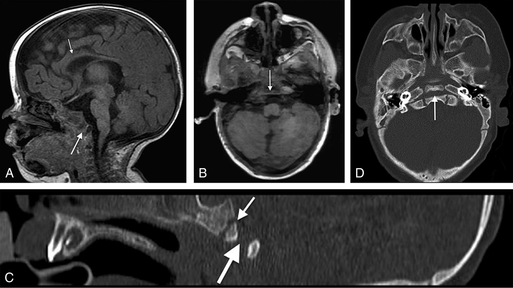
A, Midline sagittal spoiled gradient-recalled acquisition T1WI (1.5T MR imaging; TR/TE = 12/5 ms; TI = 500 ms; slice thickness = 2 mm) from a 4-month-old girl with CHARGE syndrome shows a complete coronally oriented clival cleft in the basiocciput (large arrow). Also note thinning of the corpus callosum (small arrow), cerebellar vermian hypoplasia, and brain stem volume loss. B, Axial spoiled gradient-recalled acquisition of the same patient shows a coronally oriented cleft traversing the clivus (arrow). Corresponding sagittal (C) and axial (D) CT images demonstrate the complete cleft (large arrow) as distinct from the normal spheno-occipital synchondrosis (small arrow in C).

Fig 3.
  • Download figure
  • Open in new tab
  • Download powerpoint
Fig 3.

Spoiled gradient-recalled acquisition T1WI (3T MR imaging; TR/TE = 6.6/2.5 ms; TI = 700 ms; slice thickness = 0.63 mm) from a 34-day-old boy with CHARGE syndrome shows a partial (unilateral left side) coronally oriented clival cleft in the basiocciput. A, Left parasagittal unilateral clival cleft (large arrow), mild thinning of the corpus callosum (small arrow), cerebellar vermian hypoplasia, and brain stem volume loss are illustrated. B, Right parasagittal image shows a normal appearance of the clivus without clefting, a thin corpus callosum (small arrow), and vermian and brain stem hypoplasia. C, Axial T1WI of the same patient with a left-sided cleft (large arrow) and normal spheno-occipital synchondrosis (small arrow). Corresponding sagittal and axial CT images (D and F) further demonstrate the unilateral left clival cleft (large arrow, D and F) and absence of clival clefting on the contralateral side (E). Note normal spheno-occipital synchondrosis (small arrow, D and E).

Clival Clefting/Hypoplasia and CHD7 Mutation

The clival findings were further stratified according to the CHD7 mutation. Those with CHD7 mutation (n = 6) had an equal number of complete and partial clival clefts (50%, n = 3) of each type. Those with a non-CHD7-confirmed clinical diagnosis (n = 9) were variable (partial cleft, n = 5; complete cleft, n = 2; and clival hypoplasia, n = 2).

Other imaging features pertinent to the diagnosis of CHARGE syndrome affecting the eye, olfactory apparatus, ears, and brain are classified in the On-line Table.

Discussion

CHARGE syndrome is a systemic disorder with multiorgan involvement. Specific organ abnormalities include cardiac (75%–80%, including atrial septal defect, ventricular septal defect, patent ductus arteriosus, tetralogy of Fallot, atrioventricular canal defect, and double outlet right ventricle),9 genitourinary (including hypospadias; penile agenesis; bifid scrotum; cryptorchidism; atresia of the vagina, cervix, and uterus; renal hypoplasia; solitary kidney; hydronephrosis; vesicoureteral reflux; and duplex kidneys),9 ocular (80%, including coloboma of the iris, retina-choroid, and/or disc; and microphthalmos),9 ear (80%–100%, including external ear dysmorphology; incus hypoplasia; decreased number of cochlear turns; and absent semicircular canals),9,10 olfactory (100%, including unilateral or bilateral absence or hypoplasia of the olfactory bulbs or sulci),9,11 choanal atresia (50%–60%),12 orofacial (15%–20%, including cleft lip/palate),9 brain (including corpus callosum agenesis; posterior fossa anomalies; cranial nerve dysfunction; and cerebellar vermis hypoplasia),12,13 and osseous anomalies (skull base and craniocervical junction).9,8,14 Prenatal diagnosis of CHARGE syndrome has been reported through the finding of CHD7 mutations on chorionic villus sampling and ultrasonographic findings of hydramnios, choanal atresia, tracheoesophageal fistula, and semicircular canal agenesis.15,16

Craniocervical junction abnormalities are common in CHARGE syndrome. Fujita et al8 demonstrated a high prevalence of basioccipital hypoplasia and basilar invagination in CHARGE syndrome and recommended routine assessment of these anomalies to exclude a life-threatening basilar invagination. Natung et al14 reported a case of basilar invagination, short clivus, fused cervical vertebrae, and occipitalization of the atlas. A recent case report of a coronal clival cleft has been identified in association with CHARGE syndrome.7 Recently, Hoch et al17 described a dorsally angulated clivus with posterior displacement of an ossific density in 7 of 10 patients with CHARGE syndrome. The dominant findings we have described herein, namely partial and complete coronal clival clefting, correspond to the description and imaging depictions that those authors provided and further emphasize the importance of craniocervical junction anomalies in CHARGE syndrome.

The pathogenesis of these anomalies in CHARGE syndrome is not clearly understood. They likely originate in the first trimester, concurrent with other CHARGE malformations.18 The CHD7 gene encoding chromodomain helicase DNA-binding protein 7 (CHD7 protein) may play a role through the assumption of the interaction between the neural crest and somite cells, which are responsible for formation of the basiocciput during development.19,20

The clivus is formed from 2 main components, the upper basisphenoid and the lower basiocciput. The 2 are separated by the normal spheno-occipital synchondrosis. The basiocciput is formed from 4 occipital sclerotomes.19 The clivus reaches the full developmental length by 11 years of age.21 Clival clefts occur caudal to and distinct from the spheno-occipital synchondrosis, which usually fuses between 12 and 18 years of age.19 The presence of normal anatomic variants such as clival canal/foramen, fossa navicularis magna, or basilar transverse fissure (Sauser fissure) should not be confused with a clival cleft.22⇓–24Although the etiology of coronal clival clefts is unknown, these clefts could be related to incomplete fusion of clival ossification centers, enlarged clival canals (fossa navicularis), or persistence of basilar transverse fissure.19,22⇓–24

This study has some limitations by being retrospective and having a relatively small sample size. Despite these limitations, to date, it is the largest study to describe and illustrate clival pathology and clefting in CHARGE syndrome.

Conclusions

Clival pathology is universal in CHARGE syndrome. Coronal clival clefts are extremely common, representing a useful diagnostic feature and should be considered an additional finding in CHARGE syndrome. The skull base should be scrutinized for these characteristic anomalies. Detection of a clival cleft should alert the radiologist to examine the palate, choana, eyes, ears, and olfactory centers for other signs of CHARGE syndrome.

REFERENCES

  1. 1.↵
    1. Pagon RA,
    2. Graham JM Jr.,
    3. Zonana J, et al
    . Coloboma, congenital heart disease, and choanal atresia with multiple anomalies: CHARGE association. J Pediatr 1981;99:223–27 doi:10.1016/S0022-3476(81)80454-4 pmid:6166737
    CrossRefPubMed
  2. 2.↵
    1. Verloes A
    . Updated diagnostic criteria for CHARGE syndrome: a proposal. Am J Med Genet A 2005;133A:306–08 doi:10.1002/ajmg.a.30559 pmid:15666308
    CrossRefPubMed
  3. 3.↵
    1. Blake KD,
    2. Davenport SL,
    3. Hall BD, et al
    . CHARGE association: an update and review for the primary pediatrician. Clin Pediatr (Phila) 1998;37:159–73 doi:10.1177/000992289803700302 pmid:9545604
    CrossRefPubMed
  4. 4.↵
    1. Issekutz KA,
    2. Graham JM Jr.,
    3. Prasad C, et al
    . An epidemiological analysis of CHARGE syndrome: preliminary results from a Canadian study. Am J Med Genet A 2005;133A:309–17 doi:10.1002/ajmg.a.30560 pmid:15637722
    CrossRefPubMed
  5. 5.↵
    1. Vissers LE,
    2. van Ravenswaaij CM,
    3. Admiraal R, et al
    . Mutations in a new member of the chromodomain gene family cause CHARGE syndrome. Nat Genet 2004;36:955–57 doi:10.1038/ng1407 pmid:15300250
    CrossRefPubMed
  6. 6.↵
    1. Sanlaville D,
    2. Verloes A
    . CHARGE syndrome: an update. Eur J Hum Genet 2007;15:389–99 doi:10.1038/sj.ejhg.5201778 pmid:17299439
    CrossRefPubMed
  7. 7.↵
    1. Mahdi E,
    2. Whitehead MT
    . Coronal clival cleft in CHARGE syndrome. Neuroradiol J 2017;30:574–77 doi:10.1177/1971400916678248 pmid:28059674
    CrossRefPubMed
  8. 8.↵
    1. Fujita K,
    2. Aida N,
    3. Asakura Y, et al
    . Abnormal basiocciput development in CHARGE syndrome. AJNR Am J Neuroradiol 2009;30:629–34 doi:10.3174/ajnr.A1380 pmid:19112063
    Abstract/FREE Full Text
  9. 9.↵
    1. Blake KD,
    2. Prasad C
    . CHARGE syndrome. Orphanet J Rare Dis 2006;1:34 doi:10.1186/1750-1172-1-34
    CrossRefPubMed
  10. 10.↵
    1. Morimoto AK,
    2. Wiggins RH 3rd.,
    3. Hudgins PA, et al
    . Absent semicircular canals in CHARGE syndrome: radiologic spectrum of findings. AJNR Am J Neuroradiol 2006;27:1663–71 pmid:16971610
    Abstract/FREE Full Text
  11. 11.↵
    1. Blustajn J,
    2. Kirsch CF,
    3. Panigrahy A, et al
    . Olfactory anomalies in CHARGE syndrome: imaging findings of a potential major diagnostic criterion. AJNR Am J Neuroradiol 2008;29:1266–69 doi:10.3174/ajnr.A1099 pmid:18417599
    Abstract/FREE Full Text
  12. 12.↵
    1. Adam MP,
    2. Ardinger HH,
    3. Pagon RA, et al.
    1. Lalani SR,
    2. Hefner MA,
    3. Belmont JW, et al
    . CHARGE syndrome. In: Adam MP, Ardinger HH, Pagon RA, et al., eds. GeneReviews. Seattle: University of Washington; 1993–2018 pmid:20301296
  13. 13.↵
    1. Yu T,
    2. Meiners LC,
    3. Danielsen K, et al
    . Deregulated FGF and homeotic gene expression underlies cerebellar vermis hypoplasia in CHARGE syndrome. Elife 2013;2:e01305 doi:10.7554/eLife.01305 pmid:24368733
    Abstract/FREE Full Text
  14. 14.↵
    1. Natung T,
    2. Goyal A,
    3. Handique A, et al
    . Symmetrical chorioretinal colobomata with craniovertebral junction anomalies in CHARGE syndrome: a case report with review of literature. J Clin Imaging Sci 2014;4:5 doi:10.4103/2156-7514.126046 pmid:24678437
    CrossRefPubMed
  15. 15.↵
    1. Becker R,
    2. Stiemer B,
    3. Neumann L, et al
    . Mild ventriculomegaly, mild cerebellar hypoplasia and dysplastic choroid plexus as early prenatal signs of CHARGE association. Fetal Diagn Ther 2001;16:280–83 doi:10.1159/000053928 pmid:11509849
    CrossRefPubMed
  16. 16.↵
    1. Hertzberg BS,
    2. Kliewer MA,
    3. Lile RL
    . Antenatal ultrasonographic findings in the CHARGE association. J Ultrasound Med 1994;13:238–42 doi:10.7863/jum.1994.13.3.238 pmid:7932986
    CrossRefPubMed
  17. 17.↵
    1. Hoch MJ,
    2. Patel SH,
    3. Jethanamest D, et al
    . Head and neck MRI findings in CHARGE syndrome. AJNR Am J Neuroradiol 2017;38:2357–63 doi:10.3174/ajnr.A5297 pmid:28705814
    Abstract/FREE Full Text
  18. 18.↵
    1. Sanlaville D,
    2. Etchevers HC,
    3. Gonzales M, et al
    . Phenotypic spectrum of CHARGE syndrome in fetuses with CHD7 truncating mutations correlates with expression during human development. J Med Genet 2006;43:211–17 doi:10.1136/jmg.2005.036160 pmid:16169932
    Abstract/FREE Full Text
  19. 19.↵
    1. Smoker WR
    . Craniovertebral junction: normal anatomy, craniometry, and congenital anomalies. Radiographics 1994;14:255–77 doi:10.1148/radiographics.14.2.8190952 pmid:8190952
    CrossRefPubMed
  20. 20.↵
    1. Nie X
    . Cranial base in craniofacial development: developmental features, influence on facial growth, anomaly, and molecular basis. Acta Odontol Scand 2005;63:127–35 doi:10.1080/00016350510019847 pmid:16191905
    CrossRefPubMed
  21. 21.↵
    1. Krmpotić-Nemanić J,
    2. Vinter I,
    3. Kelovizć Z, et al
    . Postnatal changes of the clivus. Ann Anat 2005;187:277–80 doi:10.1016/j.aanat.2004.11.005 pmid:16130827
    CrossRefPubMed
  22. 22.↵
    1. Inal M,
    2. Muluk NB,
    3. Ozveren MF, et al
    . The presence of clival foramen through multidetector computed tomography of the skull base. J Craniofac Surg 2015;26:e580–82 doi:10.1097/SCS.0000000000002129 pmid:26468827
    CrossRefPubMed
  23. 23.↵
    1. Beltramello A,
    2. Puppini G,
    3. El-Dalati G, et al
    . Fossa navicularis magna. AJNR Am J Neuroradiol 1998;19:1796–98 pmid:9802508
    PubMed
  24. 24.↵
    1. Hofmann E,
    2. Prescher A
    . The clivus: anatomy, normal variants and imaging pathology. Clin Neuroradiol 2012;22:123–39 doi:10.1007/s00062-011-0083-4 pmid:21710384
    CrossRefPubMed
  • Received November 11, 2017.
  • Accepted after revision February 5, 2018.
  • © 2018 by American Journal of Neuroradiology
View Abstract
PreviousNext
Back to top

In this issue

American Journal of Neuroradiology: 39 (6)
American Journal of Neuroradiology
Vol. 39, Issue 6
1 Jun 2018
  • Table of Contents
  • Index by author
  • Complete Issue (PDF)
Advertisement
Print
Download PDF
Email Article

Thank you for your interest in spreading the word on American Journal of Neuroradiology.

NOTE: We only request your email address so that the person you are recommending the page to knows that you wanted them to see it, and that it is not junk mail. We do not capture any email address.

Enter multiple addresses on separate lines or separate them with commas.
Clival Malformations in CHARGE Syndrome
(Your Name) has sent you a message from American Journal of Neuroradiology
(Your Name) thought you would like to see the American Journal of Neuroradiology web site.
CAPTCHA
This question is for testing whether or not you are a human visitor and to prevent automated spam submissions.
Cite this article
E.S. Mahdi, M.T. Whitehead
Clival Malformations in CHARGE Syndrome
American Journal of Neuroradiology Jun 2018, 39 (6) 1153-1156; DOI: 10.3174/ajnr.A5612

Citation Manager Formats

  • BibTeX
  • Bookends
  • EasyBib
  • EndNote (tagged)
  • EndNote 8 (xml)
  • Medlars
  • Mendeley
  • Papers
  • RefWorks Tagged
  • Ref Manager
  • RIS
  • Zotero
0 Responses
Respond to this article
Share
Bookmark this article
Clival Malformations in CHARGE Syndrome
E.S. Mahdi, M.T. Whitehead
American Journal of Neuroradiology Jun 2018, 39 (6) 1153-1156; DOI: 10.3174/ajnr.A5612
del.icio.us logo Twitter logo Facebook logo Mendeley logo
  • Tweet Widget
  • Facebook Like
  • Google Plus One
Purchase

Jump to section

  • Article
    • Abstract
    • ABBREVIATIONS:
    • Materials and Methods
    • Results
    • Discussion
    • Conclusions
    • REFERENCES
  • Figures & Data
  • Supplemental
  • Info & Metrics
  • Responses
  • References
  • PDF

Related Articles

  • PubMed
  • Google Scholar

Cited By...

  • Coronal Clival Cleft in CHARGE Syndrome: Fetal MRI Series
  • Persistent Trigeminal Artery: A Novel Imaging Finding in CHARGE Syndrome
  • Cerebellar Heterotopias: Expanding the Phenotype of Cerebellar Dysgenesis in CHARGE Syndrome
  • Imaging of Clival Hypoplasia in CHARGE Syndrome and Hypothesis for Development: A Case-Control Study
  • Crossref (14)
  • Google Scholar

This article has been cited by the following articles in journals that are participating in Crossref Cited-by Linking.

  • Temporal bone and intracranial abnormalities in syndromic causes of hearing loss: an updated guide
    Felice D’Arco, Adam Youssef, Evangelia Ioannidou, Sotirios Bisdas, Lorenzo Pinelli, Pablo Caro-Dominguez, Robert Nash, Ata Siddiqui, Giacomo Talenti
    European Journal of Radiology 2020 123
  • Imaging of Clival Hypoplasia in CHARGE Syndrome and Hypothesis for Development: A Case-Control Study
    C.M. de Geus, J.E.H. Bergman, C.M.A. van Ravenswaaij-Arts, L.C. Meiners
    American Journal of Neuroradiology 2018 39 10
  • CHARGE syndrome without colobomas: Ophthalmic findings
    Eniolami O. Dosunmu, Katherine M. Castleberry
    American Journal of Medical Genetics Part C: Seminars in Medical Genetics 2020 184 3
  • Phenotypic spectrum of patients with mutations in CHD7: clinical implications of endocrinological findings
    Ja Hye Kim, Yunha Choi, Soojin Hwang, Gu-Hwan Kim, Han-Wook Yoo, Jin-Ho Choi
    Endocrine Connections 2022 11 2
  • Skull base anatomy and surgical safety in isolated and CHARGE-associated bilateral choanal atresia
    Nicholas S. Fitzpatrick, Anthony C. Bartley, Elhamy Bekhit, Robert G. Berkowitz
    International Journal of Pediatric Otorhinolaryngology 2018 115
  • Ontogeny of the human fetal, neonatal, and infantile basioccipital bone: Traditional and extended eigenshape geometric morphometric analysis
    Matthew J. Zdilla, Jacob P. Pancake, Michelle L. Russell, Aaron W. Koons
    The Anatomical Record 2022 305 11
  • CHD7 regulates craniofacial cartilage development via controlling HTR2B expression
    Maximilian Breuer, Maximilian Rummler, Jaskaran Singh, Sabrina Maher, Charlotte Zaouter, Priyanka Jamadagni, Nicolas Pilon, Bettina M Willie, Shunmoogum A Patten
    Journal of Bone and Mineral Research 2024 39 4
  • An Unprecedented Complication in Pharyngeal Flap Surgery: Cerebrospinal Fluid Leakage
    Gökberk Çavuşoğlu, Murat Kara, Rahşan Göçmen
    The Cleft Palate Craniofacial Journal 2025
  • Complete transverse basilar cleft associated with hemifacial microsomia
    Laphatrada Yurasakpong, Athikhun Suwannakhan, Joe Iwanaga, R. Shane Tubbs, Arada Chaiyamoon, Nutmethee Kruepunga, Somluk Asuvapongpatana, Thanwa Sudsang
    Anatomy & Cell Biology 2024 57 3
  • Coronal Clival Cleft in CHARGE Syndrome: Fetal MRI Series
    Sara Reis Teixeira, Carmen Cerron-Vela, Nahla Khalek, Renee Wright, Matthew T. Whitehead
    American Journal of Neuroradiology 2025 46 5

More in this TOC Section

  • SyMRI & MR Fingerprinting in Brainstem Myelination
  • Comparison of Image Quality and Radiation Dose in Pediatric Temporal Bone CT Using Photon-Counting Detector CT and Energy-Integrating Detector CT
  • Dual-Layer Detector CT for PEDS Image Quality
Show more Pediatrics

Similar Articles

Advertisement

Indexed Content

  • Current Issue
  • Accepted Manuscripts
  • Article Preview
  • Past Issues
  • Editorials
  • Editor's Choice
  • Fellows' Journal Club
  • Letters to the Editor
  • Video Articles

Cases

  • Case Collection
  • Archive - Case of the Week
  • Archive - Case of the Month
  • Archive - Classic Case

More from AJNR

  • Trainee Corner
  • Imaging Protocols
  • MRI Safety Corner
  • Book Reviews

Multimedia

  • AJNR Podcasts
  • AJNR Scantastics

Resources

  • Turnaround Time
  • Submit a Manuscript
  • Submit a Video Article
  • Submit an eLetter to the Editor/Response
  • Manuscript Submission Guidelines
  • Statistical Tips
  • Fast Publishing of Accepted Manuscripts
  • Graphical Abstract Preparation
  • Imaging Protocol Submission
  • Evidence-Based Medicine Level Guide
  • Publishing Checklists
  • Author Policies
  • Become a Reviewer/Academy of Reviewers
  • News and Updates

About Us

  • About AJNR
  • Editorial Board
  • Editorial Board Alumni
  • Alerts
  • Permissions
  • Not an AJNR Subscriber? Join Now
  • Advertise with Us
  • Librarian Resources
  • Feedback
  • Terms and Conditions
  • AJNR Editorial Board Alumni

American Society of Neuroradiology

  • Not an ASNR Member? Join Now

© 2025 by the American Society of Neuroradiology All rights, including for text and data mining, AI training, and similar technologies, are reserved.
Print ISSN: 0195-6108 Online ISSN: 1936-959X

Powered by HighWire