
Article Figures & Data
Figures
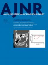
- Fig 1.
Sagittal single-shot fast spin-echo sequence of a 32-week fetus demonstrating normal midline anatomy. Note the fully formed corpus callosum (white arrows), normal tectum (black arrow), a patent cerebral aqueduct with normal intraluminal proportions (white arrowhead), and a normal fourth ventricle (asterisk). Note also the normal expected midline morphology of the third ventricle (light gray shaded area) with normal supraoptic and infundibular third ventricular recesses (black arrowheads).
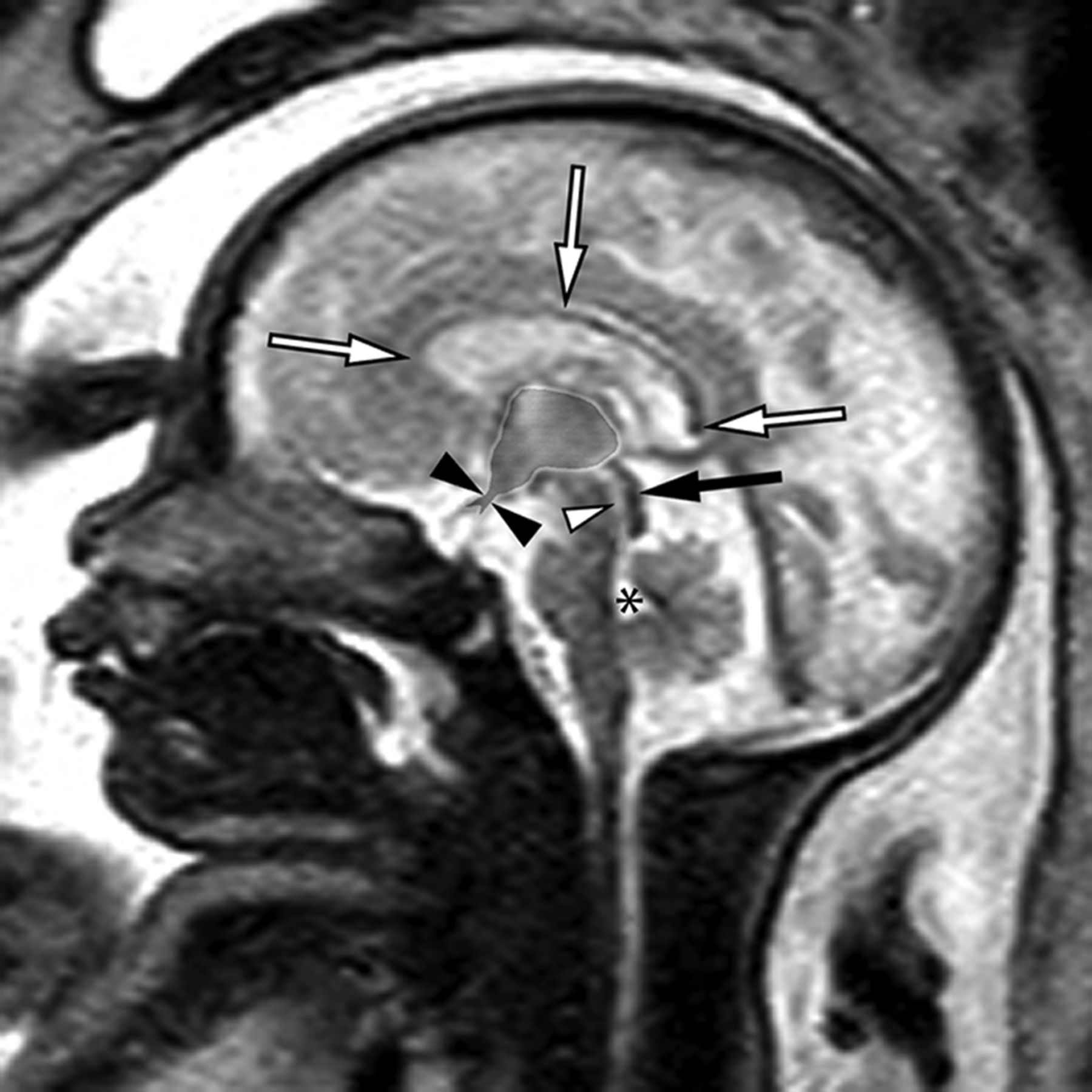
- Fig 2.
Sagittal balanced steady-state free precession sequence from fetal MR imaging (A) of a 33-week fetus and a postnatal sagittal T1-weighted sequence (B) of the same patient demonstrating stenosis of the inferior cerebral aqueduct with associated aqueductal funneling (arrow). As a result, there is marked enlarged of the lateral and third ventricles with dilation of the inferior third ventricular recesses (white arrowheads) depicted by bowing of the lamina terminalis and inferior third ventricular floor. The corpus callosum is thin and superiorly bowed (black arrowheads). Note also the normal size of the fourth ventricle.
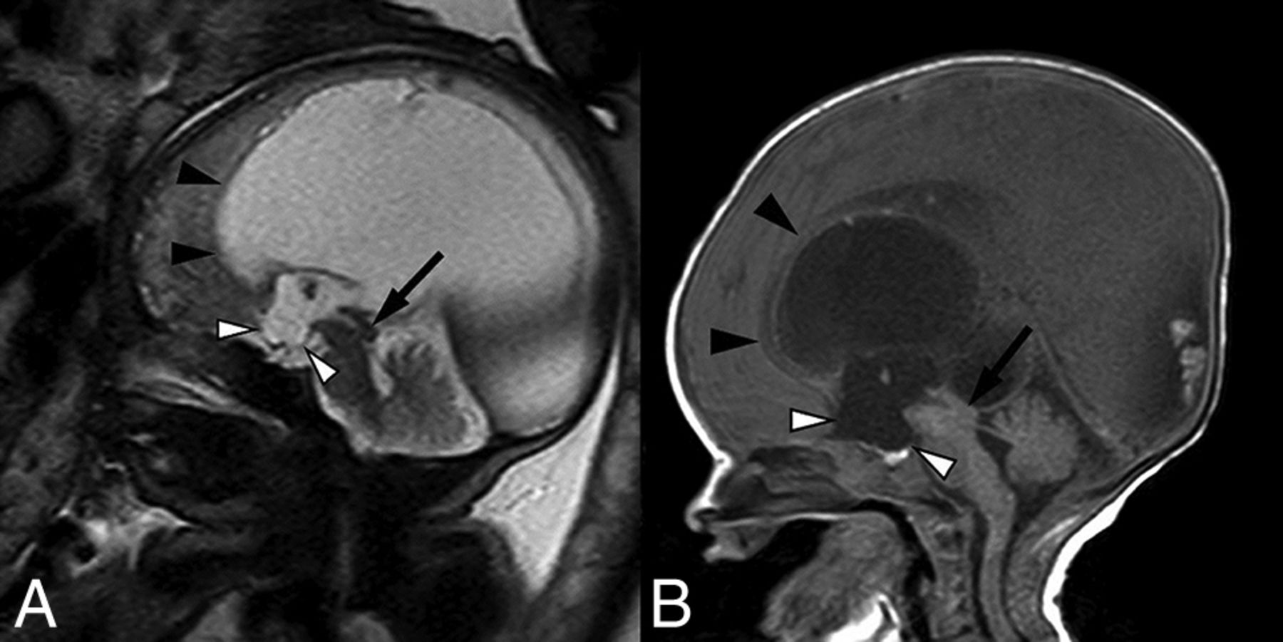
- Fig 3.
Fetal MR imaging of a 30-week fetus (A) and postnatal MR imaging correlation (B) of prenatally diagnosed aqueductal stenosis with tectal thickening and loss of intercollicular sulcus (arrows). There is subtle early prominence of the supraoptic recess of the third ventricle on fetal MR imaging (white arrowhead), which progressed to more obvious dilation of both supraoptic and infundibular recesses on postnatal imaging (white arrowheads). Note also the presence of a superiorly bowed and thinned corpus callosum (black arrowheads).
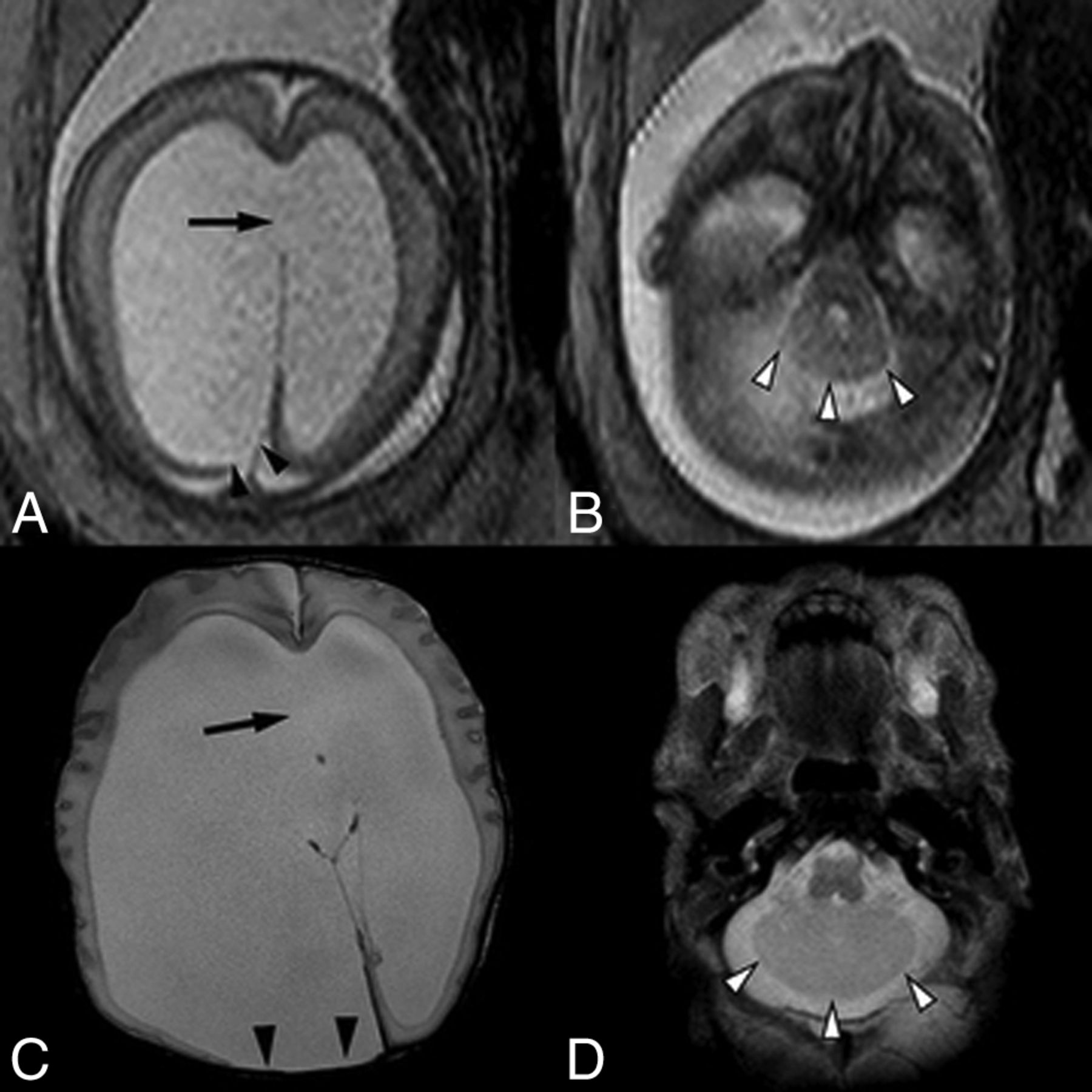
- Fig 4.
A single-shot fast spin-echo sequence in the axial planes (A and B) through the fetal head in a 23-week fetus and postnatal axial T2-weighted sequence (C and D) demonstrate asymmetric lateral ventriculomegaly with focal parenchymal disruption resulting in a posterior ventricular diverticulum (black arrowheads). Note also perforation of the septum pellucidum in A and C (arrow). Within the posterior fossa (B and D), there are a small transverse cerebellar diameter, absence of the cerebellar vermis, midline fusion of cerebellar folia, and a convex posterior cerebellar contour (white arrowheads), compatible with rhombencephalosynapsis.
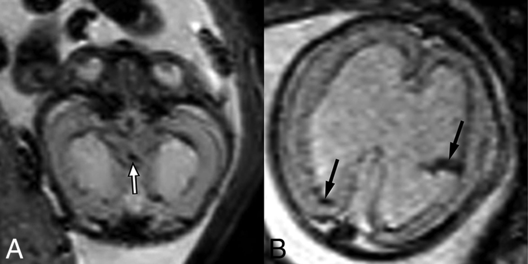
- Fig 5.
T2-weighted EPI sequences of 2 different fetuses with aqueductal stenosis demonstrating T2-hypointense hemorrhage within the cerebral aqueduct (A) in a 21-week fetus (white arrow) and within the lateral ventricles (B) in a 23-week fetus (black arrows).
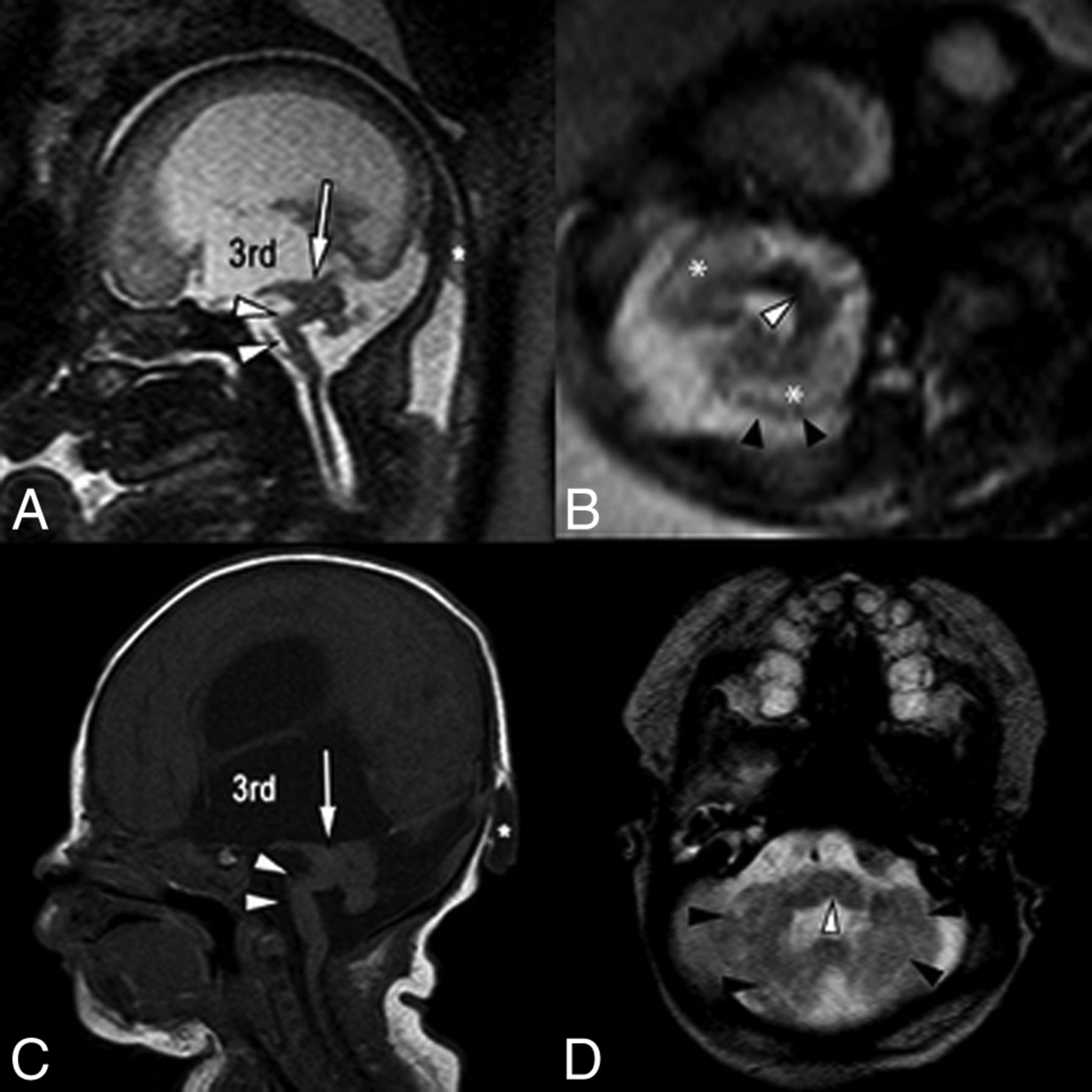
- Fig 6.
Single-shot fast spin-echo sagittal midline (A) and axial posterior fossa (B) images in a 34-week fetus with multiple findings of dystroglycanopathy suggesting Walker-Warburg syndrome. Postnatal correlation includes a sagittal T1-weighted sequence (C) and an axial T2-weighted sequence (D). Sagittal views of both pre- and postnatal MR imaging demonstrate a hypoplastic kinked brain stem (white arrowheads) and a markedly hypoplastic cerebellar vermis. Note also the dysplastic midbrain with thickening of the tectum causing stenosis of the cerebral aqueduct (arrows). Lateral and third ventricles are markedly enlarged. Incidentally noted was a small occipital cephalocele (asterisk). Axial views show cerebellar dysplasia with irregular cerebellar margins (black arrowheads) and multiple small cerebellar cysts, which account for the increased white matter T2 signal on fetal MR imaging (asterisk). Note also a midline pontine cleft (white arrowheads in B and D), another common finding in Walker-Warburg syndrome.
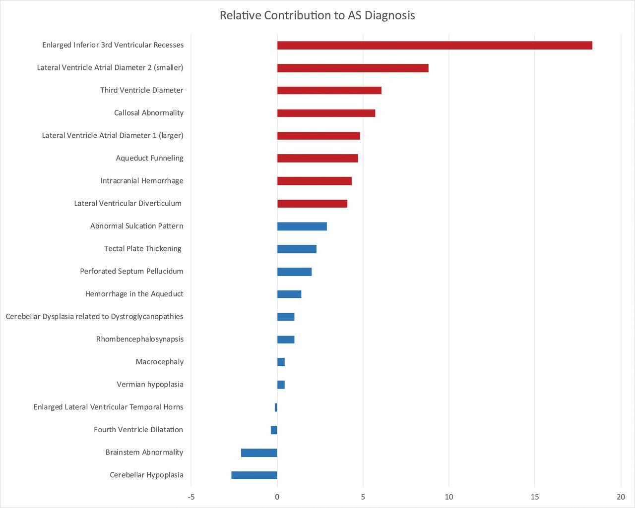
- Fig 7.
Random forest variable importance plot. This graphic shows the importance (x-axis) of each evaluated prenatal MR imaging finding (y-axis) with respect to the diagnosis of CAS. The independent contribution of each prenatal finding was estimated as the error of CAS classification by the machine-learning algorithm compared with the error that results when that finding is negated. The most important imaging findings associated with an accurate diagnosis of CAS are highlighted in red. Dominant findings include enlargement of the third ventricle inferior recesses, size of the lateral and third ventricles (especially enlargement of the smaller lateral ventricle), and an abnormally thin and/or dysgenetic corpus callosum. AS indicates aqueductal stenosis.


Tables
Direct Findings Findings Secondary to Obstructive Hydrocephalus Findings of Associated Malformations Aqueduct funneling Enlarged third ventricular recesses Abnormal sulcation Blood in the aqueduct Enlarged ventricular temporal horns Brain stem abnormality Tectal thickening Perforated septum pellucidum Cerebellar hypoplasia Lateral ventricular diverticulum Cerebellar dysplasia Callosal thinning and/or dysgenesis Rhombencephalosynapsis Macrocephaly Fourth ventricular dilation Vermian hypoplasia Note:—AS indicates aqueductal stenosis.
Categorical Variable Control (n = 32) AS (n = 43) Adjusted P Value Sensitivity (95% CI) Specificity (95% CI) PPV (95% CI) NPV (95% CI) Enlarged inferior 3rd ventricular recesses 1 (3.1%) 31 (72%) <.0023a 72 (56–85) 97 (84–100) 97 (84–100) 72 (56–85) Lateral ventricular diverticulum 1 (3.1%) 15 (35%) .0276a 35 (21–51) 97 (84–100) 94 (70–100) 53 (39–66) Callosal thinning and/or dysgenesis 16 (50%) 40 (93%) <.0023a 93 (81–99) 50 (32–68) 71 (58–83) 84 (60–97) Aqueductal funneling 0 (0%) 9 (21%) .1909 21 (10–36) 100 (89–100) 100 (60–100) 48 (36–61) Blood in the aqueduct 0 (0%) 3 (7%) 1 7.0 (1.5–19) 100 (89–100) 100 (29–100) 44 (33–57) Rhombencephalosynapsis 0 (0%) 4 (9.3%) 1 9.3 (2.6–22.1) 100 (89–100) 100 (40–100) 45 (33–57) Cerebellar dysplasia 0 (0%) 7 (16%) .4117 16 (6.8–31) 100 (89–100) 100 (59–100) 47 (35–60) Tectal plate thickening 2 (6.3%) 12 (28%) .437 28 (15–44) 94 (79–99) 86 (57–98) 49 (36–62) Intracranial hemorrhage 2 (6.3%) 15 (35%) .1035 35 (21–51) 94 (79–99) 88 (64–99) 52 (38–65) Enlarged ventricular temporal horns 20 (63%) 40 (93%) .0575 93 (81–99) 38 (21–56) 67 (53–78) 80 (52–96) Macrocephaly 4 (13%) 17 (40%) .2484 40 (25–56) 88 (71–96) 81 (58–95) 52 (38–66) Cerebellar hypoplasia 12 (38%) 5 (12%) .2714 12 (3.9–25) 63 (44–79) 29 (10–56) 34 (22–48) Vermian hypoplasia 4 (13%) 9 (21%) 1 21 (10–36) 88 (71–96) 69 (39–91) 45 (32–58) Brain stem abnormality 4 (13%) 10 (23%) 1 23 (12–39) 88 (71–96) 71 (42–92) 46 (33–59) Fourth ventricle dilation 3 (9.4%) 4 (9.3%) 1 9.3 (2.6–22) 91 (75–98) 57 (18–90) 43 (31–55) Abnormal sulcation 8 (25%) 10 (23%) 1 23 (12–39) 75 (57–89) 56 (31–78) 42 (29–56) Perforated septum pellucidum 14 (44%) 29 (67%) 1 67 (51–81) 56 (38–74) 67 (51–81) 56 (38–74) Note:—NPV indicates negative predictive value; PPV, positive predictive value; AS, aqueductal stenosis.
↵a Significant.


