
Article Figures & Data
Figures
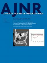
- Fig 1.
Radiologic malformations of the middle and inner ear encountered in 52 ears.
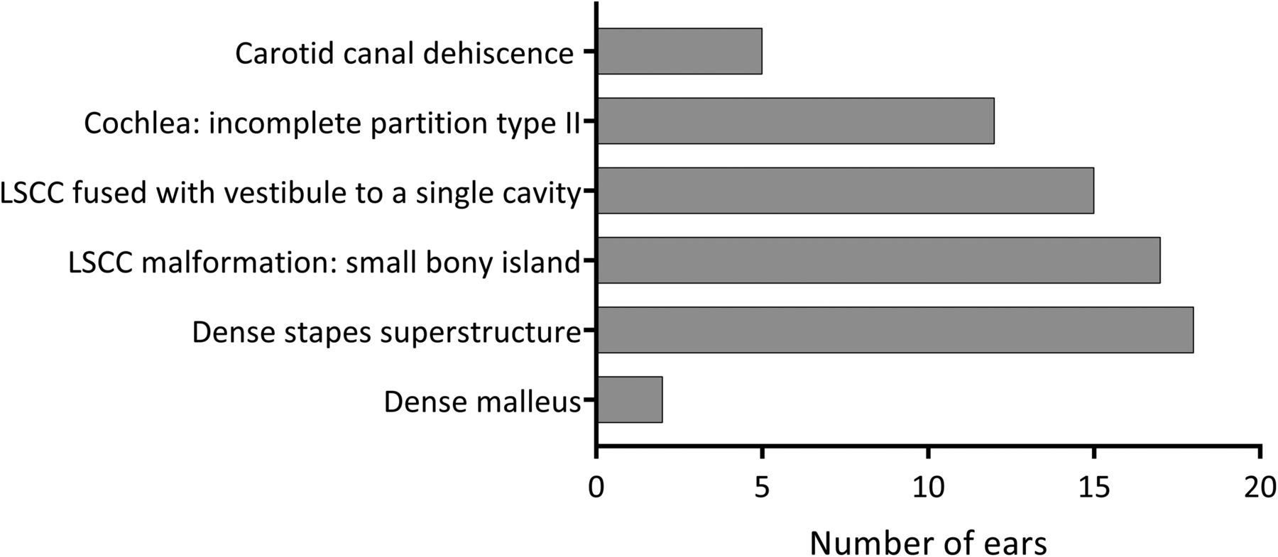
- Fig 2.
A, Axial CT image of the left mastoid shows a normal stapes superstructure for comparison with B. B, Axial CT image of the right mastoid shows a dense, thick stapes superstructure. The pure tone audiogram of this patient is shown in Fig 5B. The mastoid bones shown in A and B do not belong to the same patient.
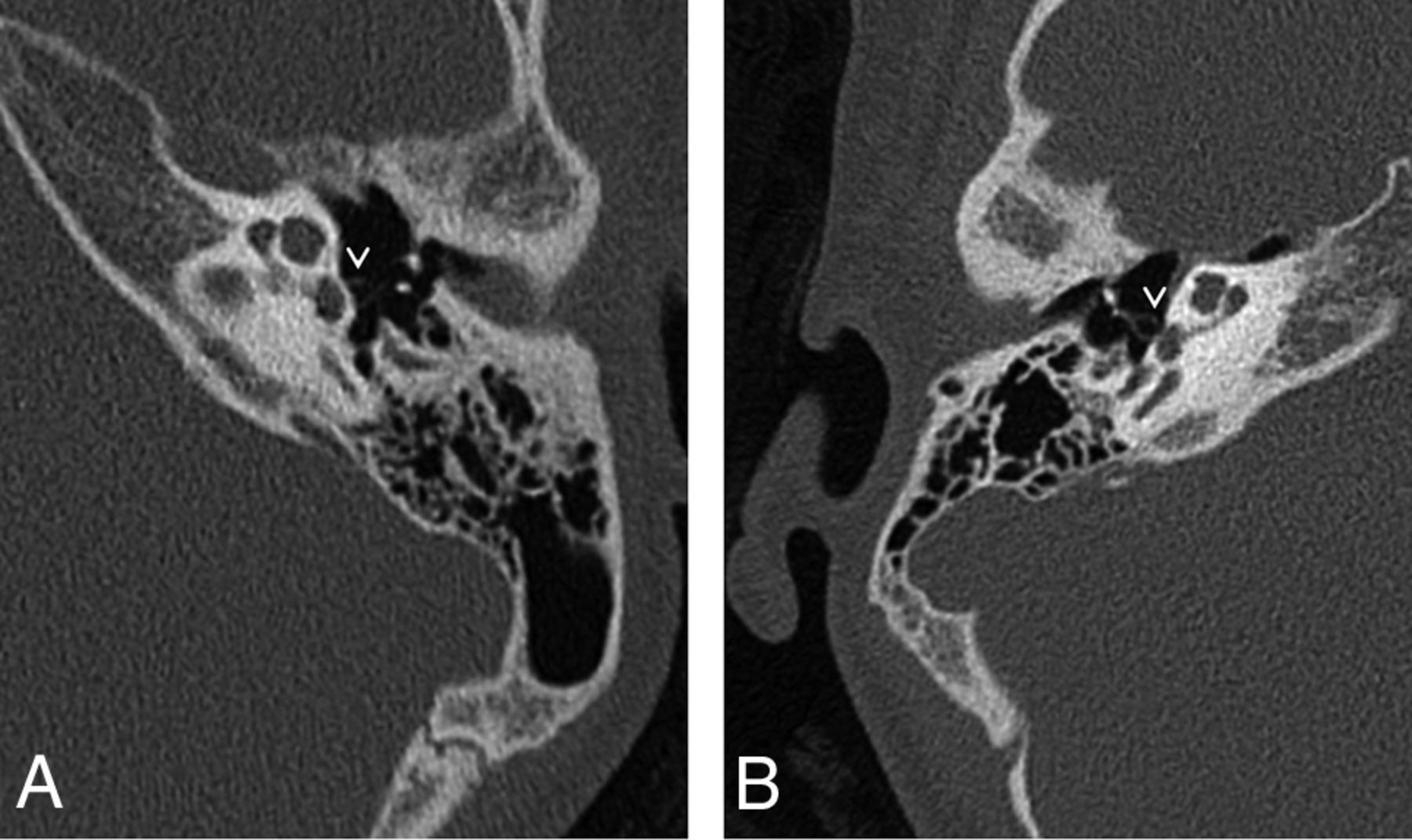
- Fig 3.
A, Axial CT image of the right mastoid shows an incomplete partition type II of the cochlea. The basal turn (BT) of the cochlea is intact; the apical and middle turn (A/M) seem confluent. B, A pure tone audiogram of the same ear shows normal hearing. The circle indicates an unmasked air-conduction threshold; the bracket, a masked bone-conduction threshold.
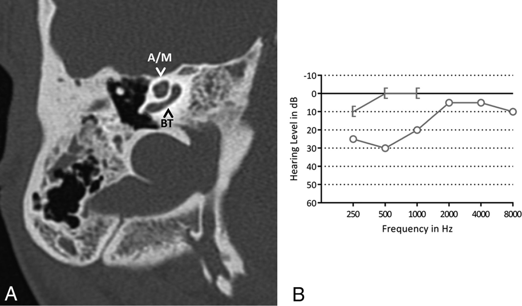
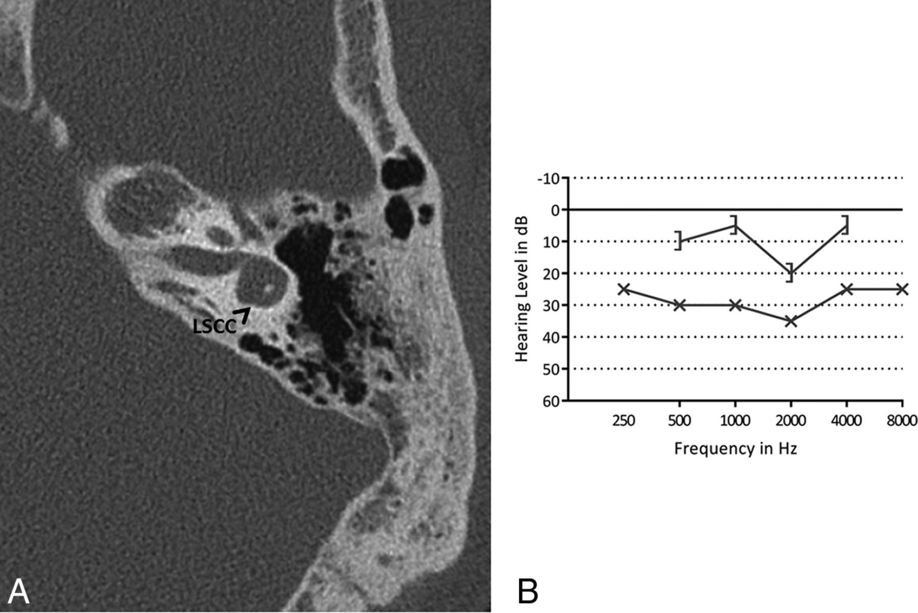
- Fig 4.
A, Axial CT image of left mastoid bone shows the lateral semicircular canal with a small bony island. B, A pure tone audiogram of the same ear shows a mild conductive hearing loss. The x indicates an unmasked air-conduction threshold; the bracket, a masked bone-conduction threshold.
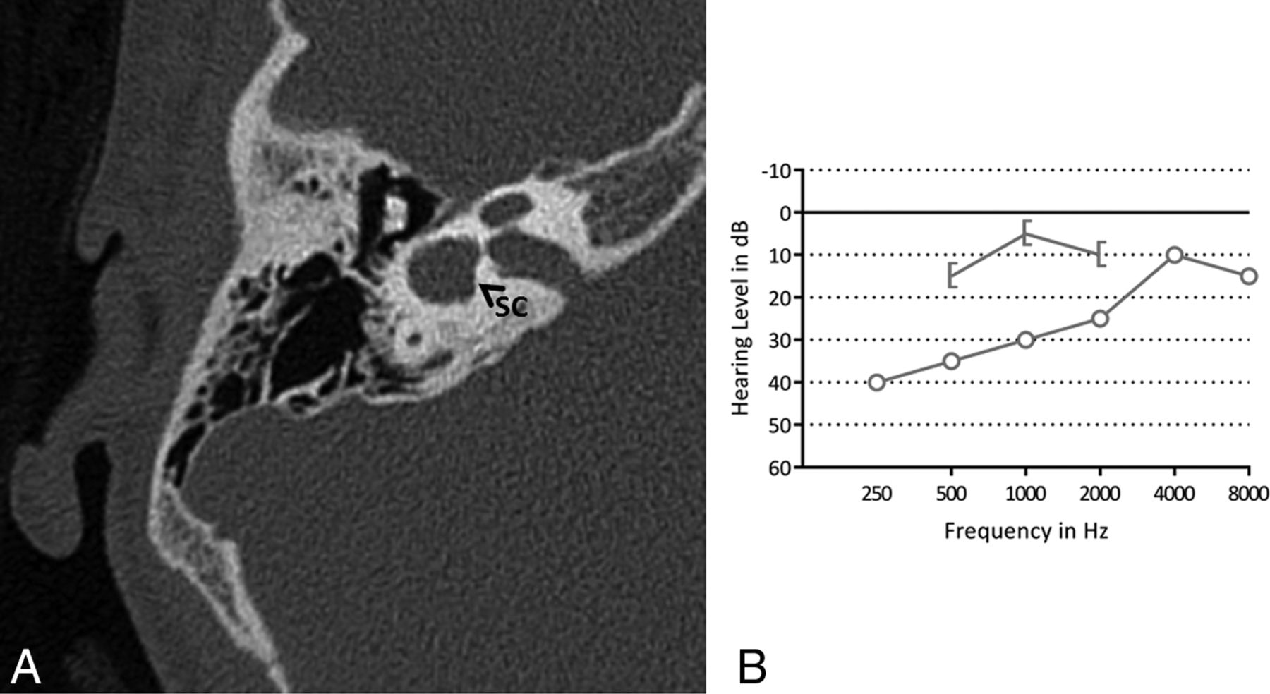
- Fig 5.
A, Axial CT image of the right mastoid bone. The bony island of the lateral semicircular canal is missing, and the canal and vestibule are composed of a single cavity (SC). B, A pure tone audiogram of the same ear shows conductive hearing loss, more pronounced in the low frequencies. The circle indicates an unmasked air-conduction threshold; the bracket, a masked bone-conduction threshold.
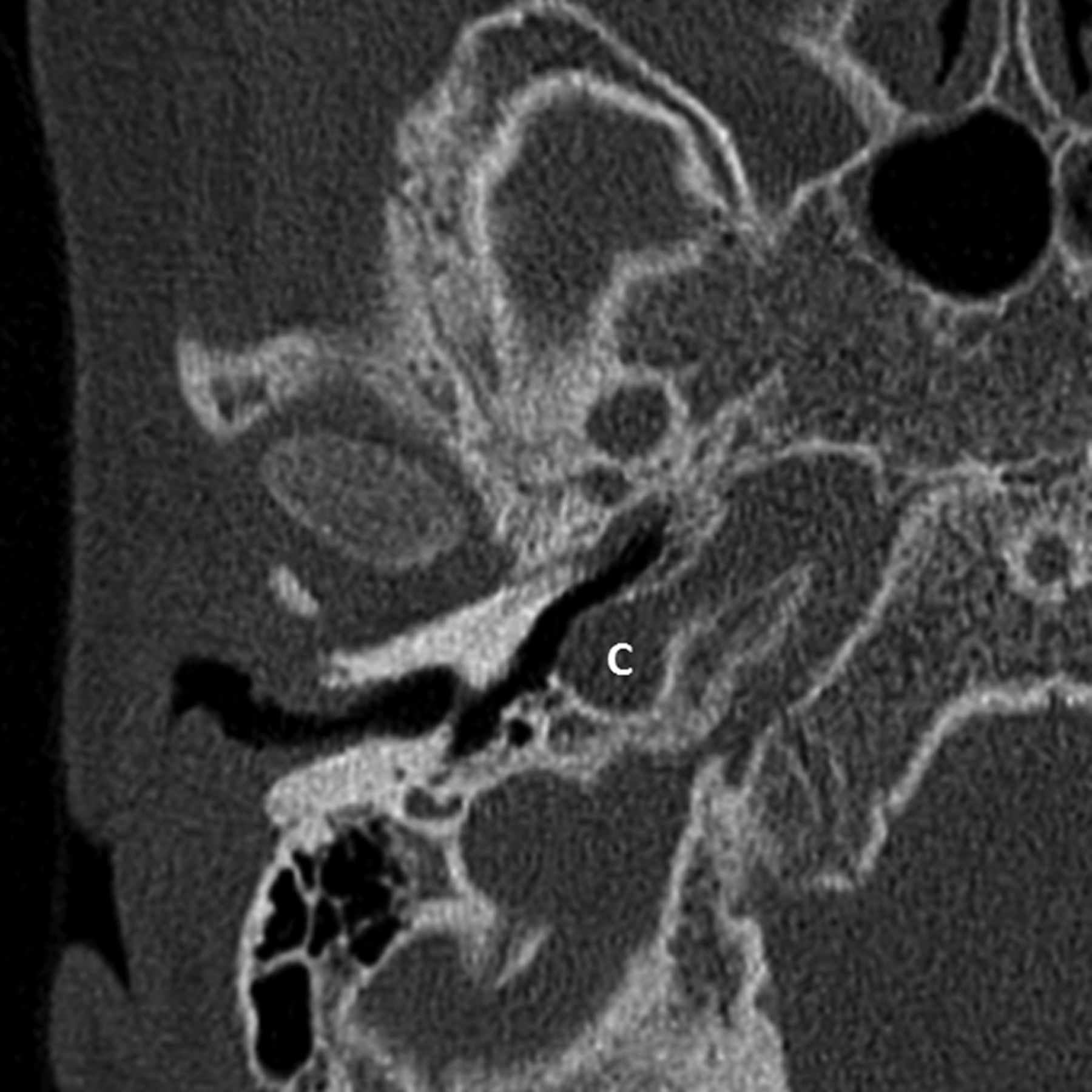
- Fig 6.
Axial CT image of the right mastoid bone showing a carotid canal dehiscence (C).


Tables
Malformation PTA Tympanometrya No HL (%) HL Type C (%) SN (%) M (%) PTA Range in dB A B C As Ad LV Dense malleus 2 (100) – – – – – – – – – – Dense stapes superstructure 7 (39) 6 (33) 1 (6) 4 (22) 21–100 3 3 1 3 – 2 IP type II 5 (42) 3 (25) 1 (8)b 3 (25) 38–100 LSCC: small bony island 7 (41) 4 (24) 3 (18)b 3 (18)b 35–100 LSCC: single cavity 6 (40) 5 (33) 2 (13) 2 (13) 25–96 Note:—HL indicates hearing loss; C, conductive; SN, sensorineural; M, mixed; PTA, pure tone average; A, normal situation; B, flat curve; C, negative peak pressure; As, small pressure peak; Ad, high pressure peak; LV, large volume; IP, incomplete partition.
↵a The number of ears does not amount to the total number of ears with a malformation due to missing data.
↵b Including 1 ear measured with visual reinforcement audiometry and brain stem evoked response audiometry.
- Table 2:
Overview of number of ears with middle and/or inner ear abnormalities reported by Loos et al16 and in the present study
Malformation Loos et al16; No. of Ears (%) (Total, 22 Ears) Present study; No. of Ears (%) (Total, 52 Ears) Total No. of Ears (%) (Total, 74 Ears) Malleus, incus, or stapes abnormalitiesa 2 (9) 3 (6b) 5 (7c) Dense stapes superstructure 10 (45) 18 (36b) 28 (39c) LSCC: single cavity 4 (18) 15 (29) 19 (26) Wide vestibule/small bony island in LSCC 14 (64) 17 (33) 31 (42) IP type II 12 (55) 12 (23) 24 (32) Carotid canal dehiscence 2 (9) 5 (10b) 7 (10c) Note:—IP indicates incomplete partition.
↵a Other than a dense stapes superstructure.
↵b Calculated from a total of 50 ears; bone structures in 2 ears in the present study could not be assessed on MRI.
↵c Calculated from a total of 72 ears; bone structures in 2 ears in the present study could not be assessed on MRI.


Jump to section
Related Articles
Cited By...
- No citing articles found.