Advertisement

Main menu

  • Home
  • Content
    • Current Issue
    • Accepted Manuscripts
    • Article Preview
    • Past Issue Archive
    • Video Articles
    • AJNR Case Collection
    • Case of the Week Archive
    • Case of the Month Archive
    • Classic Case Archive
  • Special Collections
    • AJNR Awards
    • Low-Field MRI
    • Alzheimer Disease
    • ASNR Foundation Special Collection
    • Photon-Counting CT
    • View All
  • Multimedia
    • AJNR Podcasts
    • AJNR SCANtastic
    • Trainee Corner
    • MRI Safety Corner
    • Imaging Protocols
  • For Authors
    • Submit a Manuscript
    • Submit a Video Article
    • Submit an eLetter to the Editor/Response
    • Manuscript Submission Guidelines
    • Statistical Tips
    • Fast Publishing of Accepted Manuscripts
    • Graphical Abstract Preparation
    • Imaging Protocol Submission
    • Author Policies
  • About Us
    • About AJNR
    • Editorial Board
    • Editorial Board Alumni
  • More
    • Become a Reviewer/Academy of Reviewers
    • Subscribers
    • Permissions
    • Alerts
    • Feedback
    • Advertisers
    • ASNR Home

User menu

  • Alerts
  • Log in

Search

  • Advanced search
American Journal of Neuroradiology

American Journal of Neuroradiology

ASHNR American Society of Functional Neuroradiology ASHNR American Society of Pediatric Neuroradiology ASSR
  • Alerts
  • Log in

Advanced Search

  • Home
  • Content
    • Current Issue
    • Accepted Manuscripts
    • Article Preview
    • Past Issue Archive
    • Video Articles
    • AJNR Case Collection
    • Case of the Week Archive
    • Case of the Month Archive
    • Classic Case Archive
  • Special Collections
    • AJNR Awards
    • Low-Field MRI
    • Alzheimer Disease
    • ASNR Foundation Special Collection
    • Photon-Counting CT
    • View All
  • Multimedia
    • AJNR Podcasts
    • AJNR SCANtastic
    • Trainee Corner
    • MRI Safety Corner
    • Imaging Protocols
  • For Authors
    • Submit a Manuscript
    • Submit a Video Article
    • Submit an eLetter to the Editor/Response
    • Manuscript Submission Guidelines
    • Statistical Tips
    • Fast Publishing of Accepted Manuscripts
    • Graphical Abstract Preparation
    • Imaging Protocol Submission
    • Author Policies
  • About Us
    • About AJNR
    • Editorial Board
    • Editorial Board Alumni
  • More
    • Become a Reviewer/Academy of Reviewers
    • Subscribers
    • Permissions
    • Alerts
    • Feedback
    • Advertisers
    • ASNR Home
  • Follow AJNR on Twitter
  • Visit AJNR on Facebook
  • Follow AJNR on Instagram
  • Join AJNR on LinkedIn
  • RSS Feeds

AJNR Awards, New Junior Editors, and more. Read the latest AJNR updates

LetterLetter

Multiparametric Imaging Improves Confidence in the Diagnosis of Multinodular and Vacuolating Neuronal Tumor of the Cerebrum

A. Lecler, D. Chauvet, H.A. Biassette and J. Savatovsky
American Journal of Neuroradiology February 2018, 39 (2) E32-E33; DOI: https://doi.org/10.3174/ajnr.A5425
A. Lecler
aDepartment of Radiology
  • Find this author on Google Scholar
  • Find this author on PubMed
  • Search for this author on this site
  • ORCID record for A. Lecler
D. Chauvet
bDepartment of Neurosurgery Fondation Ophtalmologique Adolphe de Rothschild Paris, France
  • Find this author on Google Scholar
  • Find this author on PubMed
  • Search for this author on this site
  • ORCID record for D. Chauvet
H.A. Biassette
cDepartment of Pathology Lariboisière Hospital, Paris-Diderot, Paris-Cité-Sorbonne University Paris, France
  • Find this author on Google Scholar
  • Find this author on PubMed
  • Search for this author on this site
  • ORCID record for H.A. Biassette
J. Savatovsky
dDepartment of Radiology Fondation Ophtalmologique Adolphe de Rothschild Paris, France
  • Find this author on Google Scholar
  • Find this author on PubMed
  • Search for this author on this site
  • ORCID record for J. Savatovsky
  • Article
  • Figures & Data
  • Info & Metrics
  • Responses
  • References
  • PDF
Loading

We read with much interest the article by Nunes et al1 in the July 2017 issue of the American Journal of Neuroradiology, in which they presented an impressive series of 33 confirmed or presumed multinodular and vacuolating neuronal tumors (MVNT) of the cerebrum, an entity that has only recently been categorized in the 2016 revision of the World Health Organization Classification of Tumors of the Central Nervous System.2⇓–4 Only a few cases of MVNT have been reported in the literature in the past 4 years with confirmed pathologic correlations.1⇓–3

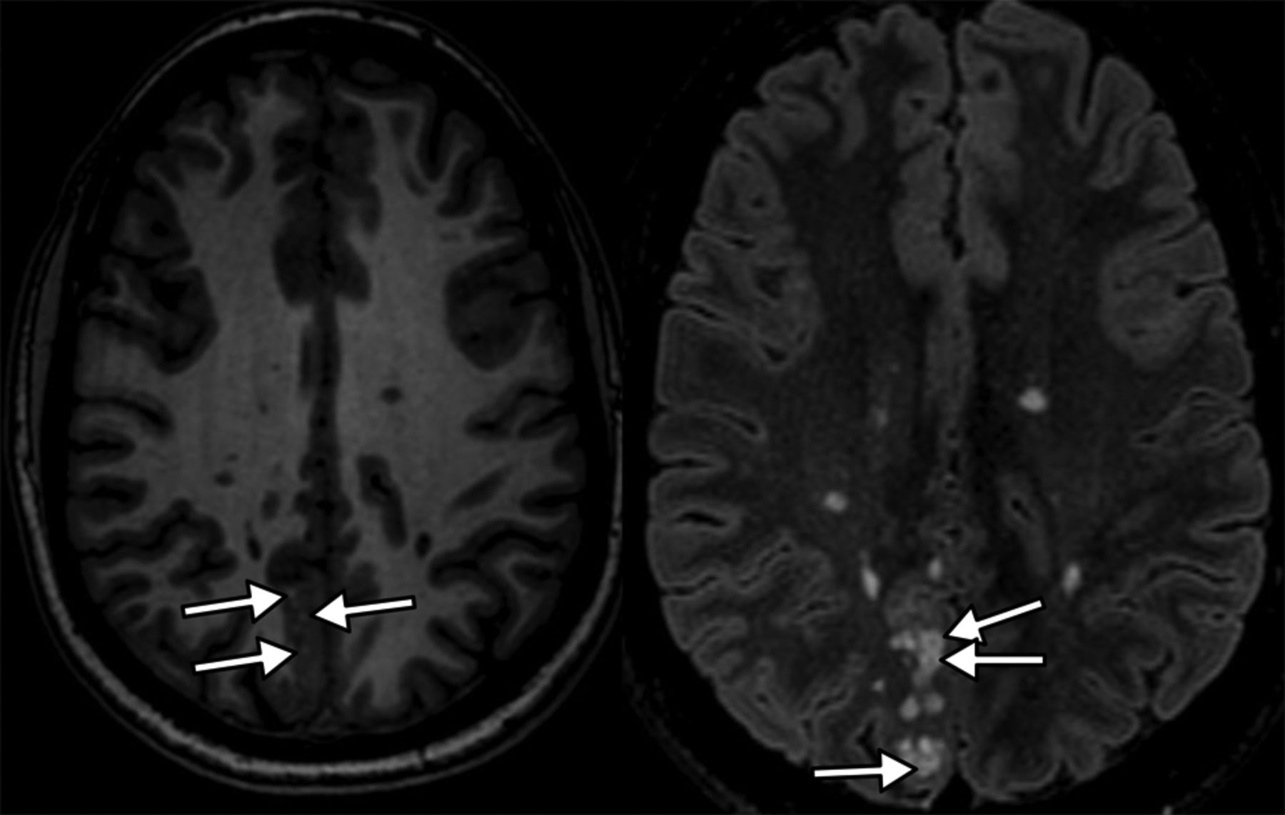
The authors presented a detailed description of the entity, which involves the deep cortical ribbon and the adjacent subcortical white matter and is distributed into small coalescent nodules, hyperintense on FLAIR and T2-weighted imaging and hypointense on T1WI. On our 3T MR imaging, we noticed that these lesions were not homogeneous but had a central FLAIR hyposignal as well as central punctiform T1 hyperintensities (white arrows, Fig 1), probably related to a high protein or solid component within the vacuolated areas. This specific feature was present in all our patients but requires reasonably high resolution to be seen. This simple sign can improve diagnostic confidence.

Fig 1.
  • Download figure
  • Open in new tab
  • Download powerpoint
Fig 1.

Axial T1 (left) and FLAIR (right) weighted imaging showing a right parietal lesion involving the deep cortical ribbon and the adjacent subcortical white matter distributed into small coalescent nodules, hyperintense on FLAIR, hypointense with punctiform hyperintensities on T1WI, and central hypointensities on FLAIR (arrow), characteristics of MVNT.

We believe that radiologists should always perform multiparametric imaging to characterize brain abnormalities consistent with an MVNT with a high-field MR imaging whenever possible. In our experience, diffusion-weighted imaging shows no restricted diffusion inside the lesion, and susceptibility-weighted imaging does not show an intratumoral susceptibility signal or intralesional hemorrhaging. Perfusion sequences such as dynamic-susceptibility contrast or arterial spin-labeling show no hyperperfusion, increasing neither cerebral blood volume nor cerebral blood flow. MR spectroscopy may show a mild decrease of NAA but no choline peak or lipid or lactate peaks, which might suggest malignancy (Fig 2). Multivoxel MR spectroscopy shows no abnormality in the surrounding brain parenchyma. All these features improve diagnostic confidence and support strong arguments in favor of this presumptive diagnosis, thus avoiding a potentially risky surgical biopsy.

Fig 2.
  • Download figure
  • Open in new tab
  • Download powerpoint
Fig 2.

MR spectroscopy shows a mild decrease of N-acetylaspartate but no choline or lipids peaks.

We think that this systematic approach best serves patients because this entity is provisionally considered a brain tumor by the World Health Organization and should be best understood using state-of-the-art MR imaging characterization. This management allows clinicians to reassure their patients who are often frightened by the term “tumor” and sometimes request surgical removal. In our experience, which is further echoed in the article, long-term follow-up MR imaging performed >10 years after the first discovery of a presumed MVNT shows perfect stability of brain abnormalities with rare or absent clinical symptoms.

We entirely agree with the authors' conclusion suggesting that MVNT is more likely a malformative lesion as opposed to a neoplasm. Clinical and histologic analysis have not yet provided clear evidence for a neoplastic process. Molecular investigations in few cases have so far failed to reveal IDH1/2, BRAF V600E, EGFR, ERBB2, KRAS, BRAF, NRAS, PIK3CA, and AKT1 mutations or obvious DNA copy number abnormalities. A point mutation involving MEK1 (MAP2K1) p. Q56P (c.167A>C) in non-small-cell lung cancers has been found in 1 case.

Thus, we believe that the name of this entity should be changed to remove the term “tumor” and replaced by the term “hamartoma,” “dysplasia,” or “malformation.” We also believe that larger series are needed to determine at what point the entity will be considered malignant.

Footnotes

  • Disclosures: Homa A. Biassette—UNRELATED: Grants/Grants Pending: DEVELAGE FP7 grant,* Comments: 2012–2015 “DEVELAGE: Pathways Common to Brain Development and Ageing: Defining Strategies for Preventive Therapy and Diagnostics.” Julien Savatovsky—UNRELATED: Grant: Association de la Recherche Sur le Syndrome Immuno-Deficitaire Acquis; Consulting Fee or Honorarium: Association de la Recherche Sur le Syndrome Immuno-Deficitaire Acquis, Madday Pharmaceutical, Bristol-Myers Squibb, GlaxoSmithKline; Support for Travel to Meetings for the Study or Other Purposes: GE Healthcare, Philips Healthcare, Bayer HealthCare; Expert Testimony: Philips Healthcare, Bayer HealthCare*; Payment for Lectures Including Service on Speakers Bureaus: Philips Healthcare, Association de la Recherche Sur le Syndrome Immuno-Deficitaire Acquis, Biogen.* *Money paid to the institution.

References

  1. 1.↵
    1. Nunes RH,
    2. Hsu CC,
    3. da Rocha AJ, et al
    . Multinodular and vacuolating neuronal tumor of the cerebrum: a new “leave me alone” lesion with a characteristic imaging pattern. AJNR Am J Neuroradiol 2017;38:1899–904 doi:10.3174/ajnr.A5281 pmid:28705817
    Abstract/FREE Full Text
  2. 2.↵
    1. Fukushima S,
    2. Yoshida A,
    3. Narita Y, et al
    . Multinodular and vacuolating neuronal tumor of the cerebrum. Brain Tumor Pathol 2015;32:131–36 doi:10.1007/s10014-014-0198-9 pmid:25146549
    CrossRefPubMed
  3. 3.↵
    1. Huse JT,
    2. Edgar M,
    3. Halliday J, et al
    . Multinodular and vacuolating neuronal tumors of the cerebrum: 10 cases of a distinctive seizure-associated lesion. Brain Pathol 2013;23:515–24 doi:10.1111/bpa.12035 pmid:23324039
    CrossRefPubMed
  4. 4.↵
    1. Louis DN,
    2. Perry A,
    3. Reifenberger G, et al
    . The 2016 World Health Organization Classification of Tumors of the Central Nervous System: a summary. Acta Neuropathol 2016;131:803–20 doi:10.1007/s00401-016-1545-1 pmid:27157931
    CrossRefPubMed
  • © 2018 by American Journal of Neuroradiology
PreviousNext
Back to top

In this issue

American Journal of Neuroradiology: 39 (2)
American Journal of Neuroradiology
Vol. 39, Issue 2
1 Feb 2018
  • Table of Contents
  • Index by author
  • Complete Issue (PDF)
Advertisement
Print
Download PDF
Email Article

Thank you for your interest in spreading the word on American Journal of Neuroradiology.

NOTE: We only request your email address so that the person you are recommending the page to knows that you wanted them to see it, and that it is not junk mail. We do not capture any email address.

Enter multiple addresses on separate lines or separate them with commas.
Multiparametric Imaging Improves Confidence in the Diagnosis of Multinodular and Vacuolating Neuronal Tumor of the Cerebrum
(Your Name) has sent you a message from American Journal of Neuroradiology
(Your Name) thought you would like to see the American Journal of Neuroradiology web site.
CAPTCHA
This question is for testing whether or not you are a human visitor and to prevent automated spam submissions.
Cite this article
A. Lecler, D. Chauvet, H.A. Biassette, J. Savatovsky
Multiparametric Imaging Improves Confidence in the Diagnosis of Multinodular and Vacuolating Neuronal Tumor of the Cerebrum
American Journal of Neuroradiology Feb 2018, 39 (2) E32-E33; DOI: 10.3174/ajnr.A5425

Citation Manager Formats

  • BibTeX
  • Bookends
  • EasyBib
  • EndNote (tagged)
  • EndNote 8 (xml)
  • Medlars
  • Mendeley
  • Papers
  • RefWorks Tagged
  • Ref Manager
  • RIS
  • Zotero
0 Responses
Respond to this article
Share
Bookmark this article
Multiparametric Imaging Improves Confidence in the Diagnosis of Multinodular and Vacuolating Neuronal Tumor of the Cerebrum
A. Lecler, D. Chauvet, H.A. Biassette, J. Savatovsky
American Journal of Neuroradiology Feb 2018, 39 (2) E32-E33; DOI: 10.3174/ajnr.A5425
del.icio.us logo Twitter logo Facebook logo Mendeley logo
  • Tweet Widget
  • Facebook Like
  • Google Plus One
Purchase

Jump to section

  • Article
    • Footnotes
    • References
  • Figures & Data
  • Info & Metrics
  • Responses
  • References
  • PDF

Related Articles

  • 3D Ultrasound for Imaging and Quantifying Carotid Ulcers
  • PubMed
  • Google Scholar

Cited By...

  • Cortically Based Brain Tumors in Children: A Decision-Tree Approach in the Radiology Reading Room
  • Crossref (8)
  • Google Scholar

This article has been cited by the following articles in journals that are participating in Crossref Cited-by Linking.

  • 2021 WHO classification of tumours of the central nervous system: a review for the neuroradiologist
    Cillian McNamara, Kshitij Mankad, Stefanie Thust, Luke Dixon, Clara Limback-Stanic, Felice D’Arco, Thomas S. Jacques, Ulrike Löbel
    Neuroradiology 2022 64 10
  • Advanced multiparametric magnetic resonance imaging of multinodular and vacuolating neuronal tumor
    A. Lecler, V. Broquet, J. Bailleux, B. Carsin, H. Adle‐Biassette, S. Baloglu, G. Forestier, F. Bonneville, E. Calvier, D. Chauvet, P. O. Comby, J. P. Cottier, F. Cotton, R. Deschamps, C. Diard‐Detoeuf, F. Ducray, C. Drissi, M. Elmaleh, J. Farras, J. Aguilar Garcia, E. Gerardin, S. Grand, D. C. Jianu, S. Kremer, H. Loiseau, N. Magne, M. Mejdoubi, A. Moulignier, M. Ollivier, S. Nagi, M. Rodallec, N. Shor, T. Tourdias, C. Vandendries, R. Anxionnat, L. Duron, J. Savatovsky
    European Journal of Neurology 2020 27 8
  • The 2021 WHO Classification for Gliomas and Implications on Imaging Diagnosis: Part 3—Summary of Imaging Findings on Glioneuronal and Neuronal Tumors
    Yae Won Park, Philipp Vollmuth, Martha Foltyn‐Dumitru, Felix Sahm, Kyu Sung Choi, Ji Eun Park, Sung Soo Ahn, Jong Hee Chang, Se Hoon Kim
    Journal of Magnetic Resonance Imaging 2023 58 6
  • A multinodular and vacuolating neuronal tumor of the cerebrum (MVNT) with glioma-like appearance
    Daniele Bagatto, Tamara Ius, Enrico Pegolo, Mauro Morassi
    Acta Neurologica Belgica 2021 121 6
  • Atypical Presentation of Multinodular and Vacuolating Neuronal Tumor of the Cerebrum in a Boy
    Austin L. Turner, Preston D’Souza, Muhittin Belirgen, Mohamad M. Al-Rahawan
    Journal of Neurosciences in Rural Practice 2019 11
  • Multinodular and Vacuolating Neuronal Tumors with Suggested Slow Progression
    Hiroshi Ito, Yukiko Nakahara, Tomihiro Wakamiya, Motofumi Koguchi, Fumitaka Yoshioka, Kohei Inoue, Atsushi Ogata, Jun Masuoka, Tatsuya Abe
    Japanese Journal of Neurosurgery 2020 29 8
  • Cortically Based Brain Tumors in Children: A Decision-Tree Approach in the Radiology Reading Room
    V. Rameh, U. Löbel, F. D’Arco, A. Bhatia, K. Mankad, T.Y. Poussaint, C.A. Alves
    American Journal of Neuroradiology 2025 46 1
  • Radiopaedia.org
    Alexandra Murphy
    2019

More in this TOC Section

  • Reply:
  • Brain AVM’s Nidus: What if We Hadn’t Understood Anything?
  • Letter to the Editor regarding “Automated Volumetric Software in Dementia: Help or Hindrance to the Neuroradiologist?”
Show more LETTERS

Similar Articles

Advertisement

Indexed Content

  • Current Issue
  • Accepted Manuscripts
  • Article Preview
  • Past Issues
  • Editorials
  • Editor's Choice
  • Fellows' Journal Club
  • Letters to the Editor
  • Video Articles

Cases

  • Case Collection
  • Archive - Case of the Week
  • Archive - Case of the Month
  • Archive - Classic Case

More from AJNR

  • Trainee Corner
  • Imaging Protocols
  • MRI Safety Corner
  • Book Reviews

Multimedia

  • AJNR Podcasts
  • AJNR Scantastics

Resources

  • Turnaround Time
  • Submit a Manuscript
  • Submit a Video Article
  • Submit an eLetter to the Editor/Response
  • Manuscript Submission Guidelines
  • Statistical Tips
  • Fast Publishing of Accepted Manuscripts
  • Graphical Abstract Preparation
  • Imaging Protocol Submission
  • Evidence-Based Medicine Level Guide
  • Publishing Checklists
  • Author Policies
  • Become a Reviewer/Academy of Reviewers
  • News and Updates

About Us

  • About AJNR
  • Editorial Board
  • Editorial Board Alumni
  • Alerts
  • Permissions
  • Not an AJNR Subscriber? Join Now
  • Advertise with Us
  • Librarian Resources
  • Feedback
  • Terms and Conditions
  • AJNR Editorial Board Alumni

American Society of Neuroradiology

  • Not an ASNR Member? Join Now

© 2025 by the American Society of Neuroradiology All rights, including for text and data mining, AI training, and similar technologies, are reserved.
Print ISSN: 0195-6108 Online ISSN: 1936-959X

Powered by HighWire