Advertisement

Main menu

  • Home
  • Content
    • Current Issue
    • Accepted Manuscripts
    • Article Preview
    • Past Issue Archive
    • Video Articles
    • AJNR Case Collection
    • Case of the Week Archive
    • Case of the Month Archive
    • Classic Case Archive
  • Special Collections
    • AJNR Awards
    • Low-Field MRI
    • Alzheimer Disease
    • ASNR Foundation Special Collection
    • Photon-Counting CT
    • View All
  • Multimedia
    • AJNR Podcasts
    • AJNR SCANtastic
    • Trainee Corner
    • MRI Safety Corner
    • Imaging Protocols
  • For Authors
    • Submit a Manuscript
    • Submit a Video Article
    • Submit an eLetter to the Editor/Response
    • Manuscript Submission Guidelines
    • Statistical Tips
    • Fast Publishing of Accepted Manuscripts
    • Graphical Abstract Preparation
    • Imaging Protocol Submission
    • Author Policies
  • About Us
    • About AJNR
    • Editorial Board
    • Editorial Board Alumni
  • More
    • Become a Reviewer/Academy of Reviewers
    • Subscribers
    • Permissions
    • Alerts
    • Feedback
    • Advertisers
    • ASNR Home

User menu

  • Alerts
  • Log in

Search

  • Advanced search
American Journal of Neuroradiology

American Journal of Neuroradiology

ASHNR American Society of Functional Neuroradiology ASHNR American Society of Pediatric Neuroradiology ASSR
  • Alerts
  • Log in

Advanced Search

  • Home
  • Content
    • Current Issue
    • Accepted Manuscripts
    • Article Preview
    • Past Issue Archive
    • Video Articles
    • AJNR Case Collection
    • Case of the Week Archive
    • Case of the Month Archive
    • Classic Case Archive
  • Special Collections
    • AJNR Awards
    • Low-Field MRI
    • Alzheimer Disease
    • ASNR Foundation Special Collection
    • Photon-Counting CT
    • View All
  • Multimedia
    • AJNR Podcasts
    • AJNR SCANtastic
    • Trainee Corner
    • MRI Safety Corner
    • Imaging Protocols
  • For Authors
    • Submit a Manuscript
    • Submit a Video Article
    • Submit an eLetter to the Editor/Response
    • Manuscript Submission Guidelines
    • Statistical Tips
    • Fast Publishing of Accepted Manuscripts
    • Graphical Abstract Preparation
    • Imaging Protocol Submission
    • Author Policies
  • About Us
    • About AJNR
    • Editorial Board
    • Editorial Board Alumni
  • More
    • Become a Reviewer/Academy of Reviewers
    • Subscribers
    • Permissions
    • Alerts
    • Feedback
    • Advertisers
    • ASNR Home
  • Follow AJNR on Twitter
  • Visit AJNR on Facebook
  • Follow AJNR on Instagram
  • Join AJNR on LinkedIn
  • RSS Feeds

AJNR Awards, New Junior Editors, and more. Read the latest AJNR updates

Research ArticleHead and Neck Imaging
Open Access

Imaging and Surgical Findings in Patients with Hemi-Laryngopharyngeal Spasm and the Potential Role of MRI in the Diagnostic Work-Up

J. Avecillas-Chasin, M.G. Kozoriz, J.R. Shewchuk, M.K.S. Heran and C.R. Honey
American Journal of Neuroradiology December 2018, 39 (12) 2366-2370; DOI: https://doi.org/10.3174/ajnr.A5851
J. Avecillas-Chasin
aFrom the Department of Surgery (J.A.-C., C.R.H.), Division of Neurosurgery
  • Find this author on Google Scholar
  • Find this author on PubMed
  • Search for this author on this site
  • ORCID record for J. Avecillas-Chasin
M.G. Kozoriz
bDepartment of Radiology (M.G.K., J.R.S., M.K.S.H.), University of British Columbia, Vancouver, British Columbia, Canada
  • Find this author on Google Scholar
  • Find this author on PubMed
  • Search for this author on this site
  • ORCID record for M.G. Kozoriz
J.R. Shewchuk
bDepartment of Radiology (M.G.K., J.R.S., M.K.S.H.), University of British Columbia, Vancouver, British Columbia, Canada
  • Find this author on Google Scholar
  • Find this author on PubMed
  • Search for this author on this site
  • ORCID record for J.R. Shewchuk
M.K.S. Heran
bDepartment of Radiology (M.G.K., J.R.S., M.K.S.H.), University of British Columbia, Vancouver, British Columbia, Canada
  • Find this author on Google Scholar
  • Find this author on PubMed
  • Search for this author on this site
  • ORCID record for M.K.S. Heran
C.R. Honey
aFrom the Department of Surgery (J.A.-C., C.R.H.), Division of Neurosurgery
cVancouver General Hospital (C.R.H.), Vancouver, British Columbia, Canada.
  • Find this author on Google Scholar
  • Find this author on PubMed
  • Search for this author on this site
  • ORCID record for C.R. Honey
  • Article
  • Figures & Data
  • Info & Metrics
  • Responses
  • References
  • PDF
Loading

Article Figures & Data

Figures

  • Tables
  • Fig 1.
    • Download figure
    • Open in new tab
    • Download powerpoint
    Fig 1.

    Grading system. The vagus nerve was rated separately for the proximal (3 mm from the brain stem) and the distal portions. A, Grade I: “no vessel contact” (white arrow). B, Grade II: “contact,” which was defined as no visible CSF between the blood vessel and the nerve but no displacement of the normal trajectory of the nerve (white arrow). C, Grade III: “compression,” which was defined as displacement of the normal trajectory of the nerve (white arrow). D, Grade I: Oblique projection showing the vagus nerve (white arrow) in contact with the PICA (black arrow). The glossopharyngeal nerve (gray arrow) is shown above the vagus nerve. E, Oblique projection of the IX/X complex close to the jugular foramen. It was possible to see the upper glossopharyngeal nerve more anteriorly and superiorly moving lateral into the foramen, and the vagus nerve branches were below this.
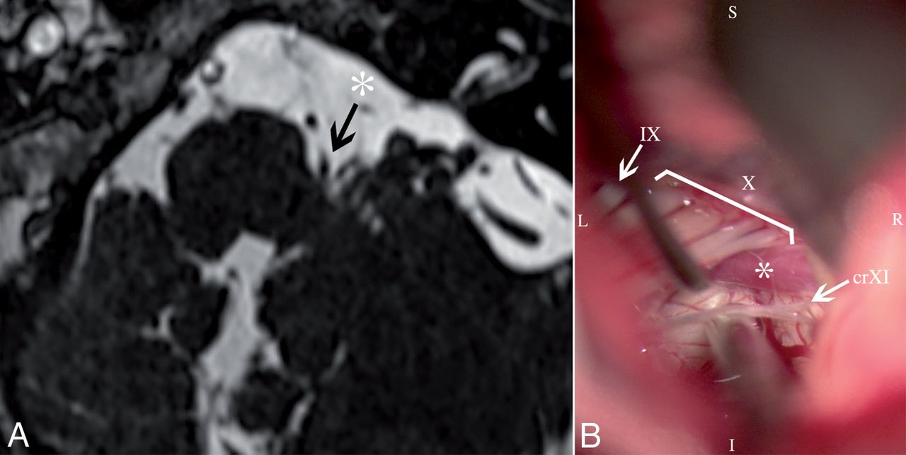
  • Fig 2.
    • Download figure
    • Open in new tab
    • Download powerpoint
    Fig 2.

    Patient 1. A, MR imaging shows neurovascular conflict of the PICA (arrow) with the proximal part of the vagus nerve. B, Intraoperative findings show the loop of the PICA (asterisk) pulsating against the caudal rootlets of the vagus nerve (X). IX indicates glossopharyngeal nerve; crXI, cranial root of the accessory nerve; S, superior; I, inferior; R, right; L, left.
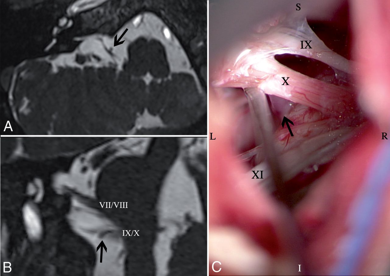
  • Fig 3.
    • Download figure
    • Open in new tab
    • Download powerpoint
    Fig 3.

    Patient 2. A, Axial view of MR imaging shows compression of the vagus nerve by the right PICA from the anterior direction (arrow). B, The coronal-oblique plane shows the vessel loop of the PICA (arrow). VII/VIII indicates the facial and vestibulocochlear nerve complex; IX/X, the glossopharyngeal and vagus nerve complex. C, Intraoperative findings confirm that the vessel loop of the PICA (arrow) is pulsating against the anterior aspect of the vagus nerve (X). IX indicates the glossopharyngeal nerve; XI, the accessory nerve; S, superior; I, inferior; R, right; L, left.
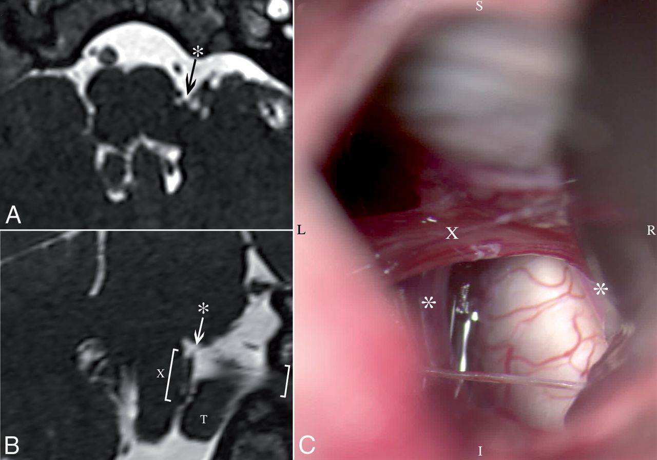
  • Fig 4.
    • Download figure
    • Open in new tab
    • Download powerpoint
    Fig 4.

    Case 3. A, Axial view of MR imaging shows neurovascular conflict of the left PICA (asterisk) and the vagus nerve. B, The coronal-oblique plane shows the loop of the left PICA (asterisk) in contact with the proximal part of the rootlets of the vagus nerve (X), which is also in contact with the left cerebellar tonsil (T). C, Intraoperative findings. After gentle retraction of the cerebellum and part of the left tonsil, the left PICA loop (asterisk) is found in contact with the proximal part of the vagus nerve (X). S indicates superior; I, inferior; R, right; L, left.

Tables

  • Figures
  • Clinical and demographic data of our patients with HELPS

    Patient No.Age/SexSymptomsOffending VesselGrading of NVC
    165/MEpisodic throat contractions and coughingLeft PICA2
    243/FEpisodic throat contractions, coughing, and vocal changesRight PICA3
    332/FEpisodic left-sided throat contractions, choking, and vocal changesLeft PICA2
    • Note:—NVC indicates neurovascular conflict.

PreviousNext
Back to top

In this issue

American Journal of Neuroradiology: 39 (12)
American Journal of Neuroradiology
Vol. 39, Issue 12
1 Dec 2018
  • Table of Contents
  • Index by author
  • Complete Issue (PDF)
Advertisement
Print
Download PDF
Email Article

Thank you for your interest in spreading the word on American Journal of Neuroradiology.

NOTE: We only request your email address so that the person you are recommending the page to knows that you wanted them to see it, and that it is not junk mail. We do not capture any email address.

Enter multiple addresses on separate lines or separate them with commas.
Imaging and Surgical Findings in Patients with Hemi-Laryngopharyngeal Spasm and the Potential Role of MRI in the Diagnostic Work-Up
(Your Name) has sent you a message from American Journal of Neuroradiology
(Your Name) thought you would like to see the American Journal of Neuroradiology web site.
CAPTCHA
This question is for testing whether or not you are a human visitor and to prevent automated spam submissions.
Cite this article
J. Avecillas-Chasin, M.G. Kozoriz, J.R. Shewchuk, M.K.S. Heran, C.R. Honey
Imaging and Surgical Findings in Patients with Hemi-Laryngopharyngeal Spasm and the Potential Role of MRI in the Diagnostic Work-Up
American Journal of Neuroradiology Dec 2018, 39 (12) 2366-2370; DOI: 10.3174/ajnr.A5851

Citation Manager Formats

  • BibTeX
  • Bookends
  • EasyBib
  • EndNote (tagged)
  • EndNote 8 (xml)
  • Medlars
  • Mendeley
  • Papers
  • RefWorks Tagged
  • Ref Manager
  • RIS
  • Zotero
0 Responses
Respond to this article
Share
Bookmark this article
Imaging and Surgical Findings in Patients with Hemi-Laryngopharyngeal Spasm and the Potential Role of MRI in the Diagnostic Work-Up
J. Avecillas-Chasin, M.G. Kozoriz, J.R. Shewchuk, M.K.S. Heran, C.R. Honey
American Journal of Neuroradiology Dec 2018, 39 (12) 2366-2370; DOI: 10.3174/ajnr.A5851
del.icio.us logo Twitter logo Facebook logo Mendeley logo
  • Tweet Widget
  • Facebook Like
  • Google Plus One
Purchase

Jump to section

  • Article
    • Abstract
    • ABBREVIATIONS:
    • MATERIALS and Methods
    • Results
    • Discussion
    • Conclusions
    • References
  • Figures & Data
  • Info & Metrics
  • Responses
  • References
  • PDF

Related Articles

  • No related articles found.
  • PubMed
  • Google Scholar

Cited By...

  • No citing articles found.
  • Crossref (7)
  • Google Scholar

This article has been cited by the following articles in journals that are participating in Crossref Cited-by Linking.

  • Hemi-laryngopharyngeal Spasm (HeLPS): Defining a New Clinical Entity
    Amanda Hu, Murray Morrison, Christopher R. Honey
    Annals of Otology, Rhinology & Laryngology 2020 129 9
  • Supraglottic Botulinum Toxin Improves Symptoms in Patients with Laryngeal Sensory Dysfunction Manifesting as Abnormal Throat Sensation and/or Chronic Refractory Cough
    Daniel Novakovic, Meet Sheth, Thomas Stewart, Katrina Sandham, Catherine Madill, Antonia Chacon, Duy Duong Nguyen
    Journal of Clinical Medicine 2021 10 23
  • Vagus Associated Neurogenic Cough Occurring Due to Unilateral Vascular Encroachment of Its Root: A Case Report and Proof of Concept of VANCOUVER Syndrome
    Christopher R. Honey, Marie T. Krüger, Murray D. Morrison, Baljinder S. Dhaliwal, Amanda Hu
    Annals of Otology, Rhinology & Laryngology 2020 129 5
  • Microvascular Decompression for Concurrent Trigeminal Neuralgia and Hemi-Laryngopharyngeal Spasm: A Case Report
    Michiru Katayama, Shusaku Noro, Bunsho Asayama, Yuki Amano, Masahiro Okuma, Kaori Honjo, Yoshinobu Seo, Hirohiko Nakamura
    Operative Neurosurgery 2023 24 2
  • Episodic symptoms caused by neurovascular conflict of vagus nerve due to hemilateral anterior inferior cerebellar artery
    Hui Lv, Peihong Li, Xiaoyan Long, Zheng Xiao, Luo Zhou
    Journal of Neurology 2023 270 12
  • The silent scream: Unraveling vagal nerve neuralgia; VANCOUVER syndrome and HeLPS
    Erfan Shahabinejad, Amirreza Shakoeizadeh, Mina Jafari, Shayesteh Haghighi, Nima Rafati, Seyyed Mohammad Alipour, Mahla Esmaeili, Behina Salarian, Farid Abbaspour, Florian Roser, Abdulrahman Albakr, Hamid Borghei-Razavi
    Clinical Neurology and Neurosurgery 2025 254
  • The vagal rhizopathies
    Christopher R. Honey
    Frontiers in Neurology 2025 15

More in this TOC Section

  • Hydrops Herniation into the Semicircular Canals
  • DIVr of NPC Predicted Outcome
  • MRI of the Parasellar Ligaments
Show more HEAD AND NECK IMAGING

Similar Articles

Advertisement

Indexed Content

  • Current Issue
  • Accepted Manuscripts
  • Article Preview
  • Past Issues
  • Editorials
  • Editor's Choice
  • Fellows' Journal Club
  • Letters to the Editor
  • Video Articles

Cases

  • Case Collection
  • Archive - Case of the Week
  • Archive - Case of the Month
  • Archive - Classic Case

More from AJNR

  • Trainee Corner
  • Imaging Protocols
  • MRI Safety Corner
  • Book Reviews

Multimedia

  • AJNR Podcasts
  • AJNR Scantastics

Resources

  • Turnaround Time
  • Submit a Manuscript
  • Submit a Video Article
  • Submit an eLetter to the Editor/Response
  • Manuscript Submission Guidelines
  • Statistical Tips
  • Fast Publishing of Accepted Manuscripts
  • Graphical Abstract Preparation
  • Imaging Protocol Submission
  • Evidence-Based Medicine Level Guide
  • Publishing Checklists
  • Author Policies
  • Become a Reviewer/Academy of Reviewers
  • News and Updates

About Us

  • About AJNR
  • Editorial Board
  • Editorial Board Alumni
  • Alerts
  • Permissions
  • Not an AJNR Subscriber? Join Now
  • Advertise with Us
  • Librarian Resources
  • Feedback
  • Terms and Conditions
  • AJNR Editorial Board Alumni

American Society of Neuroradiology

  • Not an ASNR Member? Join Now

© 2025 by the American Society of Neuroradiology All rights, including for text and data mining, AI training, and similar technologies, are reserved.
Print ISSN: 0195-6108 Online ISSN: 1936-959X

Powered by HighWire